. 2019 Feb 4:2019:4235405.
doi: 10.1155/2019/4235405. eCollection 2019.

Protective Effects of Galium verum L. Extract against Cardiac Ischemia/Reperfusion Injury in Spontaneously Hypertensive Rats

Affiliations

Protective Effects of Galium verum L. Extract against Cardiac Ischemia/Reperfusion Injury in Spontaneously Hypertensive Rats

Jovana Bradic et al. Oxid Med Cell Longev. .

Abstract

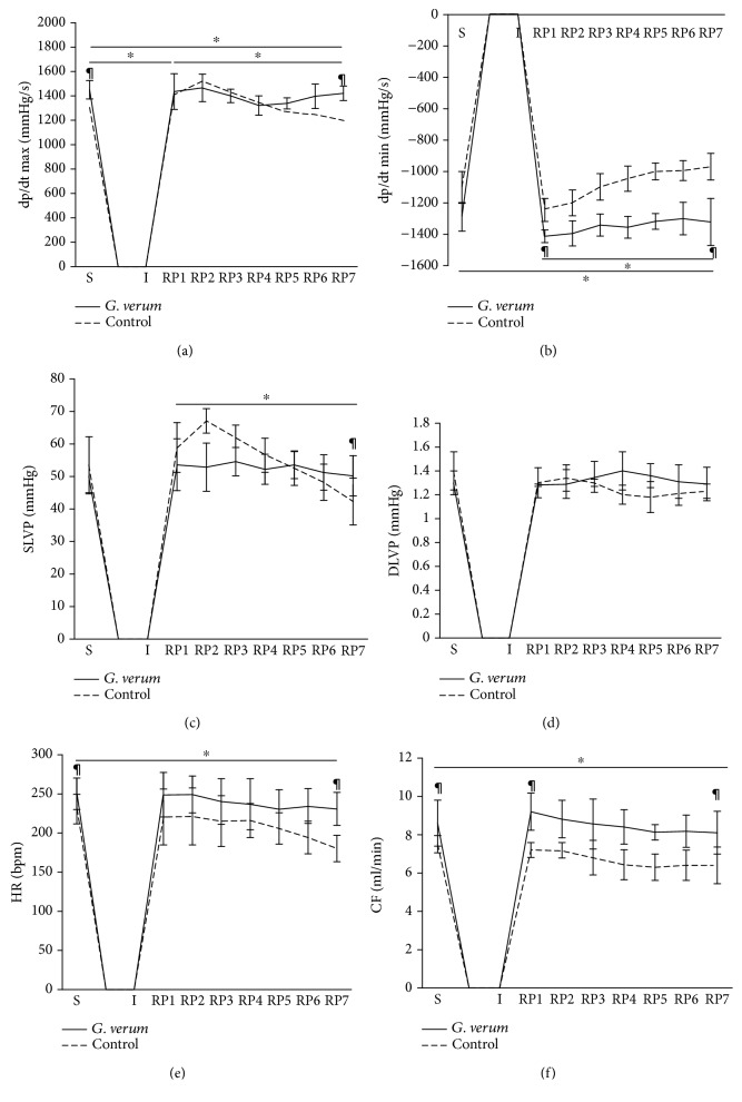
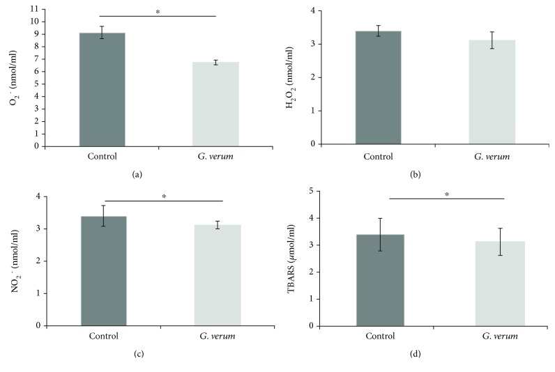
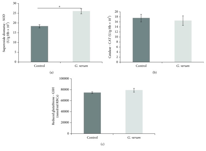
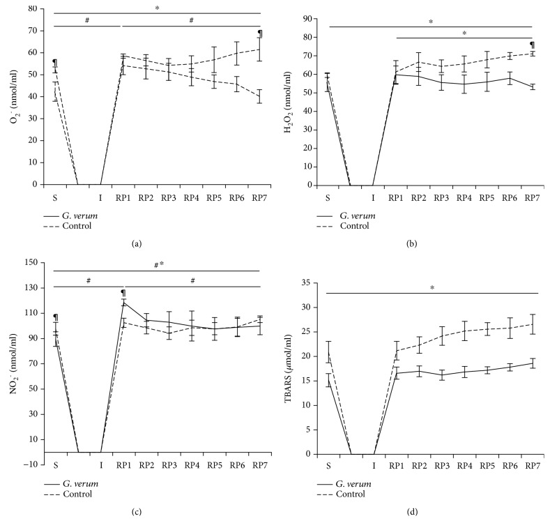
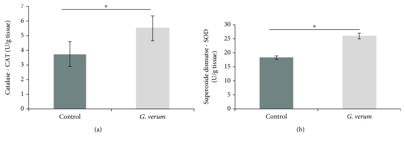
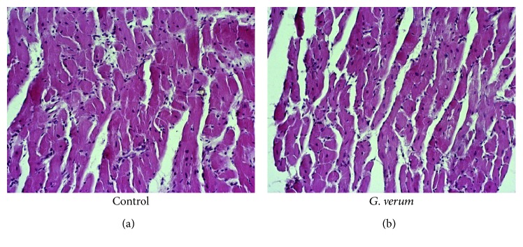
Galium verum L. (G. verum, lady's bedstraw) is a perennial herbaceous plant, belonging to the Rubiaceae family. It has been widely used throughout history due to multiple therapeutic properties. However, the effects of this plant species on functional recovery of the heart after ischemia have still not been fully clarified. Therefore, the aim of our study was to examine the effects of methanol extract of G. verum on myocardial ischemia/reperfusion (I/R) injury in spontaneously hypertensive rats (SHR), with a special emphasis on the role of oxidative stress. Rats involved in the research were divided randomly into two groups: control (spontaneously hypertensive rats (SHR)) and G. verum group, including SHR rats treated with the G. verum extract (500 mg/kg body weight per os) for 4 weeks. At the end of the treatment, in vivo cardiac function was assessed by echocardiography. Rats were sacrificed and blood samples were taken for spectrophotometric determination of systemic redox state. Hearts from all rats were isolated and retrogradely perfused according to the Langendorff technique. After a stabilization period, hearts were subjected to 20-minute ischemia, followed by 30-minute reperfusion. Levels of prooxidants were spectrophotometrically measured in coronary venous effluent, while antioxidant enzymes activity was assessed in heart tissue. Cell morphology was evaluated by hematoxylin and eosin (HE) staining. 4-week treatment with G. verum extract alleviated left ventricular hypertrophy and considerably improved in vivo cardiac function. Furthermore, G. verum extract preserved cardiac contractility, systolic function, and coronary vasodilatory response after ischemia. Moreover, it alleviated I/R-induced structural damage of the heart. Additionally, G. verum extract led to a drop in the generation of most of the measured prooxidants, thus mitigating cardiac oxidative damage. Promising potential of G. verum in the present study may be a basis for further researches which would fully clarify the mechanisms through which this plant species triggers cardioprotection.


Figures

Figure 1
Figure 1
Effects of G. verum pretreatment on ex vivo cardiac function. (a) Comparison within and between groups in the value of dp/dt max, (b) comparison within and between groups in the value of dp/dt min, (c) comparison within and between groups in the value of SLVP, (d) comparison within and between groups in the value of DLVP, (e) comparison within and between groups in the value of heart rate, and (f) comparison within and between groups in the value of coronary flow. Statistical significance at the level of p < 0.05 within the control group; #statistical significance at the level of p < 0.05 within the G. verum group; statistical significance at the level of p < 0.05 between the control and G. verum group. Data are presented as means ± SD. R1, first minute of reperfusion. R7, last minute of reperfusion.
Figure 2
Figure 2
Effects of G. verum pretreatment on the level of prooxidants determined in plasma samples. (a) Comparison between groups in the value of O2 , (b) comparison between groups in the value of H2O2, (c) comparison between groups in the value of NO2 , and (d) comparison between groups in the value of TBARS. Statistical significance at the level of p < 0.05 between the control and G. verum group. Data are presented as means ± SE.
Figure 3
Figure 3
Effects of G. verum pretreatment on parameters of antioxidant defense system determined in erythrocytes samples. (a) Comparison between groups in the activity of SOD, (b) comparison between groups in the activity of CAT, and (c) comparison between groups in the level of GSH. Statistical significance at the level of p < 0.05 between the control and G. verum group. Data are presented as means ± SE.
Figure 4
Figure 4
Effects of G. verum pretreatment on the level of cardiac prooxidants. (a) Comparison within and between groups in the value of O2 , (b) comparison within and between groups in the value of H2O2, (c) comparison within and between groups in the value of NO2 , and (d) comparison within and between groups in the value of TBARS. Statistical significance at the level of p < 0.05 within the control group; #statistical significance at the level of p < 0.05 within the G. verum group; statistical significance at the level of p < 0.05 between the control and G. verum group. Data are presented as means ± SE. R1, first minute of reperfusion. R7, last minute of reperfusion.
Figure 5
Figure 5
Effects of G. verum pretreatment on cardiac antioxidant enzymes activity. (a) Comparison between groups in the activity of CAT and (b) comparison between groups in the activity of SOD. Statistical significance at the level of p < 0.05 between the control and G. verum group. Data are presented as means ± SE.
Figure 6
Figure 6
Histopathological changes in the myocardium following reperfusion (magnification, ×200). Control group: hypertrophy of the cardiac muscle fibers, edema, necrosis, and zonal necrosis of higher number of cardiomyocytes, with hypereosinophilia, fragmentation of the fibers, and loss of nucleus. G. verum group: hypertrophy of the cardiac muscle fibers, edema, necrosis, focal necrosis of lower number of cardiomyocytes or unicellular, and less prominent degenerative changes.

References

    1. Neri M., Riezzo I., Pascale N., Pomara C., Turillazzi E. Ischemia/reperfusion injury following acute myocardial infarction: a critical issue for clinicians and forensic pathologists. Mediators of Inflammation. 2017;2017:14. doi: 10.1155/2017/7018393.7018393 - DOI - PMC - PubMed
    1. Hausenloy D. J., Yellon D. M. Myocardial ischemia-reperfusion injury: a neglected therapeutic target. The Journal of Clinical Investigation. 2013;123(1):92–100. doi: 10.1172/JCI62874. - DOI - PMC - PubMed
    1. Kalogeris T., Baines C. P., Krenz M., Korthuis R. J. Ischemia/reperfusion. Comprehensive Physiology. 2016;7(1):113–170. doi: 10.1002/cphy.c160006. - DOI - PMC - PubMed
    1. Li W., Wu N., Shu W., Jia D., Jia P. Pharmacological preconditioning and postconditioning with nicorandil attenuates ischemia/reperfusion-induced myocardial necrosis and apoptosis in hypercholesterolemic rats. Experimental and Therapeutic Medicine. 2015;10(6):2197–2205. doi: 10.3892/etm.2015.2782. - DOI - PMC - PubMed
    1. Mozaffari M. S., Liu J. Y., Abebe W., Baban B. Mechanisms of load dependency of myocardial ischemia reperfusion injury. American Journal of Cardiovascular Disease. 2013;3(4):180–196. - PMC - PubMed