. 2019 Jul;76(13):2615-2632.
doi: 10.1007/s00018-019-03059-8. Epub 2019 Mar 12.

Phosphorylated and aggregated TDP-43 with seeding properties are induced upon mutant Huntingtin (mHtt) polyglutamine expression in human cellular models

Affiliations

Phosphorylated and aggregated TDP-43 with seeding properties are induced upon mutant Huntingtin (mHtt) polyglutamine expression in human cellular models

Laurent Coudert et al. Cell Mol Life Sci. 2019 Jul.

Abstract

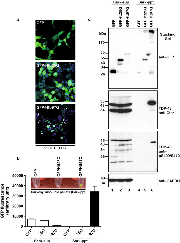
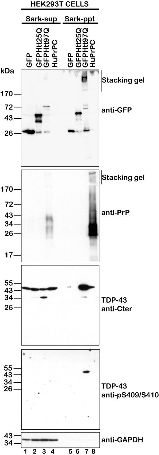
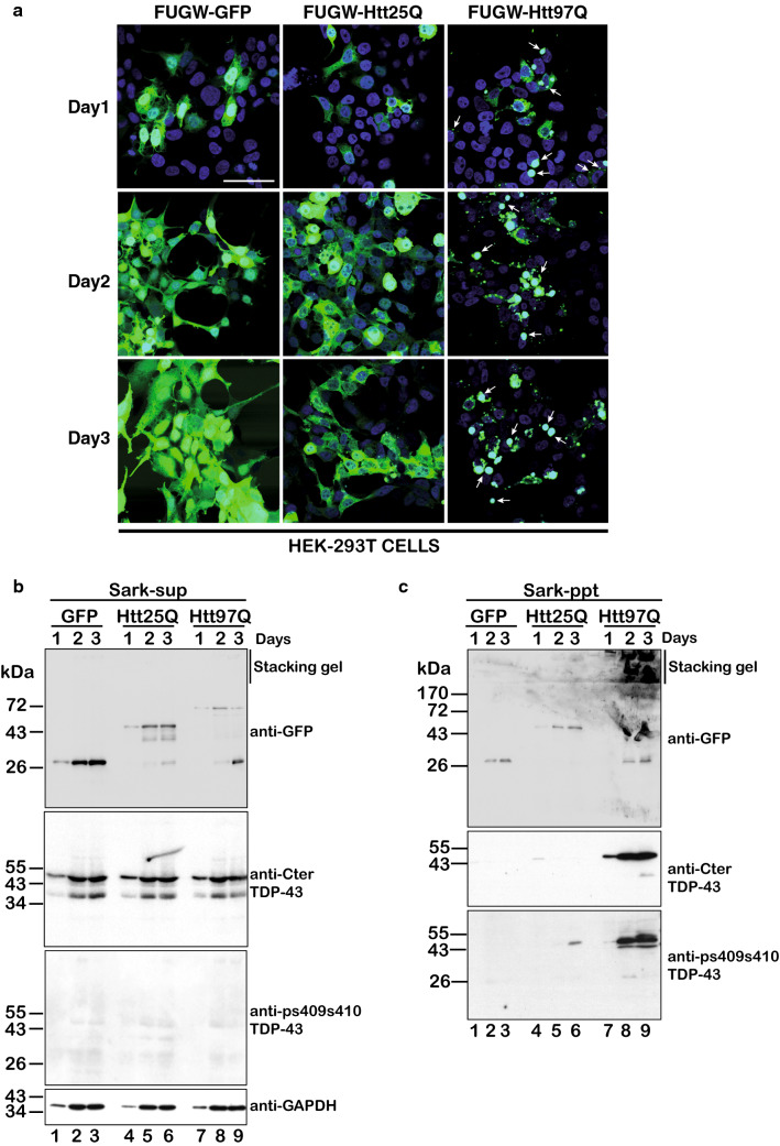
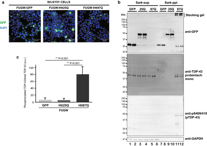
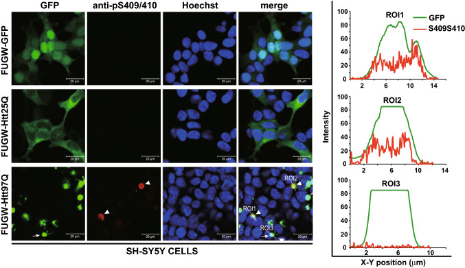
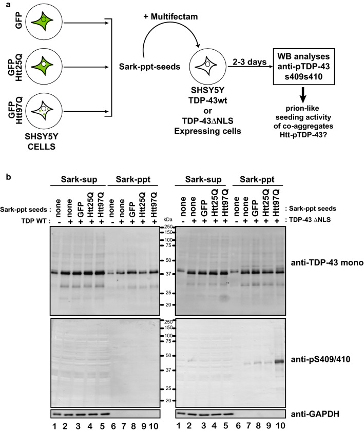
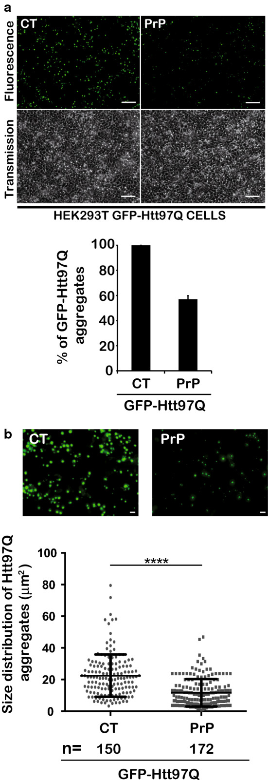
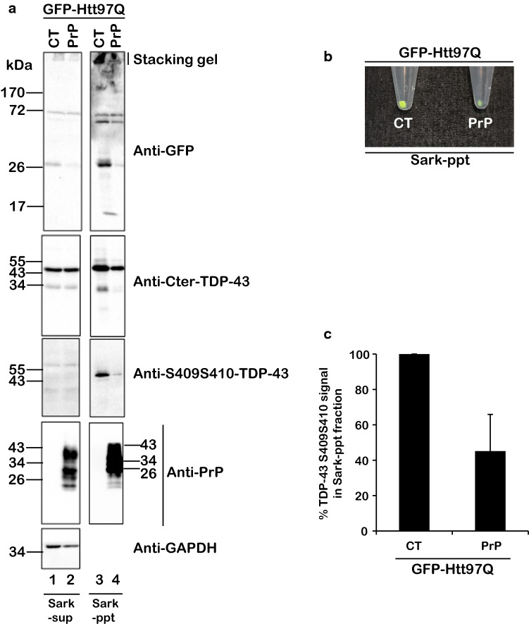
The Tar DNA-Binding Protein 43 (TDP-43) and its phosphorylated isoform (pTDP-43) are the major components associated with ubiquitin positive/Tau-negative inclusions found in neurons and glial cells of patients suffering of amyotrophic lateral sclerosis (ALS) or frontotemporal lobar degeneration-TDP-43 (FTLD-TDP). Many studies have revealed that TDP-43 is also in the protein inclusions associated with neurodegenerative conditions other than ALS and FTLD-TDP, thus suggesting that this protein may be involved in the pathogenesis of a variety of neurological disorders. In brains of Huntington-affected patients, pTDP-43 aggregates were shown to co-localize with mutant Huntingtin (mHtt) inclusions. Here, we show that expression of mHtt carrying 80-97 polyglutamines repeats in human cell cultures induces the aggregation and the phosphorylation of endogenous TDP-43, whereas non-pathological Htt with 25 polyglutamines repeats has no effect. Mutant Htt aggregation precedes accumulation of pTDP-43 and pTDP-43 co-localizes with mHtt inclusions reminding what it was previously described in brains of Huntington-affected patients. Detergent-insoluble fractions from cells expressing mHtt and containing mHtt-pTDP-43 co-aggregates can function as seeds for further TDP-43 aggregation in human cell culture. The human cellular prion protein PrPC was previously identified as a negative modulator of mHtt aggregation; here, we show that PrPC-mediated reduction of mHtt aggregation is tightly correlated with a decrease of TDP-43 aggregation and phosphorylation, thus confirming the close relationships between TDP-43 and mHtt.

Keywords: ALS; Huntingtin; Huntington; Phosphorylation; Prion; Seeds; TDP-43.


Conflict of interest statement

The authors declare that they have no competing interests.

Figures

Fig. 1
Fig. 1
Mutant Huntingtin Htt97Q expression induces aggregation and phosphorylation of endogenous TDP-43 in HEK293T cells. a HEK293T cells were transfected with FUGW–GFP, GFP–Htt25Q or GFP–Htt97Q encoding constructs and were visualized by fluorescence using a spectral confocal microscope. Blue signal corresponds to DAPI staining and green signal to GFP. Scale bar is 50 μm. b Sarkosyl-soluble supernatant (Sark-sup) and insoluble pelleted (Sark-ppt) fractions were isolated. Note the green color of the GFP–Htt97Q Sark-ppt fraction correlated with mutant Htt inclusions compared to GFP or GFP–Htt-25Q. Total GFP fluorescence associated with Sark-sup or sonicated Sark-ppt fraction was evaluated using a fluorescence reader plate. Note the strong GFP signal associated with mHtt97Q Sark-ppt fraction compared to GFP and Htt25Q controls. Oppositely, note the strong GFP signals in GFP and Htt25Q compared to mHtt97Q in Sark-sup fractions. c Sark-sup and Sark-ppt fractions, respectively, isolated from HEK293T cells expressing GFP (lanes 1 and 4), GFP–Htt25Q (lanes 2 and 5) or GFP–Htt97Q (lanes 3 and 6) were analyzed by Western blotting. Membranes were probed with antibodies directed against GFP, TDP-43 (Cter), phospho-TDP-43 S409S410 and GAPDH as loading control
Fig. 2
Fig. 2
PK-resistant prion protein PrP does not induce TDP-43 aggregation and phosphorylation. HEK293T cells were transfected with GFP, GFP–Htt25Q, GFP–Htt97Q or human PrPC encoding constructs. Sark-sup and Sark-ppt fractions were isolated from transfected cells 48 h after transfection and were analyzed by immunoblotting using antibodies directed against GFP, PrP, TDP-43-Cter, phospho-TDP-43 S409S410 or GAPDH as loading control
Fig. 3
Fig. 3
Time course of co-aggregation of Htt97Q and phosphorylated TDP-43. a HEK293T cells were transfected with GFP, GFP–Htt25Q or GFP–Htt97Q encoding constructs and were visualized by fluorescence using a spectral confocal microscope at days 1, 2 and 3 after transfection. White arrows indicate Htt97Q inclusion bodies. Blue signal corresponds to DAPI staining and green signal to GFP. Scale bar is 50 μm. At each time, cells were harvested and Sark-sup and insoluble Sark-ppt fractions were isolated by ultracentrifugation. Sark-sup (b) and Sark-ppt (c) fractions at days 1, 2 and 3 from GFP (lanes 1–3), Htt25Q (lanes 4–6) and Htt97Q (lanes 7–9) expressing cells were analyzed by immunoblotting using antibodies directed against GFP, TDP-43-Cter and phospho-TDP-43 S409S410 or GAPDH as loading control
Fig. 4
Fig. 4
Mutant Htt97Q expression induces aggregation and phosphorylation of endogenous TDP-43 in human Neuroblastoma SH-SY5Y cells. a Human neuroblastoma SH-SY5Y cells were transfected in duplicate with plasmids encoding GFP, GFP–Htt25Q or GFP–Htt97Q and were analyzed by fluorescence using a confocal microscope. Blue signal corresponds to DAPI staining and green signal to GFP. Scale bar is 50 μm. b Sark-sup and Sark-ppt fractions, respectively, from cells expressing GFP (lanes 1, 2 and 7, 8), GFP–Htt25Q (lanes 3, 4 and 9, 10) and GFP–Htt97Q (lanes 5, 6 and 11, 12) were analyzed by immunoblotting using antibodies directed against GFP, TDP-43 and phospho-TDP-43 S409S410 or GAPDH as loading control. c Quantification of phosphorylated TDP-43 formation in SH-SY5Y cells expressing GFP, GFPHtt25Q, and GFPmHtt97Q. The data are presented as a ratio of phosphorylated TDP-43 over total TDP-43. Values are given as mean ± SEM with a P < 0.001 (n = 6, one-way ANOVA)
Fig. 5
Fig. 5
Phosphorylated TDP-43 aggregates co-localize with mutant Htt97Q inclusions in SH-SY5Y cells. Human neuroblastoma SH-SY5Y cells transfected with GFP, GFP–Htt25Q or GFP–Htt97Q constructs were analyzed by immunofluorescence confocal microscopy 3 days after transfection. Cells were immunostained with anti-phospho-TDP-43 S409S410 and counterstained with Hoechst for nuclei. Scale bars are 20 μm. White arrowheads indicate co-localization of mutant Htt97Q and phosphorylated TDP-43. Region of interest (ROI; white lines) 1-3 are depicted in the merge panel. The plot profiles of GFP–mHtt97Q and phosphorylated TDP-43 (Alexa647 labeling) co-localizations (ROI1 and 2) along the ROI lines were constructed and analyzed using Image J software (see right panel). Note that mHtt97Q and pTDP-43 are well co-localized. As negative control, ROI3 shows a mHtt97Q aggregate that does not co-localize with the phosphorylated TDP-43 thus confirming that the strong signal released by GFP–mHtt97Q does not overlap with that of alexa-647 secondary antibody
Fig. 6
Fig. 6
Insoluble co-aggregates of Htt97Q and phosphorylated TDP-43 function as seeds for intracellular TDP-43 aggregation in SH-SY5Y human neuroblastoma cells. a Summary of the experiments to test whether Sark-insoluble fraction of cells expressing GFP–Htt polyQ functions as seeds for intracellular aggregation of TDP-43. b Immunoblotting of the respective Sark-sup and Sark-ppt fractions from SH-SY5Y cells expressing TDP-43 wild-type (WT, left panel) or ΔNLS (right panel) and challenged with Sark-ppt seeds isolated from GFP (lanes 3 and 8), GFP–Htt25Q (lanes 4 and 9) or GFP–Htt97Q (lanes 5 and 10) producer cells, were analyzed using antibodies directed against TDP-43 or phospho-TDP-43 S409S410. Sark-sup and Sark-ppt fractions from SH-SY5Y cells expressing TDP-43-WT or DNLS not challenged with Sark-ppt seeds were loaded as internal controls. Loading control was assessed using the anti-GAPDH antibody
Fig. 7
Fig. 7
PrPC expression negatively affects aggregation of GFP–mHtt97Q and phosphorylated TDP-43. a HEK293T cells were co-transfected with the GFP–mHtt97Q encoding constructs and the human PrPC encoding plasmid or the empty vector (CT) as negative control. Fluorescence microscopy was carried out 48 h after transfection. Number of GFP–mHtt97Q aggregates was quantified using the Image J software. Note the decrease of GFP–mHtt97Q aggregates when PrPC is expressed. The graph on bottom panel illustrates the GFP–mHtt97Q aggregates decrease upon PrPC expression. The data shown are from 3 independent experiments. Values are given as mean ± standard deviation (SD). b Size of aggregates was determined by measuring the surface of punctuated GFP–mHtt97Q inclusions using the Image J software. The graph on the bottom panel illustrates the size distribution of GFP–mHtt97Q inclusions from n = 150 and 172 aggregates from CT and PrPC conditions, respectively. Values are given as mean ± SEM with a P < 0.0001. CT corresponds to 22.50 ± 1.093 and PrPC to 11.67 ± 0.6649. Statistic and P value calculation were done using an unpaired t test. Scale bars are 100 μm (upper panels) and 20 μm (bottom panels)
Fig. 8
Fig. 8
PrP negatively affect TDP-43 phosphorylation and aggregation. a HEK293T cells co-transfected with the GFP–mHtt97Q encoding construct and with PrPC, encoding plasmid or the empty control vector (CT) were recovered and Sark-sup and Sark-ppt fractions were isolated and analyzed by Western blotting using the anti-GFP, the anti-Cter-TDP-43, the Anti-phospho-TDP43S409S410 or the anti-PrP or the anti–GAPDH for expression and loading controls. b Sark-ppt pellets after ultracentrifugation. Note the decrease of GFP-green signal in PrP condition. c Graph illustrates the decrease of phospho-TDP-43S409S410 in the Sark-ppt fraction upon PrPC expression. The data shown are from four independent experiments. Values are given as mean ± standard deviation (SD)

Similar articles

Cited by

References

    1. Brundin P, Melki R, Kopito R. Prion-like transmission of protein aggregates in neurodegenerative diseases. Nat Rev Mol Cell Biol. 2010;11(4):301–307. doi: 10.1038/nrm2873. - DOI - PMC - PubMed
    1. Goedert M, Clavaguera F, Tolnay M. The propagation of prion-like protein inclusions in neurodegenerative diseases. Trends Neurosci. 2010;33(7):317–325. doi: 10.1016/j.tins.2010.04.003. - DOI - PubMed
    1. Goedert M, Falcon B, Clavaguera F, Tolnay M. Prion-like mechanisms in the pathogenesis of tauopathies and synucleinopathies. Curr Neurol Neurosci Rep. 2014;14(11):495. doi: 10.1007/s11910-014-0495-z. - DOI - PubMed
    1. Grad LI, Fernando SM, Cashman NR. From molecule to molecule and cell to cell: prion-like mechanisms in amyotrophic lateral sclerosis. Neurobiol Dis. 2015;77:257–265. doi: 10.1016/j.nbd.2015.02.009. - DOI - PubMed
    1. Jucker M, Walker LC. Self-propagation of pathogenic protein aggregates in neurodegenerative diseases. Nature. 2013;501(7465):45–51. doi: 10.1038/nature12481. - DOI - PMC - PubMed