Review
. 2020 Jan;43(1):71-89.
doi: 10.1002/jimd.12083. Epub 2019 Apr 16.

Organelle interplay-peroxisome interactions in health and disease

Affiliations
Review

Organelle interplay-peroxisome interactions in health and disease

Michael Schrader et al. J Inherit Metab Dis. 2020 Jan.

Abstract

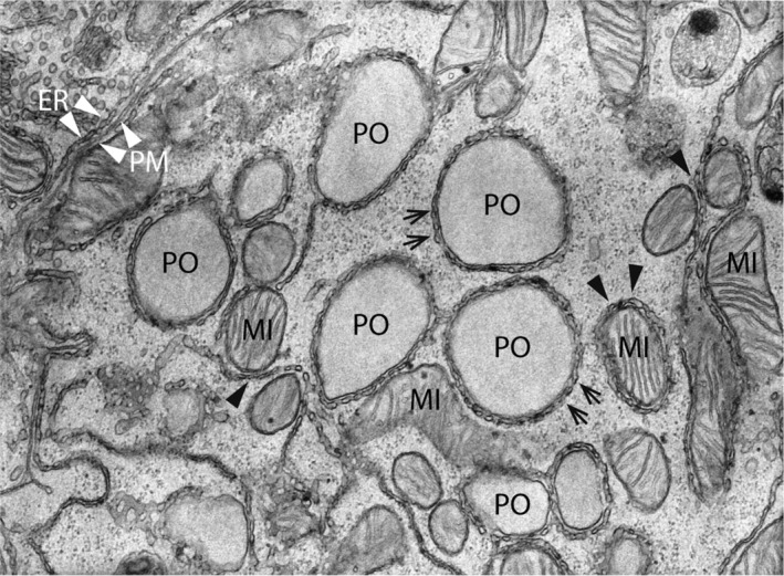
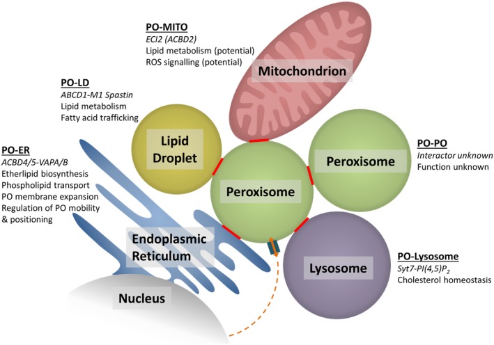
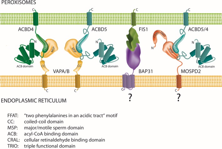
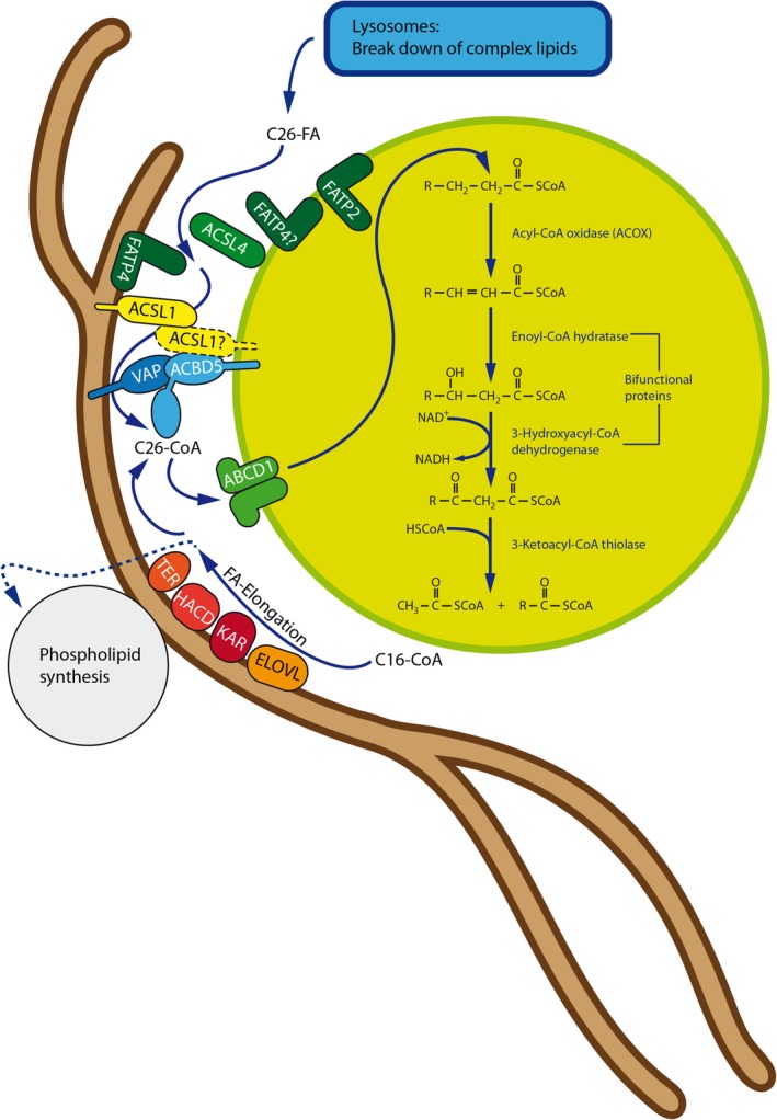
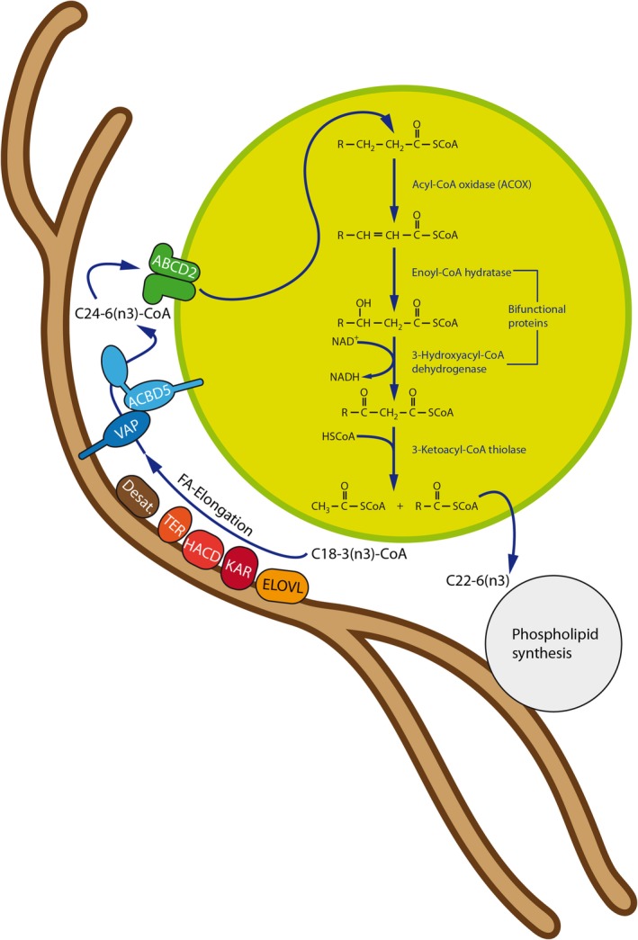
Peroxisomes are multifunctional, dynamic, membrane-bound organelles with important functions in cellular lipid metabolism, rendering them essential for human health and development. Important roles for peroxisomes in signaling and the fine-tuning of cellular processes are emerging, which integrate them in a complex network of interacting cellular compartments. Like many other organelles, peroxisomes communicate through membrane contact sites. For example, peroxisomal growth, positioning, and lipid metabolism involves contacts with the endoplasmic reticulum (ER). Here, we discuss the most recent findings on peroxisome-organelle interactions including peroxisome-ER interplay at membrane contacts sites, and functional interplay with mitochondria, lysosomes, and lipid droplets in mammalian cells. We address tether proteins, metabolic cooperation, and the impact of peroxisome interactions on human health and disease.

Keywords: ACBD5; endoplasmic reticulum; fatty acid beta-oxidation; lipid droplets; lipid metabolism; lysosomes; membrane contact sites; mitochondria; organelle interaction; peroxisomes.


Conflict of interest statement

The authors declare that they have no conflict of interest.

Figures

Figure 1
Figure 1
Electron micrograph of organelle contact sites in the rat liver. The center of the image shows five peroxisomes (PO), which are surrounded by a reticular network of smooth ER tubules (arrows). Furthermore, several mitochondria (MI) with ER MAMs can be found (black arrowheads). Note an elongated mitochondrion (center) in direct apposition to the PO‐ER contacts suggesting the existence of functionally relevant organelle triple contacts. ER—plasma membrane (PM) contacts are also observed (upper left corner; white arrowheads). Magnification: ×25 000 (kindly provided by W. Kriz, University of Heidelberg, GER)
Figure 2
Figure 2
Peroxisome‐organelle MCSs in mammalian cells and their suggested functions. Communication between PO and the nucleus is also indicated. ER, endoplasmic reticulum; LD, lipid droplet; MITO, mitochondrion; PO, peroxisome
Figure 3
Figure 3
Tethering complexes at peroxisome‐ER MCSs. All hitherto identified and potential tethering complexes connect organelle membranes via protein‐protein interactions. The C‐tail‐anchored proxisomal membrane proteins ACBD4 and ACBD5 possess FFAT‐like motifs in their middle domain which interact with N‐terminal major sperm‐binding (MSP) domains of ER‐resident VAPA and VAPB. With MOSPD2, another ER‐resident protein with an MSP domain was recently identified, which interacts with a variety of tether proteins containing FFAT motifs.36 MOSPD1 is another MSP‐domain containing protein with a proposed ER localization.37 Interaction of MOSPD proteins with ACBD4/5 has not yet been experimentally verified. The tail‐anchored membrane protein FIS1 was identified in a tethering complex with ER‐resident BAP31.38 As FIS1 also localizes to peroxisomes,39 the FIS1‐BAP31 tether may also contribute to peroxisome‐ER MCSs
Figure 4
Figure 4
Interplay between peroxisomal VLCFA degradation and ER fatty acid elongation at ACBD5‐mediated MCSs at the PO‐ER interface. At the ER membrane, palmitoyl‐CoA (C16) is elongated to form saturated VLCFA‐CoA in four subsequent steps by the enzymes ELOVL(1‐7) (elongation of very‐long chain fatty acids protein), KAR (3‐ketoacyl‐CoA reductase), HACD(1‐3) (very‐long‐chain‐3‐hydroxyacyl‐CoA dehydratase), and TER (trans‐2,3‐enoyl‐CoA reductase). VLCFAs are subsequently incorporated into membrane lipids at the ER or, when VLCFA concentrations in the membrane are already high, transferred to peroxisomal ACBD5. Long and very‐long chain fatty acid‐CoA synthetases (ACSL and FATP) generate fatty acyl‐CoA from the cytosolic free fatty acid pool. ACBD5 “senses” growing concentrations of VLCFA‐CoA at the contact site by binding to its ACB domain (probably by direct interaction with, for example, ACSL1). Bound VLCFA‐CoA is “handed over” to the peroxisomal FA import protein ABCD1 to be imported and degraded by the peroxisomal β‐oxidation pathway. Such a regulatory system may prevent excessive incorporation of VLCFA into phospholipids, which are as well generated at the ER
Figure 5
Figure 5
Peroxisome‐ER cooperation in PUFA synthesis at ACBD5‐mediated MCSs. PUFAs are synthesized at the ER by combined FA elongation and desaturation. However, peroxisomes cooperate with the ER in the synthesis of n‐3 long‐chain PUFAs such as docosahexaenoic acid (DHA) (22:6n‐3) as the ER appears to lack potent fatty acid Δ4‐desaturase activity. To this end, exploiting Δ6 desaturase activities, the ER produces 24:6 (n‐3) FA, which are subsequently chain‐shortened by one round of β‐oxidation in peroxisomes. In this scenario, ACBD5 may facilitate efficient transport of 24:6 (n‐3) CoA from the ER to peroxisomes at MCSs. PUFA, polyunsaturated fatty acid

Similar articles

Cited by

References

    1. Prinz WA. Bridging the gap: membrane contact sites in signaling, metabolism, and organelle dynamics. J Cell Biol. 2014;205:759‐769. - PMC - PubMed
    1. Simmen T, Tagaya M. Organelle communication at membrane contact sites (MCS): from curiosity to center stage in cell biology and biomedical research. Adv Exp Med Biol. 2017;997:1‐12. - PubMed
    1. Shai N, Yifrach E, van Roermund CWT, et al. Systematic mapping of contact sites reveals tethers and a function for the peroxisome‐mitochondria contact. Nat Commun. 2018;9(1):1761. - PMC - PubMed
    1. Valm AM, Cohen S, Legant WR, et al. Applying systems‐level spectral imaging and analysis to reveal the organelle interactome. Nature. 2017;546:162‐167. - PMC - PubMed
    1. Eisenberg‐Bord M, Shai N, Schuldiner M, Bohnert M. A tether is a tether is a tether: tethering at membrane contact sites. Dev Cell. 2016;39:395‐409. - PubMed

Publication types

Substances

LinkOut - more resources