. 2019 May 21;47(9):4462-4475.
doi: 10.1093/nar/gkz162.

A complex between DYRK1A and DCAF7 phosphorylates the C-terminal domain of RNA polymerase II to promote myogenesis

Affiliations

A complex between DYRK1A and DCAF7 phosphorylates the C-terminal domain of RNA polymerase II to promote myogenesis

Dan Yu et al. Nucleic Acids Res. .

Abstract

The general transcription factor P-TEFb, a master regulator of RNA polymerase (Pol) II elongation, phosphorylates the C-terminal domain (CTD) of Pol II and negative elongation factors to release Pol II from promoter-proximal pausing. We show here that P-TEFb surprisingly inhibits the myoblast differentiation into myotubes, and that P-TEFb and its two positive complexes are eliminated in this process. In contrast, DYRK1A, another CTD kinase known to control transcription of a subset of genes important for development and tissue homeostasis, is found to activate transcription of key myogenic genes. We show that active DYRK1A exists in a complex with the WD40-repeat protein DCAF7 that stabilizes and tethers DYRK1A to Pol II, so that DYRK1A-DCAF7 can co-migrate with and phosphorylate Pol II along the myogenic gene loci. Thus, DCAF7 modulates the kinase signaling output of DYRK1A on Pol II to stimulate myogenic transcription after active P-TEFb function is shut off.


Figures

Figure 1.
Figure 1.
DYRK1A and DCAF7 form a nuclear complex that promotes stability of both proteins. (A) Analysis by mass spectrometry of proteins co-immunoprecipitated with F-DYRK1A from HeLa nucleus extracts (NE) with anti-Flag mAb beads. Only the proteins specifically isolated from the F-DYRK1A-expressing cells but not the cells containing an empty vector were shown. (B) NE of HeLa cells were subjected to immunoprecipitation (IP) with either the anti-DYRK1A antibody or non-specific rabbit total IgG as a negative control. The precipitates and the NE were examined by Western-blotting (WB) for the indicated proteins. (C) NE from Hela cells expressing F-DYRK1A were adjusted to the indicated NaCl concentrations and subjected to anti-Flag IP followed by WB. (D) NE from Hela cells expressing WT or Δ84–95 F-DYRK1A were analyzed by IP/WB as in B. All IP experiments were repeated at least three times and representatives are shown. (E, F) HeLa cells expressing WT or Δ84–95 F-DYRK1A were analyzed for the presence of the indicated proteins in whole cell extracts (WCE) by WB (E) or the indicated mRNAs by qRT-PCR (F), with the signals from the empty vector (Ctl.) cells set to 1. (G) HeLa cells co-transfected with F-DCAF7 plus WT or Δ84–95 F-DYRK1A were cultured for 40 hours and then treated with cycloheximide (CHX; 50 μg/ml) for the indicated hours. WCE were analyzed by WB for the indicated proteins. (H, I) WT 293T or the 293T-based DCAF7 KO clones were analyzed by WB (H) and qRT-PCR (I) as in E and F. Error bars in F and I represent mean ± standard deviations (SD).
Figure 2.
Figure 2.
DCAF7 is required for optimal kinase and transcriptional activities of DYRK1A. (A) HeLa cells were co-transfected with the HIV-1 LTR-luciferase reporter construct containing six copies of the Gal4 upstream activation sequence (UAS) and the plasmid expressing the indicated Gal4-DYRK1A fusion. Luciferase activities in cell extracts were measured. Error bars represent mean ± SD from three independent experiments. (B) Flag-tagged WT and Δ84–95 DYRK1A were affinity-purified from NE of transfected 293T cells and analyzed in kinase reactions containing GST-CTD as the substrate. The reaction products were examined by WB for the Ser2- and Ser5-phosphorylated CTD. (C) Two independent and doxycycline (Dox)-inducible DCAF7 KD clones expressing the indicated shRNAs were co-transfected with the F-Gal4-DYRK1A-expressing plasmid (50% more was used for the Dox-plus conditions) and the 6× Gal4 UAS-HIV-1 LTR-luciferase reporter construct. Luciferase activities were measured and analyzed as in A. (D) WT F-DYRK1A affinity-purified from either the parental HeLa cells or the HeLa-based shDCAF7-2 KD cells were tested in kinase reactions as in B. (E) Recombinant GST-DYRK1A (Life Technologies) was incubated with or without F-DCAF7, which was affinity-purified from the 293T-based DYRK1A KO cells, in kinase reactions containing GST-CTD as the substrate. The reaction products were analyzed as in B. All in vitro kinase assays were repeated at least three times and representatives are shown.
Figure 3.
Figure 3.
DCAF7 interacts with RNA Pol II independently of DYRK1A and is required for the DYRK1A-Pol II interaction. (AC) NE and anti-Flag (A), anti-DCAF7 (B) or anti-Pol II (C) immuneprecipitates (IP) derived from NE of normal 293T cells (B, C) or 293T cells transfected with F-DCAF7 (A) were analyzed by WB to detect the indicated proteins. (D) WT 293T or the 293T-based DYRK1A KO cells were transfected with the F-DCAF7-expressing plasmid. Anti-Flag IP from NE of these cells were analyzed by WB as in A. (E) NE and the anti-Flag IP from 293T cells transfected with the WT or Δ84–95 F-DYRK1A-expressing plasmid were analyzed by WB as in A. (F) The 293T-based inducible DCAF7 KD cells expressing F-DYRK1A were treated with DMSO or Dox to induce the KD. NE and anti-Flag IP derived from NE were examined by WB as in A. (G) NE and anti-Flag IP from 293T cells transfected with WT or Δ84–95 F-DYRK1A, or Δ84–95&Δ566-621 F-DYRK1A-expressing plasmid were analyzed by WB. All IP experiments were repeated at least three times. (H) The anti-Flag IP examined in G were incubated with or without ATP in kinase reactions and analyzed by SDS-PAGE followed by WB.
Figure 4.
Figure 4.
DYRK1A and DCAF7 are required for optimal myoblast differentiation and expression of key myogenic genes. (A) The levels of the indicated proteins in 1 × 10⁵ C2C12 cells grown in the growth medium (GM) or differentiation medium for 1-7 days (DM1-7) were examined by WB. (B, C) The mRNA levels of the indicated genes in C2C12 cells cultured in GM or DM1-7 were measured by qRT-PCR and shown. (D) Extracts of C2C12 cells grown in GM, DM5, or containing an equal mixture of the two populations (M) were subjected to IP with the anti-DCAF7 Ab or a non-specific control IgG. The precipitates were analyzed by WB for the indicated proteins. (E, F) C2C12 cells stably expressing the indicated shRNAs were cultured in GM for 5 days and subjected to immunofluorescence staining with the mouse anti-MYH2 mAb plus goat anti-mouse AF680 (green). Nuclei are counterstained with DAPI (blue). Scale bars represent 100 μm. (G, H) The percentages of MYH(+) C2C12 cells expressing the indicated shRNAs were counted and shown. (I,J). The expression of the proteins marked on the left in cells expressing the indicated shRNAs and grown in GM or DM for 5 days was detected by WB. (K, L) The MYOG promoter-driven luciferase reporter construct was co-transfected with the indicated shRNA- and/or the MYOD-expressing plasmids. After 48 hr of transfection, luciferase activities were measured in the cell extracts. All error bars represent mean ± SD from at least three independent experiments/measurements. The asterisks indicate different levels of statistical significance as calculated by two-tailed Student's t-test. ***P < 0.001; **P < 0.01; *P < 0.05.
Figure 5.
Figure 5.
DYRK1A–DCAF7 interaction is essential for efficient expression of key myogenic genes. (A) The DYRK1A KD C2C12 cells were transfected with an empty vector (-) or the vector expressing WT or Δ84–95 F-DYRK1A and then analyzed by WB for the indicated proteins. A non-specific band is denoted with an asterisk. (BD) The cells in A were cultured in GM or DM for 3 days and the mRNAs produced from the indicated genes were detected by qRT-PCR and shown. (E) WB was used to assess the levels of endogenous DYRK1A in normal C2C12 cells and the overexpressed Δ84–95 F-DYRK1A in transfected cells. (FH) The cells in E were grown in GM and DM for 3 days and the mRNAs produced from the indicated genes were detected by qRT-PCR and shown. All error bars represent mean +/− SD from three independent measurements. The asterisks indicate different levels of statistical significance as calculated by two-tailed Student's t-test. ***P < 0.001; **P < 0.01; *P < 0.05.
Figure 6.
Figure 6.
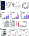
DYRK1A–DCAF7 complex exerts direct transcriptional control of key myogenic genes through binding and phosphorylating Pol II at the gene loci. (A, B) ChIP-qPCR analyses were conducted to assess the occupancy of DCAF7 (A) and DYRK1A (B) at the indicated gene promoters in C2C12 cells cultured in GM or DM for 3 days. The ChIP-qPCR signals were normalized to the input DNA, divided by the values obtained with the rabbit total IgG in control reactions and shown. (C, D) C2C12 cells stably expressing the indicated shRNAs were grown in DM for 3 days and then analyzed by ChIP-qPCR to detect the occupancy of DYRK1A (C), total Pol II (D), and the Ser5- or Ser2-phosphorylated Pol II (pSer5 or pSer2; D) at the indicated gene loci. Rabbit total IgG was used as a negative control. (E) A diagram showing the positions of the primer sets used in ChIP-qPCR reactions in F-I to map the bindings across the MYH2 gene locus. TSS: transcription start site. The vertical lines/blocks represent exons. (FI) C2C12 cells in DM for 3 days were subjected to ChIP-qPCR analysis using the primer sets in E to detect the bindings of DYRK1A (F), DCAF7 (G), total Pol II (H) and BRD4 (I) to the various positions (a to g) across the MYH2 gene locus. The ChIP-qPCR signals were normalized to the input DNA and divided by those obtained with the rabbit total IgG in control reactions. All error bars represent mean ± SD from three independent reactions. The asterisks indicate different levels of statistical significance as calculated by two-tailed Student's t-test. ***P < 0.001; **P < 0.01; *P < 0.05.
Figure 7.
Figure 7.
CDK9 inhibits expression of key myogenic genes and is inactivated during myoblast differentiation to likely allow DYRK1A–DCAF7 to activate transcription of these genes. (A) Left: C2C12 cells stably expressing the indicated shRNAs were cultured in DM for 4 days and subjected to anti-MYH2 immunofluorescence staining as in Figure 4E. Right: The percentages of MYH2(+) cells detected by immunofluorescence were counted and shown. (B) Analysis by WB of the indicated proteins in cells expressing the various shRNAs and cultured in GM or DM for 4 days. (C) The MYOG promoter-driven luciferase reporter construct was co-transfected with the indicated shRNA- and/or MYOD-expressing plasmid into C2C12 cells. Luciferase activities in cell extracts were measured and shown. (DF) qRT-PCR analyses were conducted to assess the expression of MYH2 (D), CAV3 (E) and MYOG (F) in C2C12 cells expressing the indicated shRNAs and cultured in GM or DM for the various days. (G) WCE of C2C12 cells cultured in GM (labeled as G) or DM (labeled as D) for various days were subjected to IP with either the anti-CDK9 antibody or a non-specific control IgG. The IP products and WCE were examined by WB. M represents an equal mixture of the GM and DM4 WCE. (H) Anti-CDK9 immunoprecipitates from cells grown in GM or DM for 6 days were incubated with GST-CTD in kinase reactions. The reaction mixtures were examined by WB. (I) ChIP-qPCR analysis was conducted to assess the occupancy of CDK9 at the indicated gene promoters in cells cultured in GM or DM for 3 days. The signals were processed as in Figure 6A. (J,K) C2C12 cells expressing the various shRNAs were grown in GM for the indicated number of days and the cell concentrations were determined and shown. All error bars represent mean ± SD from three independent measurements. The asterisks indicate different levels of statistical significance as calculated by two-tailed Student's t-test. ***P < 0.001; **P < 0.01; *P < 0.05.

References

    1. Hsin J.P., Manley J.L.. The RNA polymerase II CTD coordinates transcription and RNA processing. Genes Dev. 2012; 26:2119–2137. - PMC - PubMed
    1. Phatnani H.P., Greenleaf A.L.. Phosphorylation and functions of the RNA polymerase II CTD. Genes Dev. 2006; 20:2922–2936. - PubMed
    1. Corden J.L. RNA polymerase II C-terminal domain: tethering transcription to transcript and template. Chem. Rev. 2013; 113:8423–8455. - PMC - PubMed
    1. Price D.H. P-TEFb, a cyclin-dependent kinase controlling elongation by RNA polymerase II. Mol. Cell. Biol. 2000; 20:2629–2634. - PMC - PubMed
    1. Zhou Q., Li T., Price D.H.. RNA polymerase II elongation control. Annu. Rev. Biochem. 2012; 81:119–143. - PMC - PubMed

Publication types

MeSH terms