. 2019 May;43(5):2103-2117.
doi: 10.3892/ijmm.2019.4126. Epub 2019 Mar 6.

Loss of Fam20c causes defects in the acinar and duct structure of salivary glands in mice

Affiliations

Loss of Fam20c causes defects in the acinar and duct structure of salivary glands in mice

Nan Miao et al. Int J Mol Med. 2019 May.

Abstract

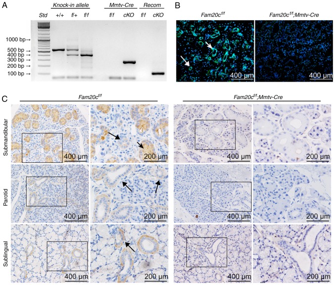
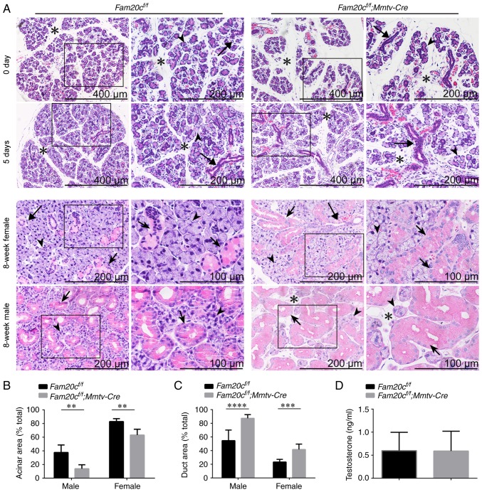
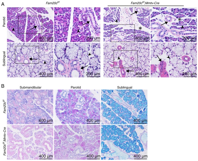
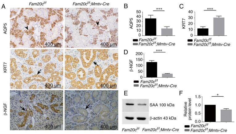
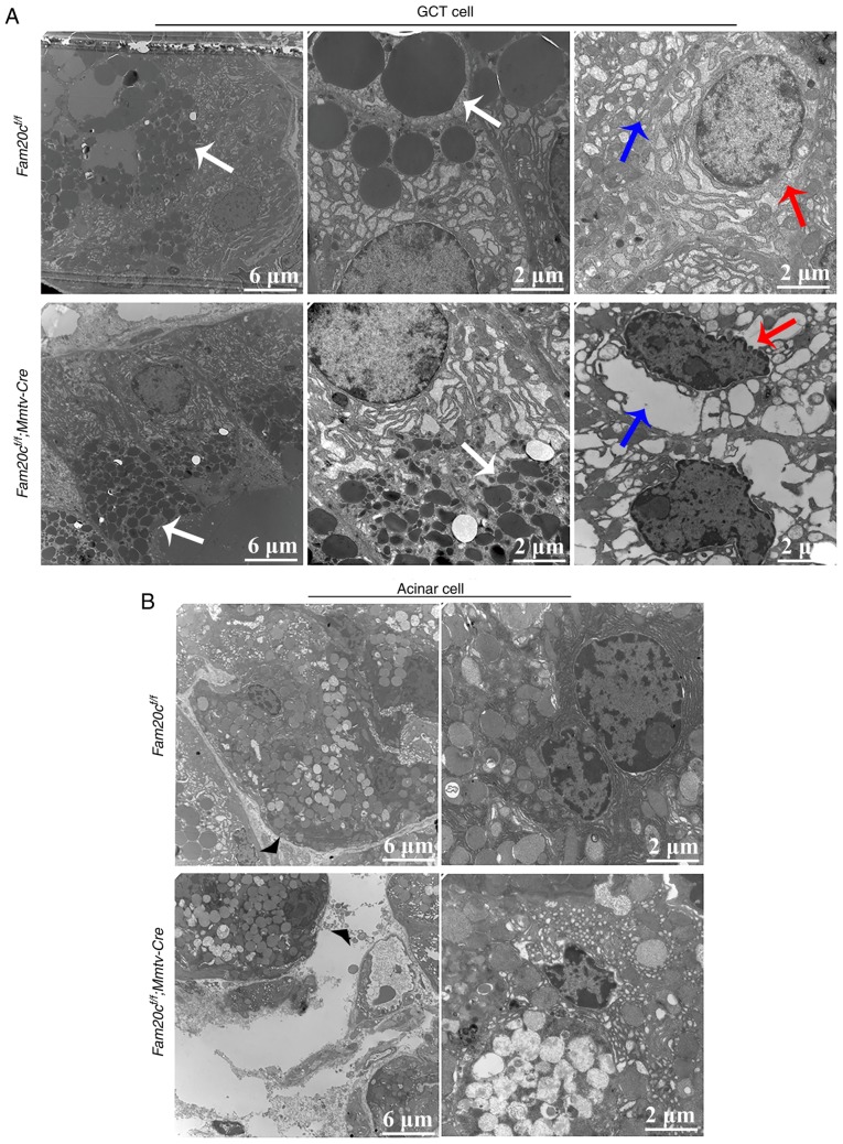
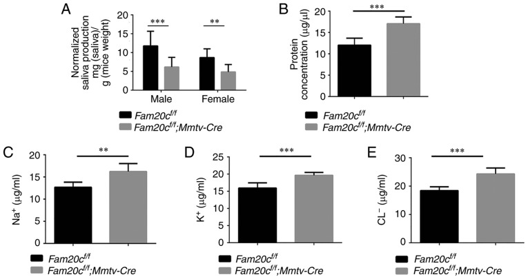
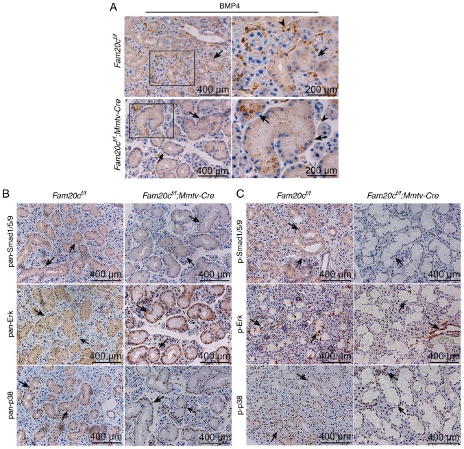
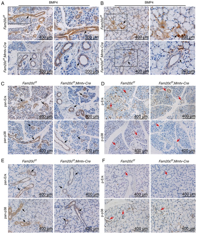
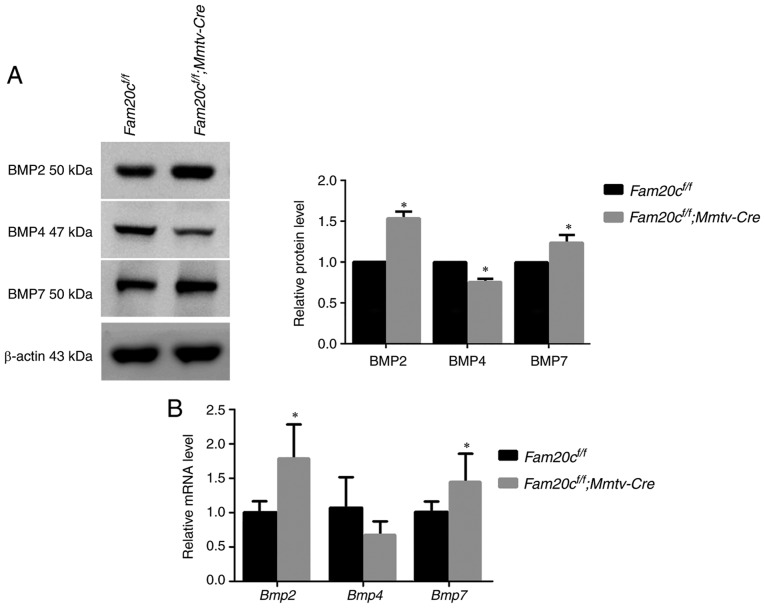
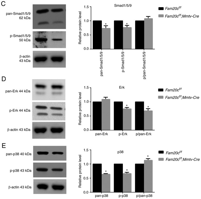
Family with sequence similarity 20‑member C (FAM20C), a recently characterized Golgi kinase, performs numerous biological functions by phosphorylating more than 100 secreted proteins. However, the role of FAM20C in the salivary glands remains undefined. The present study demonstrated that FAM20C is mainly located in the cytoplasm of duct epithelial cells in the salivary glands. Fam20cf/f; Mmtv‑Cre mice were created in which Fam20c was inactivated in the salivary gland cells and observed that the number of ducts and the ductal cross‑sectional area increased significantly, while the number of acinar cells was reduced. The granular convoluted tubules (GCTs) exhibited an accumulation of aberrant secretory granules, along with a reduced expression and altered distribution patterns of β nerve growth factor, α‑amylase and bone morphogenetic protein (BMP) 4. This abnormality suggested that the GCT cells were immature and exhibited defects in developmental and secretory functions. In accordance with the morphological alterations and the reduced number of acinar cells, FAM20C deficiency in the salivary glands significantly decreased the salivary flow rate. The Na+, Cl- and K+ concentrations in the saliva were all significantly increased due to dysfunction of the ducts. Furthermore, Fam20c deficiency significantly increased BMP2 and BMP7 expression, decreased BMP4 expression, and attenuated the canonical and noncanonical BMP signaling pathways in the salivary glands. Collectively, the results of the present study demonstrate that FAM20C is a key regulator of acinar and duct structure and duct maturation and provide a novel avenue for investigating novel therapeutic targets for oral diseases including xerostomia.


Figures

Figure 1
Figure 1
Verification of FAM20C expression and inactivation. (A) Genomic DNA was extracted from the tails of mice of each genotype and genotyping was performed with specific primers for the floxed Fam20c allele, the recombined Fam20c allele and the Mmtv-Cre allele. (B) Immunofluorescence assay of FAM20C in the SMG. The SMG from control littermates exhibited strong staining. FAM20C was expressed in the cytoplasm of the ID, SD and ED cells (long arrows) and the GCT cells (short arrows); in contrast, the cells from cKO mice did not stain. (C) Immunohistochemistry of FAM20C in the SMG, PG and SLG. In the SMG of control mice, FAM20C was expressed in the cytoplasm of the ID, SD and ED cells (long arrows) and the GCT cells (short arrows). In the PG and SLG of control mice, the immunostaining signal for FAM20C was also observed in the ductal cells, whereas the cKO mice did not stain. For images of Fam20cf/f or Fam20cf/f; Mmtv-cre mice, the second panel presents magnified images of the outlined areas from the corresponding panel. Fam20C, family with sequence similarity 20-member C; PG, parotid gland; SLG, sublingual gland; SMG, submandibular gland; GCT, granular convoluted tubule; ID, intercalated duct; SD, striated duct; ED, excretory duct; cKO, Fam20cf/f; Mmtv-Cre conditional knockout.
Figure 2
Figure 2
Morphological and structural alterations of the SMG in cKO mice. (A) Salivary glands were collected from six male or female mice at postnatal day 0, postnatal day 5 or postnatal week 8 for hematoxylin and eosin studies (n=5 in each group). At postnatal days 0 and 5, the ducts (long arrows) were more pronounced in the SMG of cKO mice. At postnatal week 8, the ratio of the ductal cross-sectional area was increased significantly (granular convoluted tubule cells: Short arrows) and the mesenchyme was increased (asterisks) in the SMG of the male and female cKO mice. The acinar cells (arrowheads) appeared normal. (B) Quantitative evaluation of the SMG acinar cross-sectional areas. **P<0.01. (C) Quantitative evaluation of the SMG duct cross-sectional areas. male, ****P<0.0001 and female, ***P<0.001. (D) ELISA of serum testosterone. The serum testosterone level was unchanged between cKO and control mice. For images of Fam20cf/f or Fam20cf/f; Mmtv-cre mice, the second panel presents magnified images of the outlined areas from the corresponding panel. SMG, submandibular gland; cKO, Fam20cf/f; Mmtv-Cre conditional knockout.
Figure 3
Figure 3
PG/SLG morphological and structural alterations in cKO mice. (A) Hematoxylin and eosin staining of the PG and SLG. There was more mesenchymal tissue (indicated by asterisks) present within the mutant PG. The number of ducts (arrows) and acinar cells (arrowheads) did not change substantially. (B) Periodic Acid-Schiff staining of the SMG, PG and SLG. The production of mucin by the SMG did not change appreciably between the cKO and control mice. Fam20C, family with sequence similarity 20-member C; PG, parotid gland; SLG, sublingual gland; SMG, submandibular gland; cKO, Fam20cf/f; Mmtv-Cre conditional knockout.
Figure 4
Figure 4
Expression of AQP5, KER7, β-NGF and SAA in the SMG. (A) Immunohistochemistry of AQP5, KER7 and β-NGF (duct cells: Arrows; acinar cells: Arrowheads). Semiquantitative immunohistochemical analysis of (B) AQP5, (C) KER7 and (D) β-NGF. In cKO mice, the expression of AQP5 and β-NGF was decreased significantly compared with the control mice. ***P<0.001. The expression of KER7 in cKO mice increased compared with the control mice. ***P<0.001. (E) The level of SAA protein was analyzed by western blotting. The expression of SAA was decreased in cKO mice compared with control mice. (F) Densitometry analysis of the western blots is presented. *P<0.05. Error bars represent the standard deviation (n=3). SAA, α-amylase; AQP5, aquaporin 5, KER7, cytokeratin 7; β-NGF, β nerve growth factor; cKO, Fam20cf/f; Mmtv-Cre conditional knockout.
Figure 5
Figure 5
Conditional knockout of Fam20c causes defective maturation of the GCTs. (A) TEM images of GCT cells from each mouse genotype. The white arrows indicate secretory granules, the red arrows indicate the cell nucleus and the blue arrows indicate the endoplasmic reticulum. In GCT cells from control mice, the apical region was filled with rounded, uniformly dense secretory granules (white arrows). In GCT cells from cKO mice, the morphology of the secretory granules appeared aberrant (white arrows). Certain GCT cells from cKO mice demonstrated nuclear pyknosis (red arrows) and hollowing of the rough endoplasmic reticulum (blue arrows). (B) TEM images of acini from each mouse genotype. In cKO mice, the cell gap was wider than that in the control mice (arrowheads). TEM, transmission electron microscope; GCT, granular convoluted tubule; cKO, Fam20cf/f; Mmtv-Cre conditional knockout; Fam20C, family with sequence similarity 20-member C.
Figure 6
Figure 6
The effects of FAM20C on salivary parameters. (A) The flow rate of saliva. The volume of saliva obtained from male (***P<0.001) and female cKO mice (**P<0.01) was significantly lower than that obtained from control mice. (B) The total protein concentration of the saliva. The total protein concentration was increased in cKO mice ***P<0.001. The (C) Na+, (D) Cl and (E) K+ ion concentrations in the saliva. The Na+, Cl and K+ concentrations were significantly increased in saliva from cKO mice compared with saliva from control mice **P<0.01. cKO, Fam20cf/f; Mmtv-Cre conditional knockou; Fam20C, family with sequence similarity 20-member C.
Figure 7
Figure 7
Conditional knockout of Fam20c leads to an altered BMP4 distribution pattern and altered BMP expression in the SMG. (A) IHC of BMP4 in the SMG. In control mice, BMP4 was localized in the cytoplasm of ducts cells (black arrows) and ECM (arrowheads). In the cKO mice, BMP4 was restricted to ducts (black arrows). For images of Fam20cf/f or Fam20cf/f; Mmtv-cre mice, the second panel presents magnified images of the outlined areas from the corresponding panel. IHC of BMP signaling pathways components in the SMG. IHC reaction to (B) pan-Smad1/5/9, (C) p-Smad1/5/9, pan-ERK and pan-p38 were detected in the cytoplasm of duct cells (black arrows) while the signals against p-Erk and p-p38 were observed in the nucleus (red arrows). IHC, immunohistochemistry; BMP, bone morphorgenic protein; Smad, mothers against decapentaplegic homolog 9; p-, phosphorylated; ERK, extracellular signal regulated kinase; Fam20C, cKO, Fam20cf/f; Mmtv-Cre conditional knockout; family with sequence similarity 20-member C.
Figure 8
Figure 8
Conditional knockout of Fam20c leads to alterations in the distribution pattern of BMP4 and the expression of BMP signaling pathway components in the PG and SLG IHC of BMP4 in the (A) PG and the (B) SLG. In control mice, BMP4 was localized to the cytoplasm of duct cells (black arrows). For images of Fam20cf/f or Fam20cf/f; Mmtv-cre mice, the second panel exhibits magnified images of the outlined areas from the corresponding panel. IHC of BMP signaling pathways in the PG and the SLG. (C) IHC of pan-ERK and pan-p38 in the PG. (D) IHC of p-ERK and p-p38 in the PG. (E) IHC of pan-ERK and pan-p38 in the SLG. (F) IHC of p-ERK and p-p38 in the SLG. IHC reaction to pan-ERK and pan-p38 were detected to the cytoplasm of duct cells (black arrows), however the p-Erk and p-p38 were observed in the PG (red arrows) but not in the SLG (red arrows). IHC, immunohistochemistry; BMP, bone morphorgenic protein; p-, phosphorylated; ERK, extracellular signal regulated kinase; PG, parotid gland; SLG, sublingual gland; Fam20C, family with sequence similarity 20-member C.
Figure 9
Figure 9
The canonical and noncanonical BMP signaling pathways are attenuated in the SMG of cKO mice. (A) Canonical and noncanonical BMP signaling pathways components were examined by western blotting with antibodies against BMP2, BMP4 and BMP7. (B) Comparison of the relative mRNA expression levels of Bmp2, Bmp4 and Bmp7 between normal control mice and cKO mice. *P<0.05. (C) Canonical and noncanonical BMP signaling pathways components were examined by western blotting, with antibodies against pan-Smad1/5/9 and p-Smad1/5/9, with antibodies against (D) pan-Erk and (E) p-Erk, and with antibodies against pan-p38 and p-p38. β-actin was used as the internal control. *P<0.05 vs. the Fam20cf/f mice and the error bars represent the standard deviation (n=3). BMP, bone morphogenic protein; Smad, mothers against decapentaplegic homolog 9; p-, phosphorylated; ERK, extracellular signal regulated kinase; cKO, Fam20cf/f; Mmtv-Cre conditional knockout.
Figure 9
Figure 9
The canonical and noncanonical BMP signaling pathways are attenuated in the SMG of cKO mice. (A) Canonical and noncanonical BMP signaling pathways components were examined by western blotting with antibodies against BMP2, BMP4 and BMP7. (B) Comparison of the relative mRNA expression levels of Bmp2, Bmp4 and Bmp7 between normal control mice and cKO mice. *P<0.05. (C) Canonical and noncanonical BMP signaling pathways components were examined by western blotting, with antibodies against pan-Smad1/5/9 and p-Smad1/5/9, with antibodies against (D) pan-Erk and (E) p-Erk, and with antibodies against pan-p38 and p-p38. β-actin was used as the internal control. *P<0.05 vs. the Fam20cf/f mice and the error bars represent the standard deviation (n=3). BMP, bone morphogenic protein; Smad, mothers against decapentaplegic homolog 9; p-, phosphorylated; ERK, extracellular signal regulated kinase; cKO, Fam20cf/f; Mmtv-Cre conditional knockout.

Similar articles

Cited by

References

    1. Fox PC. Autoimmune diseases and Sjogren's syndrome: An autoimmune exocrinopathy. Ann NY Acad Sci. 2007;1098:15–21. doi: 10.1196/annals.1384.003. - DOI - PubMed
    1. Shiboski CH, Hodgson TA, Ship JA, Schiødt M. Management of salivary hypofunction during and after radiotherapy. Oral Surg Oral Med Oral Pathol Oral Radiol Endod. 2007;103(Suppl):S66.e61–e19. doi: 10.1016/j.tripleo.2006.11.013. - DOI - PubMed
    1. Nederfors T. Xerostomia and hyposalivation. Adv Dent Res. 2000;14:48–56. doi: 10.1177/08959374000140010701. - DOI - PubMed
    1. Napeñas JJ, Brennan MT, Fox PC. Diagnosis and treatment of xerostomia (dry mouth) Odontology. 2009;97:76–83. doi: 10.1007/s10266-008-0099-7. - DOI - PubMed
    1. Tucker AS. Salivary gland development. Semin Cell Dev Biol. 2007;18:237–244. doi: 10.1016/j.semcdb.2007.01.006. - DOI - PubMed

MeSH terms

Substances