

Clinical and Histopathologic Characteristics of Melanocytic Lesions on the Volar Skin Without Typical Dermoscopic Patterns
- PMID: 30865233
- PMCID: PMC6506895
- DOI: 10.1001/jamadermatol.2018.5926
Clinical and Histopathologic Characteristics of Melanocytic Lesions on the Volar Skin Without Typical Dermoscopic Patterns
Abstract
Importance: It is challenging to differentiate melanoma from melanocytic nevus on the volar skin in the absence of typical dermoscopic patterns.
Objective: To identify the frequency and clinical and dermoscopic characteristics of melanocytic lesions on the volar skin not displaying a parallel furrow pattern, lattice-like pattern, fibrillar pattern, or parallel ridge pattern on results of dermoscopy.
Design, setting, and participants: In this retrospective cohort study, a total of 504 melanocytic lesions on the volar skin were evaluated in the Shinshu University Hospital department of dermatology between January 1, 2000, and December 31, 2012. Dermoscopic images were independently assessed by 3 dermoscopists for the presence of established dermoscopic criteria. Statistical analysis was performed from October 1, 2017, to April 30, 2018.
Main outcomes and measures: Frequency of dermoscopic criteria and corresponding clinical (patient age and size and location of lesion) and histopathologic features.
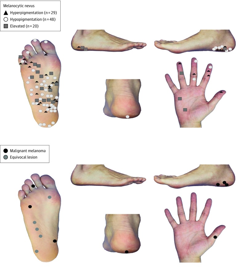
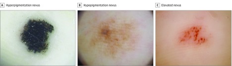
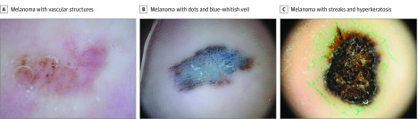
Results: Of 504 lesions, 110 (21.8%) (melanocytic nevus, 97; melanoma, 8; and equivocal melanocytic lesion, 5) from 108 patients (68 female and 40 male patients; mean age, 40.1 years [range, 1-86 years]) did not show a parallel furrow pattern, lattice-like pattern, fibrillar pattern, or parallel ridge pattern. Among them, the mean patient age was significantly higher for melanoma than for melanocytic nevus (65.3 vs 38.0 years; P < .001), as was mean maximum lesion diameter (11.8 vs 5.7 mm; P < .001). Melanomas and equivocal melanocytic lesions tended to be distributed on weight-bearing areas of the foot sole, such as the heel, while nevi were spread over non-weight-bearing regions. Dermoscopically, 95 melanocytic nevi (97.9%) were symmetrical in 1 or 2 axes while melanomas were not. A total of 91 melanocytic nevi (93.8%) had 1 or 2 colors per lesion, and 4 melanomas (50.0%) had more than 2 colors. Vascular structures were seen in 3 melanocytic nevi (3.1%) and 3 melanomas (37.5%). Blue-white structures were seen in 18 melanocytic nevi (18.6%) and 3 melanomas (37.5%). Dots and globules were seen in 22 melanocytic nevi (22.7%) and 4 melanomas (50.0%). Vascular structures, blue-white structures, and dots and globules were irregularly distributed in the melanomas. Ulcer, hyperkeratosis, and irregular streaks were observed only in melanomas.
Conclusions and relevance: More than one-fifth of melanocytic lesions on the volar skin did not display typical dermoscopic patterns. Asymmetry, numerous colors (≥3), and other melanoma-specific dermoscopic findings were more frequently observed for melanomas. Clinical information, including patient age and lesion size and location, was helpful in differentiating melanoma from melanocytic nevus. Further prospective clinical studies are warranted to clarify the diagnostic accuracy of dermoscopy combined with clinical information.
Conflict of interest statement
Figures
References
Publication types
MeSH terms
LinkOut - more resources
Full Text Sources
Other Literature Sources
Medical
Miscellaneous
