

Oxygen concentration alters mitochondrial structure and function in in vitro fertilized preimplantation mouse embryos
- PMID: 30865267
- PMCID: PMC7967792
- DOI: 10.1093/humrep/dez011
Oxygen concentration alters mitochondrial structure and function in in vitro fertilized preimplantation mouse embryos
Erratum in
-
Oxygen concentration alters mitochondrial structure and function in in vitro fertilized preimplantation mouse embryos.Hum Reprod. 2020 Jun 1;35(6):1476. doi: 10.1093/humrep/deaa047. Hum Reprod. 2020. PMID: 32436960 Free PMC article. No abstract available.
Abstract
Study question: Does the oxygen concentration in the culture medium [either physiologic (5%) or atmospheric (20%)] affect mitochondrial ultrastructure and function in preimplantation mouse embryos generated by IVF?
Summary answer: Embryos cultured in 20% oxygen show increased mitochondrial abnormalities compared to embryos cultured in 5% oxygen.
What is known already: ART are widely used and have resulted in the birth of more than 8 million children. A variety of media and oxygen concentrations are used to culture embryos. Embryos cultured under physiological O2 tension (5%) reach the blastocyst stage faster and have fewer alterations in gene expression when compared with embryos cultured under atmospheric oxygen conditions (20%). The mechanisms by which oxygen tension affects preimplantation development remain unclear, but mitochondria are believed to play an important role. The aim of this study was to evaluate how mitochondrial ultrastructure and function in IVF embryos were affected by culture under physiologic (5%) or atmospheric (20%) oxygen concentrations.
Study design, size, duration: Zygotes, 2-cell, 4-cell, morula and blastocyst were flushed out of the uterus after natural fertilization and used as controls. IVF was performed in CF1 x B6D2F1 mice and embryos were cultured in Potassium simplex optimized medium (KSOM) with amino acids (KAA) under 5% and 20% O2 until the blastocyst stage. Embryo development with the addition of antioxidants was also tested.
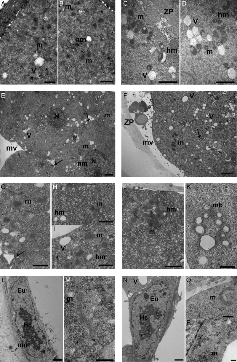
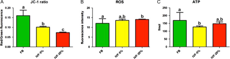
Participants/materials, setting, methods: Mitochondrial function was assessed by measuring mitochondrial membrane potential, reactive oxygen species (ROS) production, ATP levels, and the expression of selected genes involved in mitochondrial function. Mitochondria ultrastructure was evaluated by transmission electron microscopy (TEM).
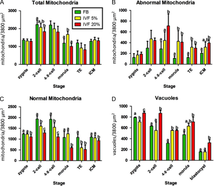
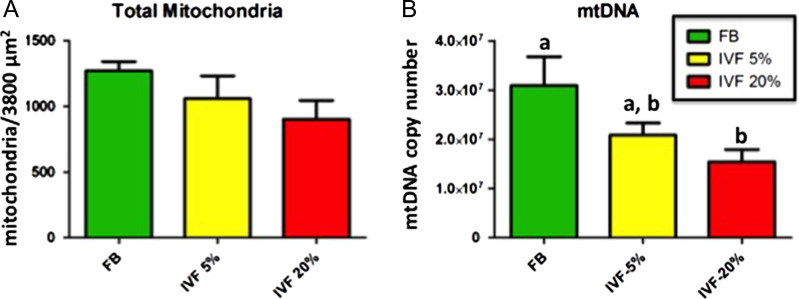
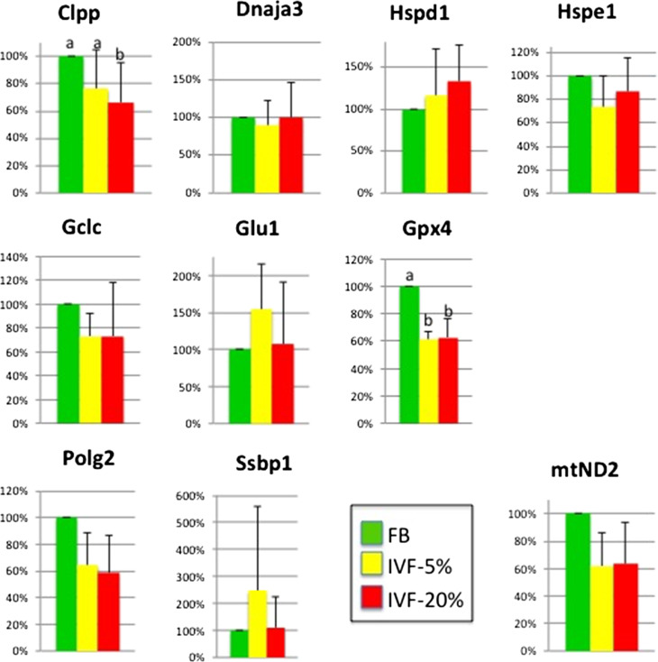
Main results and the role of chance: Embryos cultured under 20% O2 had fewer mitochondria and more vacuoles and hooded (abnormal) mitochondria compared to the other groups (P < 0.05). At the blastocyst stage the mitochondria of IVF embryos cultured in 20% O2 had lower mtDNA copy number, a denser matrix and more lamellar cristae than controls. Overall IVF-generated blastocysts had lower mitochondrial membrane potential, higher ROS levels, together with changes in the expression of selected mitochondrial genes (P < 0.05). ATP levels were significantly lower than controls only under 5% O2, with the 20% O2 IVF group having intermediate levels. Unexpectedly, adding antioxidant to the culture medium did not improve development.
Large scale data: N/A.
Limitations, reasons for caution: Findings in mice embryos might be different from human embryos.
Wider implications of the findings: This study suggests that changes in the mitochondria may be part of the mechanism by which lower oxygen concentration leads to better embryo development and further emphasize the importance of mitochondria as a locus of reprogramming.
Study funding/competing interest(s): This study was funded by R01 HD 082039 to PFR, the Department of Life, Health and Environmental Sciences, University of L'Aquila, Italy (RIA 2016-2018) and the Department of Anatomy, Histology, Forensic Medicine and Orthopaedics, La Sapienza University of Rome, Italy (University grants 2016-2017). The authors declare no competing interests.
Keywords: IVF; ROS; embryo culture; mitochondria; oxygen levels.
© The Author(s) 2019. Published by Oxford University Press on behalf of the European Society of Human Reproduction and Embryology. All rights reserved. For Permissions, please e-mail: journals.permissions@oup.com.
Figures
References
-
- Acton BM, Jurisicova A, Jurisica I, Casper RF. Alterations in mitochondrial membrane potential during preimplantation stages of mouse and human embryo development. Mol Hum Reprod 2004;10:23–32. - PubMed
-
- Adam AAG, Takahashi Y, Katagiri S, Nagano M. In vitro culture of mouse preantral follicles using membrane inserts and developmental competence of in vitro ovulated oocytes. J Reprod Dev 2004;50:579–586. - PubMed
-
- Cebral E, Carrasco I, Vantman D, Smith R. Preimplantation embryotoxicity after mouse embryo exposition to reactive oxygen species. Biocell 2007;31:51–59. - PubMed
-
- Chronopoulou E, Harper JC. IVF culture media: past, present and future. Hum Reprod Update 2015;21:39–55. - PubMed
Publication types
MeSH terms
Substances
Grants and funding
LinkOut - more resources
Full Text Sources
Other Literature Sources
Medical
Research Materials
