. 2019 Mar 13;14(3):e0199799.
doi: 10.1371/journal.pone.0199799. eCollection 2019.

Pasakbumin A controls the growth of Mycobacterium tuberculosis by enhancing the autophagy and production of antibacterial mediators in mouse macrophages

Affiliations

Pasakbumin A controls the growth of Mycobacterium tuberculosis by enhancing the autophagy and production of antibacterial mediators in mouse macrophages

Hyo-Ji Lee et al. PLoS One. .

Abstract

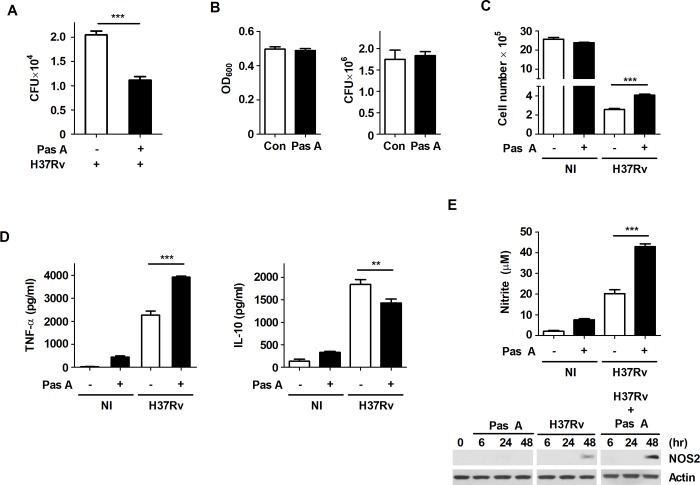
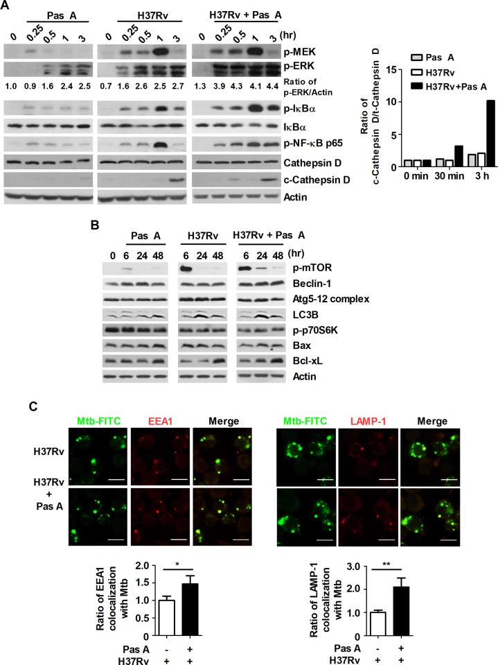
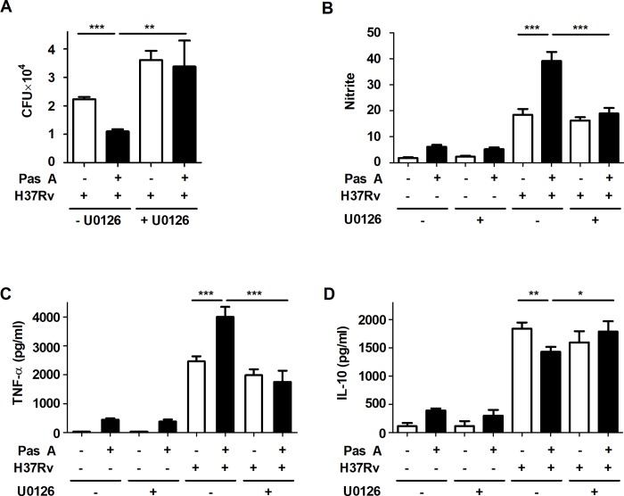
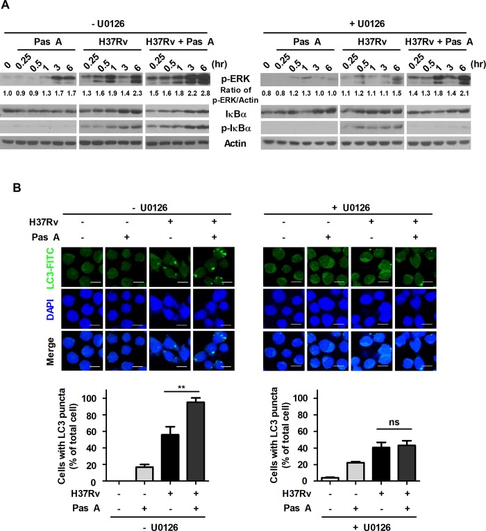
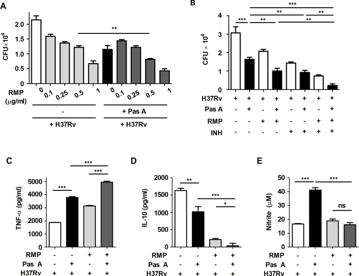
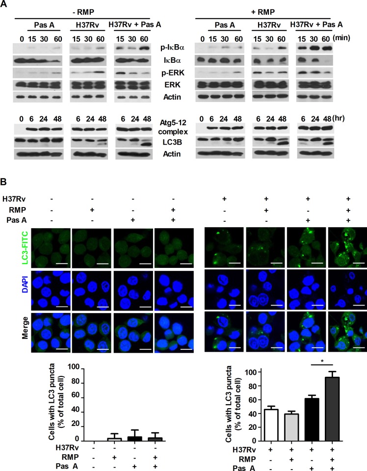
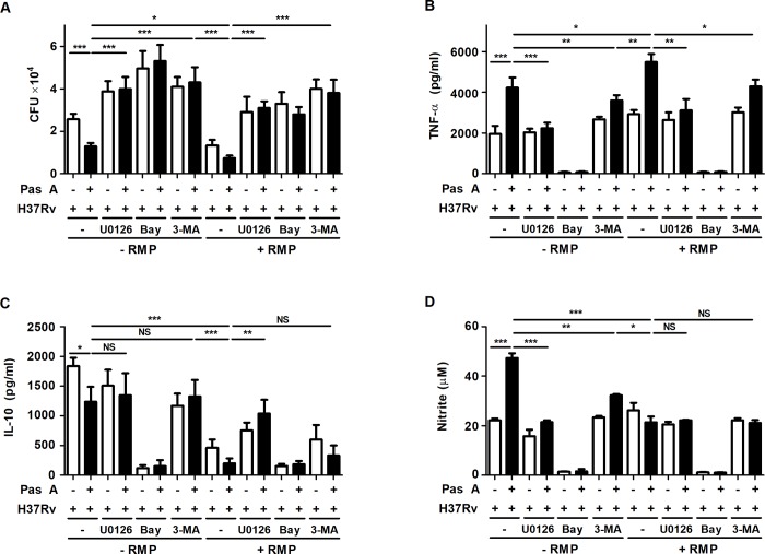
Tuberculosis (TB) is a chronic infectious disease caused by Mycobacterium tuberculosis (Mtb) and remains a major health problem worldwide. Thus, identification of new and more effective drugs to treat emerging multidrug-resistant TB (MDR-TB) and to reduce the side effects of anti-TB drugs, such as liver toxicity and other detrimental changes, is urgently needed. In this study, to develop a novel candidate drug for effective TB treatment with few side effects in the host, we selected pasakbumin A isolated from Eurycoma longifolia (E. longifolia) Jack, which protected host cells against Mtb infection-induced death. Pasakbumin A significantly inhibited intracellular Mtb growth by inducing the autophagy via the ERK1/2-mediated signaling pathway in Mtb-infected macrophages. We further investigated whether pasakbumin A could be used as a potential adjuvant for TB treatment. Treatment with pasakbumin A and anti-TB drug rifampicin (RMP) potently suppressed intracellular Mtb killing by promoting autophagy as well as TNF-α production via the ERK1/2- and NF-κB-mediated signaling pathways in Mtb-infected cells. Our results suggest that pasakbumin A could be developed as a novel anti-TB drug or host-directed therapeutic (HDT) strategy to protect against host cell death and improve host defense mechanisms against Mtb infection in macrophages.


Conflict of interest statement

The authors have declared that no competing interests exist.

Figures

Fig 1
Fig 1. Pasakbumin A controls intracellular Mtb growth by increasing the production of NO and pro-inflammatory cytokine in H37Rv-infected macrophages.
Raw264.7 macrophages were stimulated with pasakbumin A (Pas A, 10 μM) for 48 h after infection with H37Rv (at MOIs of 1 or 5). (A) Intracellular bacterial survival was determined by counting the number of CFUs at 3-weeks after inoculation. (B) The cultures were grown in 7H9 medium supplemented with 10% ADC containing 0.2% glycerol at 37°C for 72 h with or without pasakbumin A (Pas A, 10 μM). Left, bacterial growth was measured as OD600 at 72 h after Mtb inoculation. Right, colony-forming units (CFUs) were measured by plating bacterial dilutions onto 7H10 agar supplemented with 10% OADC containing 0.5% glycerol at 72 h. Error bars represents the standard deviation of the mean. (C) Cell viability was assessed using trypan-blue exclusion assay. (D) Culture supernatants were measured for the production of TNF-α and IL-10 by ELISA in non-infected (NI) or H37Rv-infected cells. (E) Bar graph in the left panel represents NO production (as indicated by the nitrite level) by diazotization (Griess method) assay. Right panel represents NOS2 protein by western blot analysis. Actin served as a loading control. The full length of all western blots has been provided in the S1 Fig. Statistical significance is indicated as **, p<0.01 and ***, p<0.001.
Fig 2
Fig 2. Pasakbumin A activates the NF-κB- and ERK1/2-mediated signaling pathways and induces autophagy in Mtb-infected macrophages.
Raw264.7 macrophages were infected with H37Rv (at a MOI of 5), and then treated with pasakbumin A (Pas A, 10 μM) for the indicated time points. Western blot analysis showed the expression for various proteins of (A) the NF-κB, MAPK signaling pathways and (B) the expression of apoptosis- and autophagy-related proteins in pasakbumin A-treated macrophages during H37Rv infection. The band intensity was quantified, and the ratio of p-ERK, LC3-II and cleaved cathepsin D band intensity was shown in bottom of the blot. (C) Raw264.7 cells were infected with FITC-labeled H37Rv, and then treated with pasakbumin A (Pas A, 10 μM) for 3 h. The colocalization of FITC-labeled Mtb and EEA1 or LAMP-1 was monitored by confocal microscopy. Bar graphs represent as fold of change over FITC-labeled Mtb infected cells to indicate the number of FITC-labeled Mtb in EEA1+ or LAMP-1+ compartment. Bar scale, 20 μM. (A) The full length of all western blots has been provided in the S2 Fig. (B) Cropped membrane from different gels was used in western blot assay.
Fig 3
Fig 3. Pasakbumin A controls the intracellular Mtb growth and production of inflammatory mediators through ERK-mediated signaling.
Raw264.7 cells were pre-treated with U0126 (10 μM) for 1 h and then stimulated with pasakbumin A (Pas A, 10 μM) during H37Rv infection (MOI of 1 or 5). (A) Intracellular bacterial survival was determined by counting the number of CFUs at 3-weeks after inoculation. (B-D) Culture supernatants were measured for the production of (B) NO, (C) TNF-α and (D) IL-10 at 48 h. The experiments were performed in triplicate. *, p<0.05; **, p<0.01 and ***, p<0.001.
Fig 4
Fig 4. Pasakbumin A induces autophagy by activating ERK1/2-mediated signaling.
Raw264.7 cells were pre-treated with U0126 (10 μM) for 1 h and then stimulated with pasakbumin A (Pas A, 10 μM) during H37Rv infection (MOI of 1 or 5). (A) The changes in the phosphorylated and total protein levels of ERK1/2 and IκBα were assessed using western blot analysis. Cropped membrane from different gels was used in western blot assay. The band intensity was quantified, and the ratio of p-ERK band intensity was shown in bottom of the blot. (B) Immunofluorescence staining of LC3 in Mtb-infected cells treated as described above. The number of LC3-positive puncta was counted under a microscope, and the percentage of cells containing LC3-positive puncta relative to the total cell number was calculated. Bar scale, 20 μM. Statistical significance is indicated as *, p<0.05 and ns, not significant (p>0.05).
Fig 5
Fig 5. Combination treatment with pasakbumin A and an anti-TB drug improves the antibacterial immunity in Mtb-infected macrophages.
(A) Raw264.7 macrophages were infected with H37Rv (at MOI of 1 or 5) for 4 h and treated with pasakbumin A (Pas A, 10 μM) alone or Pas A combined with an anti-TB drug, rifampicin (RMP), for 48 h in a dose-dependent manner. Intracellular bacterial survival was determined by counting the number of CFUs at 3-weeks after inoculation. (B) Raw264.7 macrophages were infected with H37Rv for 4 h and treated with Pas A (10 μM) alone, Pas A and RMP (0.5 μg/ml), Pas A and isoniazid (INH, 0.1 μg/ml) or Pas A and RMP/INH for 48 h. Intracellular bacterial survival was determined by counting the number of CFUs. (C, D) Culture supernatants were measured for the production of (C) TNF-α and (D) IL-10 with ELISA at 48 h. (E) NO production was detected in cell culture supernatants. Statistical significance is indicated as *, p<0.05; **, p<0.01, ***, p<0.001 and ns, not significant (p<0.05).
Fig 6
Fig 6. Treatment of pasakbumin A combined with RMP accelerates autophagy and the activation of NF-κB- and ERK1/2-mediated signaling compared to that of cells treated with pasakbumin A alone during Mtb infection.
(A) Raw264.7 macrophages were infected with H37Rv (at a MOI of 5), and then treated with pasakbumin A (Pas A, 10 μM) in presence or absence of RMP for the indicated time points. (A) The expression levels of autophagy-related proteins as well as phosphorylated and total protein for various components of the NF-κB and MAPK signaling pathways were examined by western blot assay. Cropped membrane from different gels was used in western blot assay. (B) Raw264.7 macrophages were infected with H37Rv (at a MOI of 5) for 4 h, and then treated with pasakbumin A (Pas A, 10 μM) in presence or absence of RMP for 48 h. The LC3-positive macrophages after treatment with Pas A with or without RMP during Mtb infection were detected via immunofluorescence staining. The number of LC3-positive puncta was counted under a microscope, and the percentage of cells containing LC3-positive puncta relative to the total cell number was calculated. Bar scale, 20 μM. Statistical significance is indicated as *, p<0.05.
Fig 7
Fig 7. Treatment of pasakbumin A combined with RMP effectively controls intracellular Mtb growth by accelerating autophagy via NF-κB- and ERK1/2-mediated signaling.
Raw264.7 macrophages were pre-treated with ERK1/2 inhibitor (U0126, 10 μM), NF-κB inhibitor (bay 11–7082, 10 μM) or autophagy inhibitor (3-MA, 5 mM) for 1 h and infected with H37Rv for 4 h. After 4 h, cells were treated with pasakbumin A (Pas A, 10 μM) in presence or absence of RMP for 48 h. (A) Intracellular bacterial survival was determined by counting the number of CFUs at 3-weeks after inoculation. (B, C) Culture supernatants were measured for the production of (B) TNF-α and (C) IL-10 with ELISA at 48 h. (D) NO production was detected in cell culture supernatants. Statistical significance is indicated as *, p<0.05; **, p<0.01, ***, p<0.001 and ns, not significant (p<0.05).

Similar articles

Cited by

References

    1. Zumla A, George A, Sharma V, Herbert RH, Baroness Masham of I, Oxley A, et al. The WHO 2014 global tuberculosis report—further to go. Lancet Glob Health. 2015;3(1):e10–2. 10.1016/S2214-109X(14)70361-4 . - DOI - PubMed
    1. Pieters J. Mycobacterium tuberculosis and the macrophage: maintaining a balance. Cell Host Microbe. 2008;3(6):399–407. 10.1016/j.chom.2008.05.006 . - DOI - PubMed
    1. Mortaz E, Adcock IM, Tabarsi P, Masjedi MR, Mansouri D, Velayati AA, et al. Interaction of Pattern Recognition Receptors with Mycobacterium Tuberculosis. J Clin Immunol. 2015;35(1):1–10. 10.1007/s10875-014-0103-7 - DOI - PMC - PubMed
    1. Domingo-Gonzalez R, Prince O, Cooper A, Khader SA. Cytokines and Chemokines in Mycobacterium tuberculosis Infection. Microbiol Spectr. 2016;4(5). 10.1128/microbiolspec.TBTB2-0018-2016 - DOI - PMC - PubMed
    1. North RJ, Jung YJ. Immunity to tuberculosis. Annu Rev Immunol. 2004;22:599–623. 10.1146/annurev.immunol.22.012703.104635 . - DOI - PubMed

Publication types

MeSH terms