. 2019 Mar 13;14(3):e0213666.
doi: 10.1371/journal.pone.0213666. eCollection 2019.

NAP (davunetide) preferential interaction with dynamic 3-repeat Tau explains differential protection in selected tauopathies

Affiliations

NAP (davunetide) preferential interaction with dynamic 3-repeat Tau explains differential protection in selected tauopathies

Yanina Ivashko-Pachima et al. PLoS One. .

Abstract

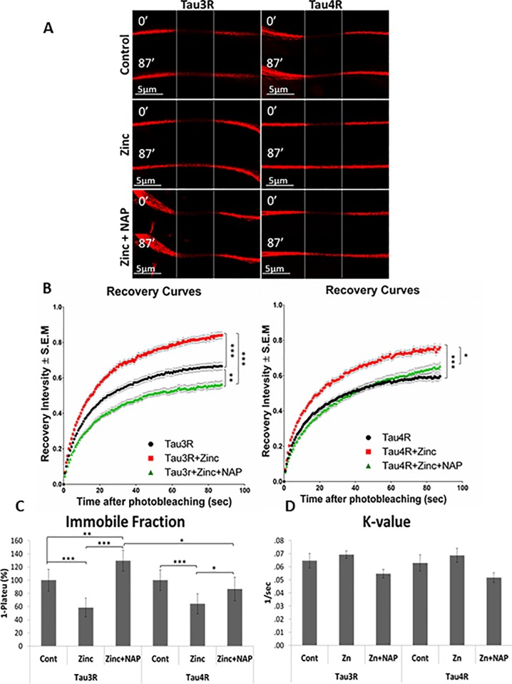
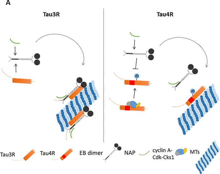
The microtubule (MT) associated protein Tau is instrumental for the regulation of MT assembly and dynamic instability, orchestrating MT-dependent cellular processes. Aberration in Tau post-translational modifications ratio deviation of spliced Tau isoforms 3 or 4 MT binding repeats (3R/4R) have been implicated in neurodegenerative tauopathies. Activity-dependent neuroprotective protein (ADNP) is vital for brain formation and cognitive function. ADNP deficiency in mice causes pathological Tau hyperphosphorylation and aggregation, correlated with impaired cognitive functions. It has been previously shown that the ADNP-derived peptide NAP protects against ADNP deficiency, exhibiting neuroprotection, MT interaction and memory protection. NAP prevents MT degradation by recruitment of Tau and end-binding proteins to MTs and expression of these proteins is required for NAP activity. Clinically, NAP (davunetide, CP201) exhibited efficacy in prodromal Alzheimer's disease patients (Tau3R/4R tauopathy) but not in progressive supranuclear palsy (increased Tau4R tauopathy). Here, we examined the potential preferential interaction of NAP with 3R vs. 4R Tau, toward personalized treatment of tauopathies. Affinity-chromatography showed that NAP preferentially interacted with Tau3R protein from rat brain extracts and fluorescence recovery after photobleaching assay indicated that NAP induced increased recruitment of human Tau3R to MTs under zinc intoxication, in comparison to Tau4R. Furthermore, we showed that NAP interaction with tubulin (MTs) was inhibited by obstruction of Tau-binding sites on MTs, confirming the requirement of Tau-MT interaction for NAP activity. The preferential interaction of NAP with Tau3R may explain clinical efficacy in mixed vs. Tau4R pathologies, and suggest effectiveness in Tau3R neurodevelopmental disorders.


Conflict of interest statement

Professor Gozes also serves as the Chief Scientific Officer of Coronis Neurosciences, developing CP201 for the ADNP syndrome, under patent protection. This does not alter our adherence to PLOS ONE policies on sharing data and materials.

Figures

Fig 1
Fig 1. NAP preferentially associates with 3R-tau.
(A) Western blot analysis of equal amounts of protein extracts from newborn (NB) rat brain cortex and 60-day-old (60d) rat brain cortex. Significantly larger amount of Tau3R was detected in the one-day-old cortical extract compared to the 60-day cortical extract, and Tau4R was recognized in the 60-day cortical extract only. (B) Bio-SafeTM Coomassie protein staining of the different fractions (F–flow-through, W—first wash, Wl—last wash, E1/2/3 –elution fractions by order) of the NAP affinity column fractions (left panel). Western blot analysis of elution fractions (E1) obtained by NAP-affinity chromatography with the same protein extracts of rat brain cortex (right panel). Tau3R, total-Tau, and tubulin were identified in the NAP-binding fraction of the newborn rat cortical brain extract, but essentially neither Tau nor tubulin was identified in the elution fraction of the 60-day-old cortical brain extract (three independent experiments). Please see S1 Fig for loading controls.
Fig 2
Fig 2. NAP induces increased recruitment of human Tau3R to MTs under zinc toxic condition in comparison to Tau4R.
(A) Representative images of photo-bleaching and fluorescence recovery of mCherry-tagged Tau3R and 4R in differentiated N1E-115 cells treated with extracellular zinc (400μM, 2hrs) with or without NAP treatment (10-12M, 2hrs). N1E-115 cells expressing m-Cherry-Tau3R/4R without any treatment represented the control. (B) FRAP recovery curves of normalized data (see “Materials and Methods”). (C) The graph represents percentages (±SEM) of the fitted data (from three independent experiments) of immobile fractions relative to control– 100% (data were collected on 87 sec after photobleaching). Normalized FRAP data were fitted with one-exponential functions (GraphPad Prism 6), and statistical analysis was performed by Two Way ANOVA (SigmaPlot 11). Statistical significance is presented by *P<0.05, **P<0.01, *** P<0.001. Tau3R: Control n = 58, zinc n = 85, zinc + NAP n = 58; Tau4R: Control n = 56, zinc n = 47, zinc + NAP n = 60.
Fig 3
Fig 3. NAP interaction with tubulin, but not with Tau, is inhibited by paclitaxel.
(A) Bio-SafeTM Coomassie protein staining of the different fractions (F–flow-through, W—first wash, Wl—last wash, E1/2/3 –elution fractions by order) obtained from NAP-affinity column loaded with protein extracts of newborn rat cerebral cortex with 4 mg paclitaxel, dissolved in 80μl DMSO, or equal volume of DMSO, alone. Almost no tubulin is evident in the elution fractions in the paclitaxel column, in contrast to the control one. (B, C) Western analysis of elution fractions obtained similarly as in panel (A). (B) Tubulin is not detected in the elution fractions of the pre-incubated paclitaxel column in comparison to the control column. (C) Tau3R is observed in the elution fractions of both the pre-incubated paclitaxel column and the control column. (D) Graphic depiction of the hypothesis that NAP, bound to an affinity column, interacts with tubulin throughout mediation of Tau.
Fig 4
Fig 4. NAP protective activity is inhibited by paclitaxel.
Cell viability test performed by the MTS assay (measures mitochondrial activity, see “Materials and Methods”). Differentiated N1E-115 cells are exposed to different concentrations of NAP (10-15M, 10-12M, 10-9M) and paclitaxel (5, 6 and 7 μM) for 2 hrs. Average of mitochondrial activity results (MTS reduction) are displayed in relation to the control (non-treated cell) value– 0.1411±0.02989. Statistical analysis of the data was performed using one-way ANOVA with Tukey post hoc test, n = 5. Statistical significance is presented relative to control as *P<0.05, **P<0.01, *** P<0.001; to “w/o NAP” (cells treated with paclitaxel, alone) as #P<0.05, ##P<0.01, ###P<0.001.
Fig 5
Fig 5. Amino-acid sequence of the Tau isoform 2 (NP_005901, 441aa) (Tau4R).
The translated protein sequence of exon 10, which is spliced in Tau3R, is marked by red. Functional motifs, predicted by ELM analysis [30] (S1 Table and Table 1) are indicated.
Fig 6
Fig 6. Increased phosphorylation of Tau4R reduces NAP effect on Tau-EB-tubulin interaction.
Differentiated human neuroblastoma SH-SY5Y cells were transfected with expression plasmids encoding GFP-Tau3R or GFP-Tau4R. Cells expressing GFP only were used as negative controls. Immunoprecipitation (IP) was performed using GFP antibodies with and without NAP (see “Materials and Methods”). (A) Flow-through (F), first and third washes (W1 and W3) fractions were collected and analyzed by immunoblot with GFP antibody (IB: GFP). (B) Elution fractions (E) were collected and analyzed by immunoblotting (IB) with the appropriate antibodies, as listed on the figure.
Fig 7
Fig 7. Suggested explanation for the preference of NAP binding to Tau3R over Tau4R.
Graphic depiction of our hypothesis suggesting the preference of NAP to interact with Tau3R based on the findings of the ELM prediction analysis and experimental results presented in Fig 6. The second MT-binding repeat of Tau4R (spliced in Tau3R) includes cyclin A-docking motif and thus may enable Tau binding to cyclin A, essential for the activation of Cdk1/2 [31]. Cks1 may also associate with Cdk and cyclin A to form more efficient cyclin A-Cdk-Csk1 phosphorylation complex. Tau4R phosphorylation by Cdk1/2 differs from Cdk5 (a conventional Tau kinase) [46] and may disturb/attenuate Tau-EB interaction, which has been previously indicated as a crucial for NAP interaction with MTs [16, 35]. The EB dimer structure was constructed according to a published review [42].

References

    1. Witte H, Neukirchen D, Bradke F. Microtubule stabilization specifies initial neuronal polarization. The Journal of cell biology. 2008;180(3):619–32. 10.1083/jcb.200707042 - DOI - PMC - PubMed
    1. Lee VM, Balin BJ, Otvos L Jr., Trojanowski JQ. A68: a major subunit of paired helical filaments and derivatized forms of normal Tau. Science. 1991;251(4994):675–8. . - PubMed
    1. Trojanowski JQ, Schuck T, Schmidt ML, Lee VM. Distribution of tau proteins in the normal human central and peripheral nervous system. The journal of histochemistry and cytochemistry: official journal of the Histochemistry Society. 1989;37(2):209–15. 10.1177/37.2.2492045 . - DOI - PubMed
    1. Morris M, Maeda S, Vossel K, Mucke L. The many faces of tau. Neuron. 2011;70(3):410–26. 10.1016/j.neuron.2011.04.009 - DOI - PMC - PubMed
    1. Lee VM, Goedert M, Trojanowski JQ. Neurodegenerative tauopathies. Annual review of neuroscience. 2001;24:1121–59. 10.1146/annurev.neuro.24.1.1121 . - DOI - PubMed

Publication types

MeSH terms