. 2019 Mar 7;24(5):929.
doi: 10.3390/molecules24050929.

Bee Venom and Its Peptide Component Melittin Suppress Growth and Migration of Melanoma Cells via Inhibition of PI3K/AKT/mTOR and MAPK Pathways

Affiliations

Bee Venom and Its Peptide Component Melittin Suppress Growth and Migration of Melanoma Cells via Inhibition of PI3K/AKT/mTOR and MAPK Pathways

Haet Nim Lim et al. Molecules. .

Abstract

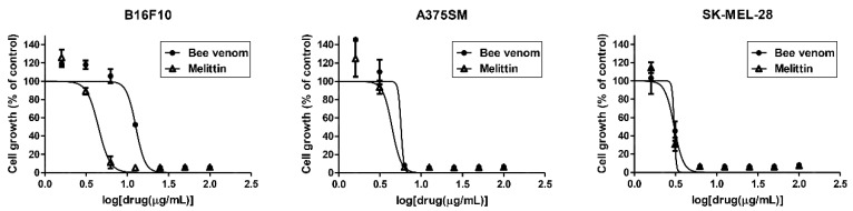
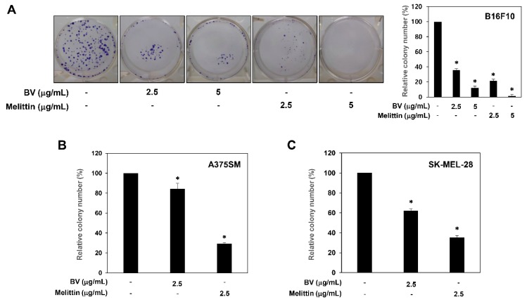
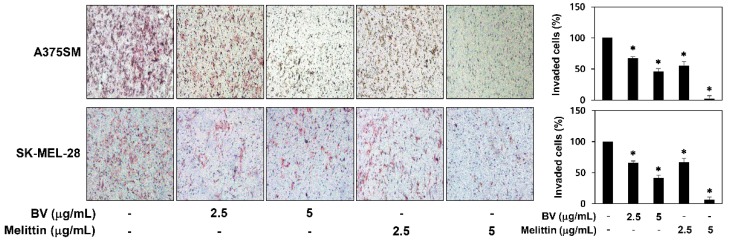
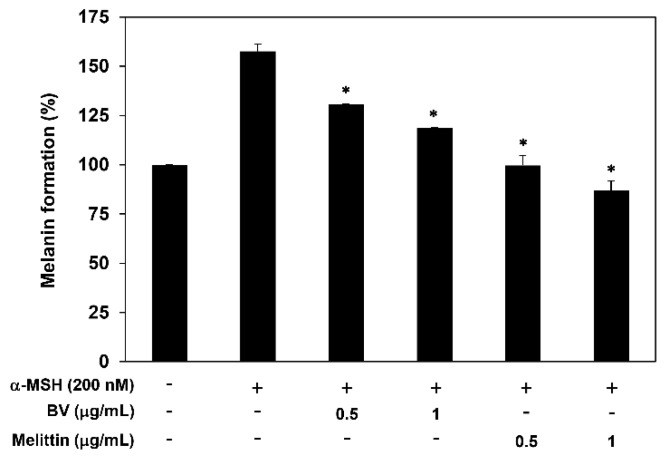
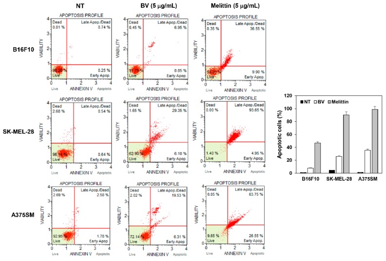
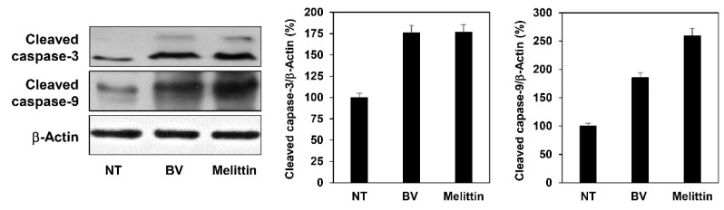
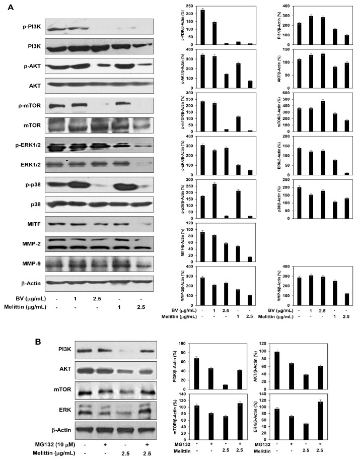
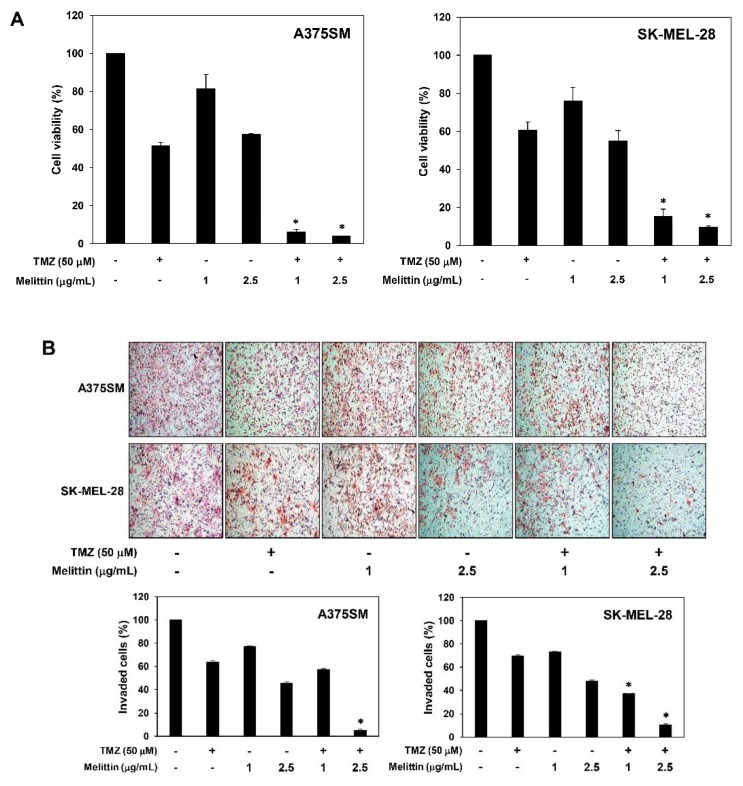
Malignant melanoma is the deadliest form of skin cancer and highly chemoresistant. Melittin, an amphiphilic peptide containing 26 amino acid residues, is the major active ingredient from bee venom (BV). Although melittin is known to have several biological activities such as anti-inflammatory, antibacterial and anticancer effects, its antimelanoma effect and underlying molecular mechanism have not been fully elucidated. In the current study, we investigated the inhibitory effect and action mechanism of BV and melittin against various melanoma cells including B16F10, A375SM and SK-MEL-28. BV and melittin potently suppressed the growth, clonogenic survival, migration and invasion of melanoma cells. They also reduced the melanin formation in α-melanocyte-stimulating hormone (MSH)-stimulated melanoma cells. Furthermore, BV and melittin induced the apoptosis of melanoma cells by enhancing the activities of caspase-3 and -9. In addition, we demonstrated that the antimelanoma effect of BV and melittin is associated with the downregulation of PI3K/AKT/mTOR and MAPK signaling pathways. We also found that the combination of melittin with the chemotherapeutic agent temozolomide (TMZ) significantly increases the inhibition of growth as well as invasion in melanoma cells compared to melittin or TMZ alone. Taken together, these results suggest that melittin could be potentially applied for the prevention and treatment of malignant melanoma.

Keywords: AKT; MAPK; bee venom; melanoma; melittin; temozolomide.


Conflict of interest statement

The authors declare no conflict of interest.

Figures

Figure 1
Figure 1
The effect of BV and melittin on the growth of melanoma cell lines. Cells were treated with various concentrations of BV and melittin for 72 h, and cell growth was measured using the MTT colorimetric assay. Data were presented as percentage relative to DMSO-treated control (% of control). Each value represents the mean ± SE from three independent experiments.
Figure 2
Figure 2
The effect of BV and melittin on the colony forming ability of melanoma cell lines. (A) B16F10, (B) A375SM and (C) SK-MEL-28 cells were treated with BV and melittin for 8–10 days. The cell colonies were detected by crystal violet staining and then counted. * p < 0.05 versus the control. Each value represents the mean ± SE from three independent experiments.
Figure 3
Figure 3
The effect of BV and melittin on the migration of melanoma cell lines. The migratory potential of melanoma cells was analyzed using wound healing assay. Cells were treated with BV and melittin for 24 h. Dotted black lines indicate the edge of the gap at 0 h. * p < 0.05 versus the control. Each value represents the mean ± SE from three independent experiments.
Figure 4
Figure 4
The effect of BV and melittin on the invasion of melanoma cell lines. The invasiveness of melanoma cells was analyzed using Matrigel-coated polycarbonate filters. Cells were treated with BV and melittin for 24 h. Cells penetrating the filters were stained and counted under an optical microscope. * p < 0.05 versus the control. Each value represents the mean ± SE from three independent experiments.
Figure 5
Figure 5
The effect of BV and melittin on the melanogenesis of α-MSH-stimulated B16F10 cells. Cells were treated with BV and melittin in the presence or absence of α-MSH for 72 h, and the cellular melanin contents were determined. * p < 0.05 versus the α-MSH control. Each value represents the mean ± SE from three independent experiments.
Figure 6
Figure 6
The effect of BV and melittin on the apoptotic cell death of melanoma cell lines. Cells were treated with BV and melittin for 24 h. Apoptotic cells were determined by flow cytometric analysis following annexin V-FITC and propidium iodide (PI) dual labeling. Each value represents the mean ± SE from three independent experiments.
Figure 7
Figure 7
The effect of BV and melittin on the expression of apoptosis regulatory proteins in A375SM melanoma cells. Cells were treated with BV and melittin for 24 h, and the expression levels of cleaved caspase-3 and cleaved caspase-9 were detected by Western blotting. The levels of β-actin were used as an internal control. Each value represents the mean ± SE from three independent experiments.
Figure 8
Figure 8
The effect of BV and melittin on the regulation of PI3K/AKT/mTOR and MAPK pathways. A375SM melanoma cells were treated with (A) BV, melittin and (B) MG132, and the protein levels were detected by Western blot analysis using specific antibodies. The levels of β-actin were used as an internal control. Each value represents the mean ± SE from three independent experiments.
Figure 9
Figure 9
The effect of the combination of melittin with TMZ on the growth and invasion of melanoma cell lines. (A) Cells were treated with melittin and TMZ for 72 h, and cell growth was measured using the MTT colorimetric assay. (B) Cells were treated with melittin and TMZ for 24 h. Cells penetrating the Matrigel-coated polycarbonate filters were stained and counted under an optical microscope. * p < 0.05 versus the single agent treatment. Each value represents the mean ± SE from three independent experiments.

Similar articles

Cited by

References

    1. Miller K.D., Siegel R.L., Lin C.C., Mariotto A.B., Kramer J.L., Rowland J.H., Stein K.D., Alteri R., Jemal A. Cancer treatment and survivorship statistics. CA Cancer J. Clin. 2016;66:271–289. doi: 10.3322/caac.21349. - DOI - PubMed
    1. Cummins D.L., Cummins J.M., Pantle H., Silverman M.A., Leonard A.L., Chanmugam A. Cutaneous malignant melanoma. Mayo Clin. Proc. 2006;81:500–507. doi: 10.4065/81.4.500. - DOI - PubMed
    1. Kato M., Liu W., Akhand A.A., Hossain K., Takeda K., Takahashi M., Nakashima I. Ultraviolet radiation induces both full activation of ret kinase and malignant melanocytic tumor promotion in RFP-RET-transgenic mice. J. Investig. Dermatol. 2000;115:1157–1158. doi: 10.1046/j.1523-1747.2000.0202a-2.x. - DOI - PubMed
    1. Libra M., Malaponte G., Navolanic P.M., Gangemi P., Bevelacqua V., Proietti L., Bruni B., Stivala F., Mazzarino M.C., Travali S., et al. Analysis of BRAF mutation in primary and metastatic melanoma. Cell Cycle. 2005;4:1382–1384. doi: 10.4161/cc.4.10.2026. - DOI - PubMed
    1. Hocker T.L., Singh M.K., Tsao H. Melanoma genetics and therapeutic approaches in the 21st century: Moving from the benchside to the bedside. J. Investig. Dermatol. 2008;128:2575–2595. doi: 10.1038/jid.2008.226. - DOI - PubMed

MeSH terms