Review
. 2019 Mar 7;20(5):1170.
doi: 10.3390/ijms20051170.

Dual-Specificity Phosphatases in Neuroblastoma Cell Growth and Differentiation

Affiliations
Review

Dual-Specificity Phosphatases in Neuroblastoma Cell Growth and Differentiation

Caroline E Nunes-Xavier et al. Int J Mol Sci. .

Abstract

Dual-specificity phosphatases (DUSPs) are important regulators of neuronal cell growth and differentiation by targeting proteins essential to neuronal survival in signaling pathways, among which the MAP kinases (MAPKs) stand out. DUSPs include the MAPK phosphatases (MKPs), a family of enzymes that directly dephosphorylate MAPKs, as well as the small-size atypical DUSPs, a group of low molecular-weight enzymes which display more heterogeneous substrate specificity. Neuroblastoma (NB) is a malignancy intimately associated with the course of neuronal and neuroendocrine cell differentiation, and constitutes the source of more common extracranial solid pediatric tumors. Here, we review the current knowledge on the involvement of MKPs and small-size atypical DUSPs in NB cell growth and differentiation, and discuss the potential of DUSPs as predictive biomarkers and therapeutic targets in human NB.

Keywords: MAP kinase phosphatases; MAP kinases; atypical dual-specificity phosphatases; dual-specificity phosphatases; neuroblastoma; neuronal differentiation.


Conflict of interest statement

The authors declare no conflict of interest.

Figures
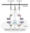
Figure 1
Figure 1
Schematic depiction of the major pathways involved in signaling through the ALK-MYCN axis in neuroblastoma (NB). ALK signals downstream mainly through the RAS-ERK1/2 MAP kinase (MAPK) pathway, as well as through the PI3K/AKT and JAK/STAT pathways, resulting in increased MYCN transcription and cell growth (straight dashed lines). Signaling through Trk neurotrophin receptors is also shown. MYCN transcriptional activity positively feeds the pathway by promoting ALK transcription (curved blue dotted lines), whereas transcriptional activity mediated by the MAPK nuclear effectors negatively feed-back the MAPK pathways by promoting the transcription of MAPK phosphatases (MKP) genes (curved red dotted line). Straight solid lines indicate direct dephosphorylation of protein substrates by MKPs or by other dual-specificity phosphatases (DUSPs). Dephosphorylation of MAPKs by MKPs is well documented, whereas evidence on the dephosphorylation of STATs or Trks by other DUSPs is limited. See text for more details.
Figure 2
Figure 2
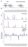
(A) Schematic depiction of the domain composition of MKPs and small-atypical DUSPs. KIM—kinase interaction motif; PTP—protein tyrosine phosphatase. (B) Upper panel: mRNA expression of MKPs and small-atypical DUSPs in adrenal gland. Data from GTEx (Genotype-Tissue Expression) data sets. RNA-seq data is reported as median RPKM (reads per kilobase per million reads mapped) (https://www.proteinatlas.org/). Middle panel: mRNA expression of MKPs and small-atypical DUSPs in SH-SY5Y human NB cells; data from the Human Protein Atlas. RNA-seq data is reported as median TPM (transcripts per kilobase of exon per million reads) (https://www.proteinatlas.org/). Bottom panel: mRNA expression analysis of MKPs and small-atypical DUSPs from SH-SY5Y, SMS-KCNR, and IMR-32 human NB cell lines treated with retinoic acid (RA). Cell lines were kept untreated or were treated for 10 days with RA, mRNA was extracted and RT-qPCR was performed using a set of MKP and small-atypical DUSP primers, as described in Reference [67]. Relative mRNA expression values are shown in Log2 as fold change +SD of treated cells versus untreated cells, from at least two independent experiments. Mean fold change above 2 or below −2 was considered significant. MKP mRNA expression data from bottom panel has been previously published [67].
Figure 3
Figure 3
Immunostaining of selected MKPs from tissue sections from two NB tumors (#1: Stage I, unknown MYCN status, low-risk; #2: Stage IV, metastatic, MYCN amplification, high-risk). Representative staining patterns are shown. DUSP5 and DUSP7 mostly showed high-moderate immunoreactivity, whereas DUSP9 mostly displayed low immunoreactivity (magnification is ×100). Immunostaining was performed as described in [67].

References

    1. Siegel R.L., Miller K.D., Jemal A. Cancer statistics, 2018. CA Cancer J. Clin. 2018;68:7–30. doi: 10.3322/caac.21442. - DOI - PubMed
    1. Ward E., DeSantis C., Robbins A., Kohler B., Jemal A. Childhood and adolescent cancer statistics, 2014. CA Cancer J. Clin. 2014;64:83–103. doi: 10.3322/caac.21219. - DOI - PubMed
    1. Maris J.M., Hogarty M.D., Bagatell R., Cohn S.L. Neuroblastoma. Lancet. 2007;369:2106–2120. doi: 10.1016/S0140-6736(07)60983-0. - DOI - PubMed
    1. Matthay K.K., Maris J.M., Schleiermacher G., Nakagawara A., Mackall C.L., Diller L., Weiss W.A. Neuroblastoma. Nat. Rev. Dis. Primers. 2016;2:16078. doi: 10.1038/nrdp.2016.78. - DOI - PubMed
    1. Tsubota S., Kadomatsu K. Origin and initiation mechanisms of neuroblastoma. Cell Tissue Res. 2018;372:211–221. doi: 10.1007/s00441-018-2796-z. - DOI - PubMed

MeSH terms

Substances