Review
. 2019 Mar 7;11(3):232.
doi: 10.3390/v11030232.

Kuru, the First Human Prion Disease

Affiliations
Review

Kuru, the First Human Prion Disease

Paweł P Liberski et al. Viruses. .

Abstract

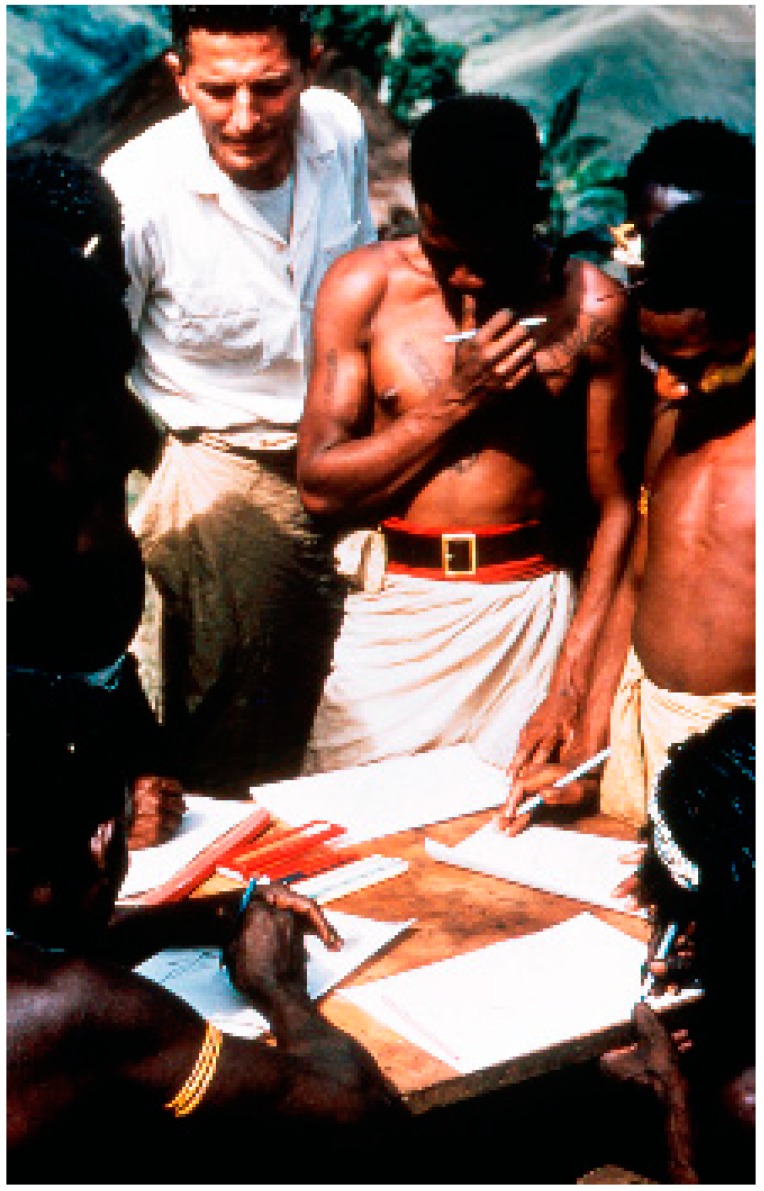
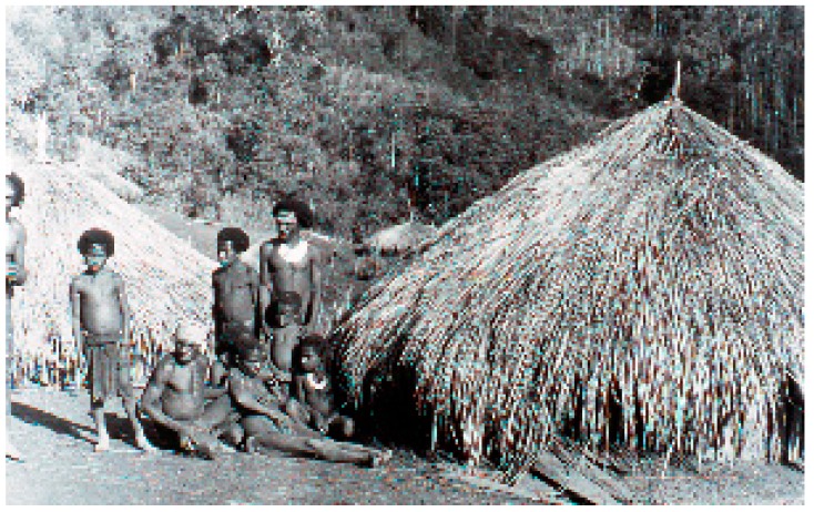
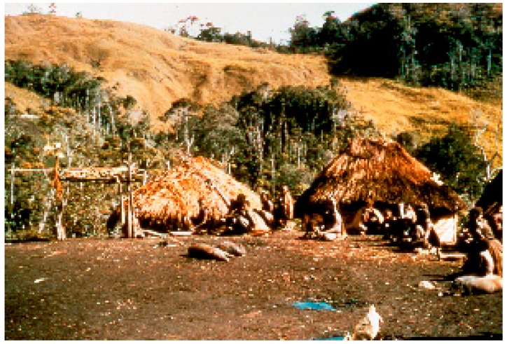
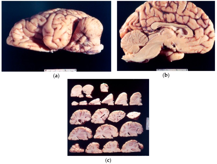
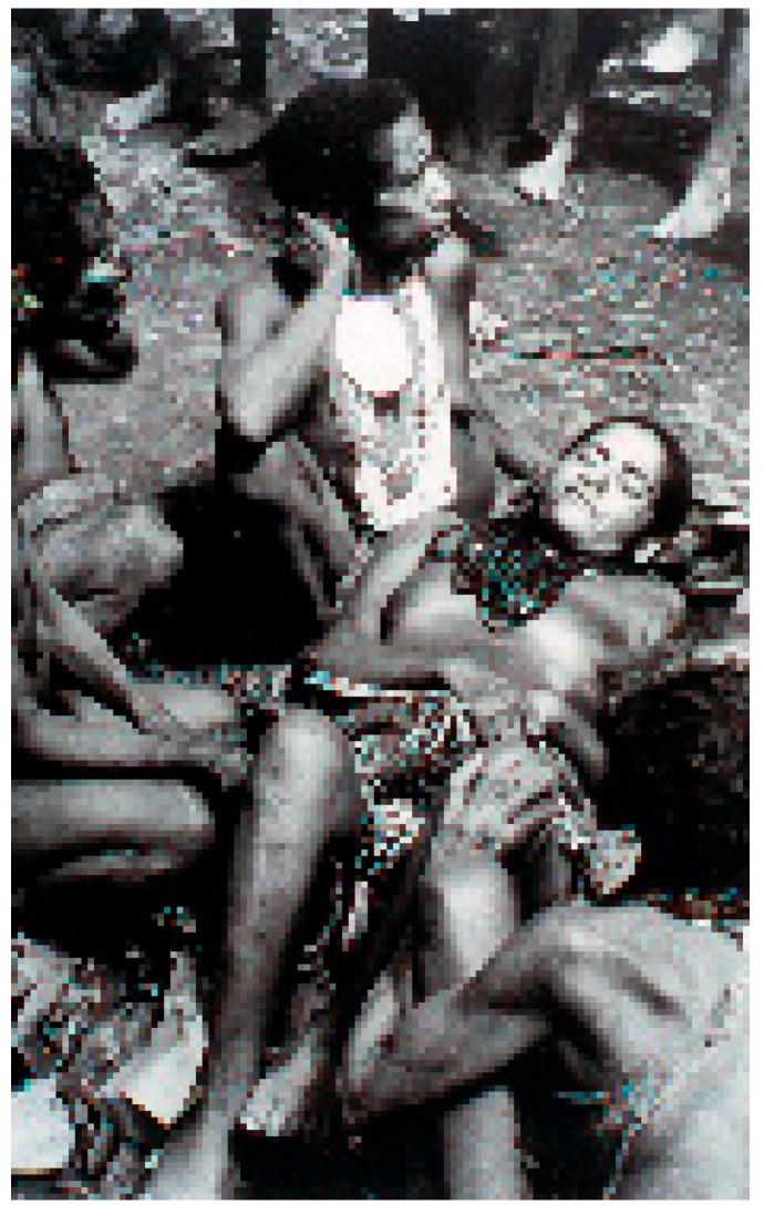
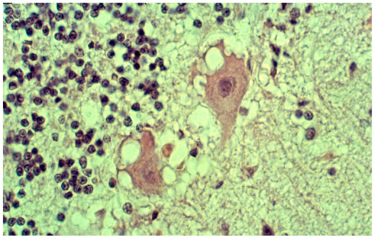
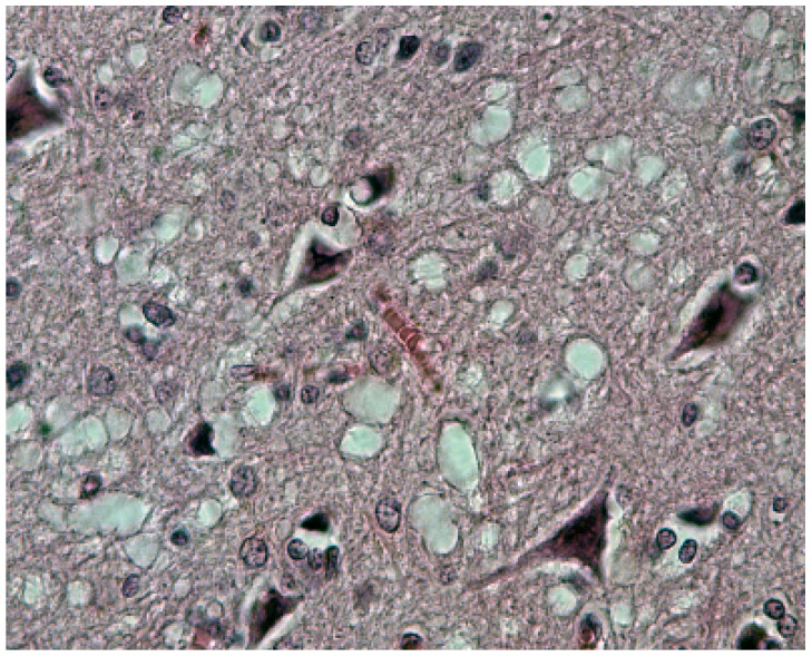
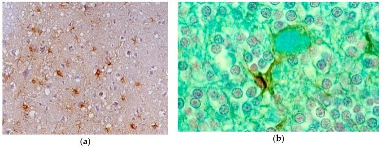
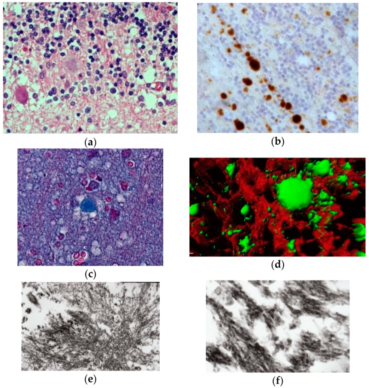
Kuru, the first human prion disease was transmitted to chimpanzees by D. Carleton Gajdusek (1923⁻2008). In this review, we summarize the history of this seminal discovery, its anthropological background, epidemiology, clinical picture, neuropathology, and molecular genetics. We provide descriptions of electron microscopy and confocal microscopy of kuru amyloid plaques retrieved from a paraffin-embedded block of an old kuru case, named Kupenota. The discovery of kuru opened new vistas of human medicine and was pivotal in the subsequent transmission of Creutzfeldt⁻Jakob disease, as well as the relevance that bovine spongiform encephalopathy had for transmission to humans. The transmission of kuru was one of the greatest contributions to biomedical sciences of the 20th century.

Keywords: Carleton Gajdusek; Kuru; neuropathology; prion diseases.


Conflict of interest statement

The authors declare no conflicts of interest.

Figures

Figure 1
Figure 1
Dr. D. Carleton Gajdusek (lhr-57-ng-332). Courtesy of D. Carleton Gajdusek.
Figure 2
Figure 2
Dr. Vin Zigas (dcg-57-ng-336). Courtesy of D. Carleton Gajdusek.
Figure 3
Figure 3
A general view of a Fore hamlet. A kuru victim is sitting in the first row between two supporters (dcg-ng-bw-12). Courtesy of D. Carleton Gajdusek.
Figure 4
Figure 4
A general view of a Fore hamlet (dcg-57-ng-186). Courtesy of D. Carleton Gajdusek.
Figure 5
Figure 5
(ac) Macroscopic picture of the chimp brain (67-10825-2-1261214; 67-10825-5-1261214; 67-10825-5-126121-24). Courtesy of D. Carleton Gajdusek.
Figure 6
Figure 6
A group of several Fore men (dcg-57-ng-118). Courtesy of D. Carleton Gajdusek.
Figure 7
Figure 7
A group of kuru victims. Courtesy of D. Carleton Gajdusek.
Figure 8
Figure 8
A boy afflicted with kuru showing atethoid movements (dcg-ng-bw-5). Courtesy of D. Carleton Gajdusek.
Figure 9
Figure 9
A group of children with kuru (dcg-bw-9). Courtesy of D. Carleton Gajdusek.
Figure 10
Figure 10
A group of women afflicted with kuru (dcg-57-ng-573). Courtesy of D. Carleton Gajdusek.
Figure 11
Figure 11
A woman affected with kuru (dcg-ng-57-346A). Courtesy of D. Carleton Gajdusek.
Figure 12
Figure 12
A dead woman with kuru (dcg-ng-bw-11). Courtesy of D. Carleton Gajdusek.
Figure 13
Figure 13
A vacuolated “moth-eaten” neuron. Courtesy of D. Carleton Gajdusek.
Figure 14
Figure 14
Typical spongiform change (41-93-2a-40-3). Courtesy of D. Carleton Gajdusek.
Figure 15
Figure 15
(a) Proliferation of GFAP-positive astrocytes against the background of spongiform change; (b) amyloid plaque stained with Alcian blue surrounded by an astrocyte. Courtesy of D. Carleton Gajdusek.
Figure 16
Figure 16
(a) Typical kuru plaque stained silver. Courtesy of D. Carleton Gajdusek; (b) a linear row of kuru plaques probably attached to a neurite; (c) a kuru plaque stained with Alzian blue (41-93-2a-alcian-20-17-alcian). Courtesy of D. Carleton Gajdusek; (d) A kuru plaque and PrPSc deposits surrounded by glial cells in the human cerebellum. Confocal laser microscopy, prion protein (clone 12F10, Alexa Fluor 488)—green, GFAP (polyclonal rabbit, AlexaFluor 546)—red, magnification 600×, digital zoom 2.1×. Three-dimensional reconstruction with surface rendering. (e) Electron microscopic image of the kuru plaque, material reversed from paraffin, 13,000×; (f); higher magnification to show bundles of kuru plaque-fibrils, 50,000×.

Comment in

Similar articles

Cited by

References

    1. Asher D.M., Gibbs C.J., Jr., Sulima M.P., Bacote A., Amyx H., Gajdusek D.C. Transmission of human spongiform encephalopathies to experimental animals: Comparison of chimpanzee and Squirrel monkey. In: Brown P., editor. Transmissible Spongiform Encephalopathies—Impact on Animal and Human Health. Volume 80. Karger; Basel, Switzerland: 1993. pp. 9–13. - PubMed
    1. Asher D.M. Kuru: Memories of the NIH years. Phil. Trans. R. Soc. B. 2008;363:3618–3625. doi: 10.1098/rstb.2008.4002. - DOI - PMC - PubMed
    1. Gajdusek D.C. Kuru: An appraisal of five years of investigation. Eugen. Q. 1962;9:69–74. doi: 10.1080/19485565.1962.9987505. - DOI - PubMed
    1. Gibbs C.J., Jr., Gajdusek D.C. Infection as the etiology of spongiform encephalopathy (Creutzfeldt-jakob disease) Science. 1969;165:1023–1025. doi: 10.1126/science.165.3897.1023. - DOI - PubMed
    1. Hornabrook R.W. Kuru. Papua New Guinea Med. J. 1975;4:203–206. doi: 10.1016/S0140-6736(70)90356-9. - DOI - PubMed

LinkOut - more resources