Review
. 2019 Jun;39(6):939-958.
doi: 10.1177/0271678X19835552. Epub 2019 Mar 13.

The post-cardiac arrest syndrome: A case for lung-brain coupling and opportunities for neuroprotection

Affiliations
Review

The post-cardiac arrest syndrome: A case for lung-brain coupling and opportunities for neuroprotection

Nguyen Mai et al. J Cereb Blood Flow Metab. 2019 Jun.

Abstract

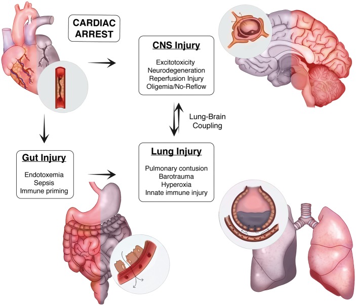
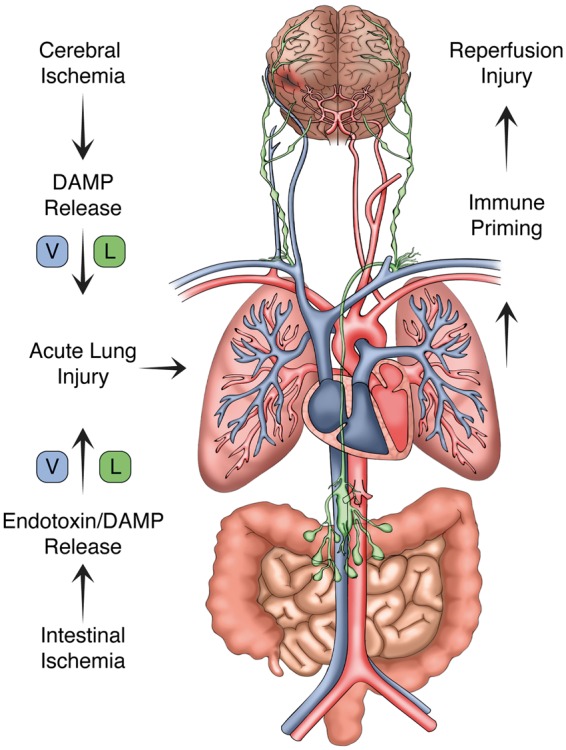
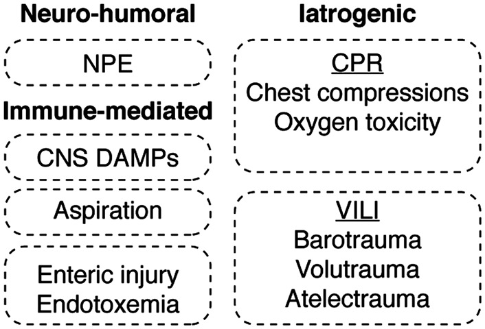
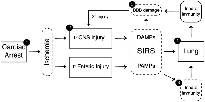
Systemic inflammation and multi-organ failure represent hallmarks of the post-cardiac arrest syndrome (PCAS) and predict severe neurological injury and often fatal outcomes. Current interventions for cardiac arrest focus on the reversal of precipitating cardiac pathologies and the implementation of supportive measures with the goal of limiting damage to at-risk tissue. Despite the widespread use of targeted temperature management, there remain no proven approaches to manage reperfusion injury in the period following the return of spontaneous circulation. Recent evidence has implicated the lung as a moderator of systemic inflammation following remote somatic injury in part through effects on innate immune priming. In this review, we explore concepts related to lung-dependent innate immune priming and its potential role in PCAS. Specifically, we propose and investigate the conceptual model of lung-brain coupling drawing from the broader literature connecting tissue damage and acute lung injury with cerebral reperfusion injury. Subsequently, we consider the role that interventions designed to short-circuit lung-dependent immune priming might play in improving patient outcomes following cardiac arrest and possibly other acute neurological injuries.

Keywords: Cardiac arrest; acute lung injury; blood–brain barrier; damage-associated molecular patterns; innate immune priming; ischemic neurodegeneration; neuroprotection; neutrophil; pathogen-associated molecular patterns; post-cardiac arrest syndrome; sepsis; systemic inflammatory response syndrome.


Figures

Figure 1.
Figure 1.
Lung–brain coupling in the post-cardiac arrest syndrome. Acute myocardial ischemia and associated ventricular arrhythmia induce end organ ischemic injury by perturbing systemic perfusion. In the CNS, ischemia induces cellular injury directly through both acute and delayed mechanisms. In the periphery, ischemia damages the physical, biochemical, and immune defenses present within the intestinal mucosae resulting in the translocation of gastrointestinal flora and systemic release of pro-inflammatory molecules and microorganisms. With the return of spontaneous circulation and tissue reperfusion, hematogenous spread of DAMPs from the CNS and PAMPs from the gut trigger acute lung inflammation. These systemic factors, combined with a variety of iatrogenic insults to the lung during the early phase of treatment, contribute to a feed-forward mechanism of reperfusion injury that heightens post-ischemic neuroinflammation.
Figure 2.
Figure 2.
The lymphatic system and lung–brain immune priming. With the return of spontaneous circulation, both the brain and gut release damage-associated molecular patterns (DAMPs) and pathogen-associated molecular patterns (PAMPs) into the circulation. CNS DAMPs conveyed via cerebral venous return or indirectly via cervical lymphatic channels induce acute lung inflammation. PAMPs generated within the gut are delivered to the pulmonary circulation through analogous venous and lymphatic channels. As shown, the thoracic duct provides lymphatic drainage from the abdominal cavity and lower extremities terminating at the angle of the left subclavian and internal jugular veins. Following the return of spontaneous circulation, these channels contribute to acute lung inflammation, which in turn trigger systemic immune priming and secondary CNS reperfusion injury.
Figure 3.
Figure 3.
Mechanisms of acute lung injury in PCAS. The mechanisms leading to acute lung injury in the post-cardiac arrest syndrome are divided into neuro-humoral, immune-mediated and iatrogenic etiologies. Neurogenic pulmonary edema (NPE) reflects an increase in pulmonary interstitial and alveolar fluid that stems from sympathetically mediated pathways. The lung is also subject to immune priming caused by the ischemia-induced release of both DAMPs and PAMPs from post-ischemic tissues combined with the inflammatory effects of aspiration pneumonitis. Equally important, iatrogenic factors encountered in the course of cardiopulmonary resuscitation (CPR), as well as those associated with ventilator-induced lung injury (VILI), are shown.
Figure 4.
Figure 4.
Neurotherapeutic targets in PCAS. The identification of lung-brain coupling in the post-cardiac arrest syndrome provides an additional point of intervention to reduce post-ischemic neurodegeneration. These include shortening the time to ROSC and optimization of supportive measures during the prehospital phase of care (1) and devising acute interventions (2) that can be combined with targeted temperature management to induce early neuroprotection (3). The delayed administration of agents that block required ligand-receptor immune interactions in the periphery could prevent systemic priming. Finally, interventions that reduce acute lung injury (4) or preserve blood-brain barrier (BBB) integrity (5) would, in theory, reduce systemic leukocyte activation, reduce cerebral edema, maintain CNS immune privilege, and hasten the resolution of neuroinflammation.
Figure 5.
Figure 5.
Therapeutic approaches to target lung–brain coupling in PCAS. Developing effective neuroprotective strategies for the post-cardiac arrest syndrome may require utilizing unconventional delivery routes. While traditional IV delivery of BBB-penetrant small molecules remains a viable approach, intranasal administration of both peptides and small compounds has shown promise in conveying anti-inflammatory and neuroprotective effects on the CNS. Also, targeting the alimentary tract through either oral or IP delivery of a therapeutic could mitigate against mesenteric injury and the production and release of priming antigens produced by the enteric flora. To reduce the effects of acute lung injury on CNS reperfusion, inhalation of therapeutic compounds and biologics during the pre-hospital phase of care could modulate lung–brain coupling early in the course of disease.

References

    1. Kilgannon JH, Jones AE, Shapiro NI, et al. Association between arterial hyperoxia following resuscitation from cardiac arrest and in-hospital mortality. JAMA 2010; 303: 2165–2171. - PubMed
    1. Albert CM, Mittleman MA, Chae CU, et al. Triggering of sudden death from cardiac causes by vigorous exertion. N Engl J Med 2000; 343: 1355–1361. - PubMed
    1. Wood MA, Stambler BS, Damiano RJ, et al. Lessons learned from data logging in a multicenter clinical trial using a late-generation implantable cardioverter-defibrillator. The Guardian ATP 4210 Multicenter Investigators Group. J Am Coll Cardiol 1994; 24: 1692–1699. - PubMed
    1. Go AS, Mozaffarian D, Roger VL, et al. Heart disease and stroke statistics – 2014 update: a report from the American Heart Association. Circulation 2014; 129: e28–e292. - PMC - PubMed
    1. Nadkarni VM, Larkin GL, Peberdy MA, et al. First documented rhythm and clinical outcome from in-hospital cardiac arrest among children and adults. JAMA 2006; 295: 50–57. - PubMed

Publication types

LinkOut - more resources