

Lysosomal dysfunction in proteinopathic neurodegenerative disorders: possible therapeutic roles of cAMP and zinc
- PMID: 30866990
- PMCID: PMC6417073
- DOI: 10.1186/s13041-019-0439-2
Lysosomal dysfunction in proteinopathic neurodegenerative disorders: possible therapeutic roles of cAMP and zinc
Abstract
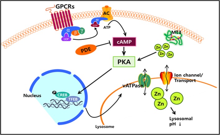
A number of neurodegenerative diseases, including Alzheimer's disease, Parkinson's disease, and amyotrophic lateral sclerosis, share intra- and/or extracellular deposition of protein aggregates as a common core pathology. While the species of accumulating proteins are distinct in each disease, an increasing body of evidence indicates that defects in the protein clearance system play a crucial role in the gradual accumulation of protein aggregates. Among protein degradation systems, the endosome-autophagosome-lysosome pathway (EALP) is the main degradation machinery, especially for large protein aggregates. Lysosomal dysfunction or defects in fusion with vesicles containing cargo are commonly observed abnormalities in proteinopathic neurodegenerative diseases. In this review, we discuss the available evidence for a mechanistic connection between components of the EALP-especially lysosomes-and neurodegenerative diseases. We also focus on lysosomal pH regulation and its significance in maintaining flux through the EALP. Finally, we suggest that raising cAMP and free zinc levels in brain cells may be beneficial in normalizing lysosomal pH and EALP flux.
Keywords: EALP; Lysosome; MT3; Zinc; cAMP.
Conflict of interest statement
Ethics approval and consent to participate
Not applicable
Consent for publication
Not applicable
Competing interests
The author declares that they have no competing interests.
Publisher’s Note
Springer Nature remains neutral with regard to jurisdictional claims in published maps and institutional affiliations.
Figures
References
-
- Komatsu M, Waguri S, Chiba T, Murata S, Iwata J, Tanida I, et al. Loss of autophagy in the central nervous system causes neurodegeneration in mice. Nature. 2006;441:880–884. - PubMed
-
- Jin S. Autophagy, mitochondrial quality control, and oncogenesis. Autophagy. 2006;2:80–84. - PubMed
-
- De Duve C, Wattiaux R. Functions of lysosomes. Annu Rev Physiol. 1966;28:435–492. - PubMed
-
- Rajawat YS, Hilioti Z, Bossis I. Aging: central role for autophagy and the lysosomal degradative system. Ageing Res Rev. 2009;8:199–213. - PubMed
-
- Eskelinen EL. New insights into the mechanisms of macroautophagy in mammalian cells. Int Rev Cell Mol Biol. 2008;266:207–247. - PubMed
Publication types
MeSH terms
Substances
LinkOut - more resources
Full Text Sources
Other Literature Sources
Medical
