. 2019 Mar 12;10(1):83.
doi: 10.1186/s13287-019-1163-7.

A scalable solution for isolating human multipotent clinical-grade neural stem cells from ES precursors

Affiliations

A scalable solution for isolating human multipotent clinical-grade neural stem cells from ES precursors

Dasa Bohaciakova et al. Stem Cell Res Ther. .

Abstract

Background: A well-characterized method has not yet been established to reproducibly, efficiently, and safely isolate large numbers of clinical-grade multipotent human neural stem cells (hNSCs) from embryonic stem cells (hESCs). Consequently, the transplantation of neurogenic/gliogenic precursors into the CNS for the purpose of cell replacement or neuroprotection in humans with injury or disease has not achieved widespread testing and implementation.

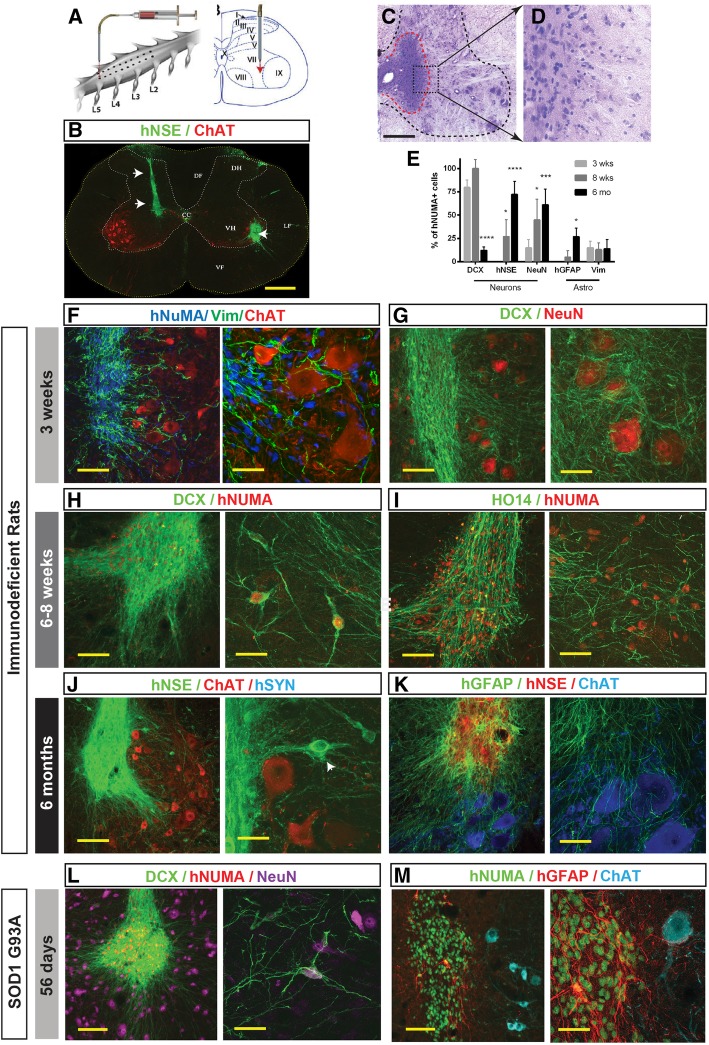
Methods: Here, we establish an approach for the in vitro isolation of a highly expandable population of hNSCs using the manual selection of neural precursors based on their colony morphology (CoMo-NSC). The purity and NSC properties of established and extensively expanded CoMo-NSC were validated by expression of NSC markers (flow cytometry, mRNA sequencing), lack of pluripotent markers and by their tumorigenic/differentiation profile after in vivo spinal grafting in three different animal models, including (i) immunodeficient rats, (ii) immunosuppressed ALS rats (SOD1G93A), or (iii) spinally injured immunosuppressed minipigs.

Results: In vitro analysis of established CoMo-NSCs showed a consistent expression of NSC markers (Sox1, Sox2, Nestin, CD24) with lack of pluripotent markers (Nanog) and stable karyotype for more than 15 passages. Gene profiling and histology revealed that spinally grafted CoMo-NSCs differentiate into neurons, astrocytes, and oligodendrocytes over a 2-6-month period in vivo without forming neoplastic derivatives or abnormal structures. Moreover, transplanted CoMo-NSCs formed neurons with synaptic contacts and glia in a variety of host environments including immunodeficient rats, immunosuppressed ALS rats (SOD1G93A), or spinally injured minipigs, indicating these cells have favorable safety and differentiation characteristics.

Conclusions: These data demonstrate that manually selected CoMo-NSCs represent a safe and expandable NSC population which can effectively be used in prospective human clinical cell replacement trials for the treatment of a variety of neurodegenerative disorders, including ALS, stroke, spinal traumatic, or spinal ischemic injury.

Keywords: Amyotrophic lateral sclerosis (ALS); Bioinformatic tools to study xenografts; Human embryonic stem cell (hESC); Neural stem cell (NSC); Spinal cord; Spinal traumatic injury.


Conflict of interest statement

Ethics approval and consent to participate

Animal studies

All animal studies were approved by the University of California, San Diego Institutional Animal Care and Use Committee (Protocol No.: S01193) and by the Institutional Animal Care and Use Committee of the Czech Academy of Sciences and were in compliance with The Association for Assessment of Laboratory Animal Care guidelines for animal use.

Use of human ES lines

This study was approved by the University of California, San Diego (UCSD) Internal Review Board (IRB), (approval ID#101323ZX).

Consent for publication

Not applicable.

Competing interests

The authors declare that they have no competing interests.

Publisher’s Note

Springer Nature remains neutral with regard to jurisdictional claims in published maps and institutional affiliations.

Figures

Fig. 1
Fig. 1
Strategy for generation, expansion, and characterization of human embryonic stem cell-derived neural stem cells. a Schematic diagram depicting the experimental design of in vitro ES-NSCs generation and in vitro and in vivo post-grafting characterization. b, c Morphology of NSCs derived by FACS Sorting (FACS-NSCs) and clonal morphology manual selection (CoMo-NSCs) at passage 10 and 15. d, e Expression of NSC-specific and ESC-specific markers determined by flow cytometry at passages less than 15 (d) and greater than 15 (e). Data are represented as mean ± SEM. f Differential gene expression plot showing the log-fold change and average transcripts per million (TPM) of each gene when comparing CoMo-NSCs and FACS-NSCs. Gray dots represent genes that are not significantly different between the two groups; the absence of black dots seen in the plot indicate that there were no genes that were significantly different between the two groups. g Heat map of log2(TPM+1) values of genes that distinguish ESCs from FACS-NSCs across ESC, FACS-NSC, and CoMo-NSC samples. The selection of genes is described in the methods (scale bars: b, c 50 μm)
Fig. 2
Fig. 2
In vitro proliferating clonal morphology-derived NSCs (CoMo-NSCs) show consistent morphology and expression of markers characteristic of immature NSCs. a, b Characteristic stable morphology of CoMo-NSCs in low (a) and high (b) density. c Expression of selected NSC markers (Sox1, Sox2, Nestin, CD24, Pax6, and CD44) evaluated by flow cytometry at different passages. Data are represented as mean ± SEM. df Expression of NSC-specific markers (Sox2, Nestin, Plzf, Dach-1, N-cadherin) and tight junction protein ZO-1 in undifferentiated CoMo-NSCs as determined by indirect immunofluorescence (scale bars: a, b 25 μm; df 10 μm)
Fig. 3
Fig. 3
CoMo-NSCs generate astrocytes and functional neurons upon in vitro differentiation. a, b Changing morphology of differentiating CoMo-NSCs towards large flat cell type after 40 days treatment with astrocyte-inducting media. cf Expression of human-specific GFAP (hGFAP) and CD44 in CoMo-NSCs-derived astrocytes and human fetal brain-derived astrocytes. A comparable expression pattern for both markers can be seen. g Representative flow cytometry plots from fixed/permeabilized cells at day 21 of astrocyte differentiation. CoMo-NSC-derived astrocytes were nearly all CD44+, with a fraction expressing GFAP. Primary fetal astrocytes (ScienCell) were used as a positive control, compared to CoMo-NSCs as a negative control. All cells lacked signal when analyzed in the absence of antibodies (data not shown). h Expression of synapsin promoter-driven GFP and appearance of neuronal morphology in CoMo-NSC-derived neurons at 6 weeks after induction using BDNF, GDNF, and cAMP. il Expression of neuronal markers (DCX, MAP2, human-specific axonal neurofilament HO14 and NeuN) in CoMo-NSC-derived neurons at 6 weeks after induction. mp Patch-clamp recording in Syn-GFP neurons in vitro: voltage-clamp recording in Syn-GFP + neurons with fast inward (Na+) and persistent outward (K+) currents in depolarized membrane potentials (characteristic of neuronal cells) can be seen (o). In current-clamp recording (membrane potential − 65 mV), action potentials are triggered by depolarizing current pulses (p) (scale bars: a, b 100 μm; c, e 10 μm; h 200 μm; ik 50 μm; l 25 μm)
Fig. 4
Fig. 4
Spinally grafted clonal-derived NSCs show a long-term engraftment, no tumor formation, and time-dependent expression of human-specific markers characteristic of immature and mature neurons and glial cells. a Single suspension of NSCs was injected bilaterally into central gray matter of lumbar spinal cord segments in immunodeficient or G93A ALS rat using glass capillary. b Grafted cells were identified by expression of human-specific markers such as hNSE (green; white arrows). c, d H&E staining of lumbar spinal cord section at 6 months after NSCs grafting show well engrafted cells (red dotted area) with no detectable tumor formation. e Quantitative analysis of neuronal and glial differentiation at 3 weeks, 8 weeks, and 6 months after spinal NSC grafting in immunodeficient rats. Data are expressed as percent of double-stained hNUMA/DCX, hNUMA/hNSE, hNUMA/NeuN, hNUMA/GFAP, and hNUMA/Vim relative to hNUMA+ cells. Data are presented as mean ± SD. f, g At 3 weeks after grafting, a marker characteristic for proliferating immature glial precursors (Vimentin) and early post-mitotic neurons (DCX) are seen in grafted hNUMA+ cells. Extensive axo-dendritic sprouting of DCX+ positive processes surrounding the host interneurons and α-motoneurons can be seen (g). h, i At 6–8 weeks after NSCs transplantation, a more advanced cell migration and neuronal maturation were seen. Numerous double hNUMA/DCX+ neurons residing outside of the graft core were identified in the gray matter (h). Similarly, extensive axonal sprouting of HO14+ human axons was seen in the host gray matter (i). j, k At 6 months after NSCs grafting the appearance of mature neuronal and glial markers was identified throughout the graft. A high intensity of human-specific NSE was seen in grafted areas with several hNSE+ neurons identified outside of the graft (j, white arrow). Staining with human-specific GFAP antibody showed a high density of GFAP+ network with numerous hGFAP+ processes found in the ventral gray matter between α-motoneurons of the host (k). l, m Analysis of grafted NSCs at 56 days after grafting in G93A ALS rat lumbar spinal cord. A high density of double hNUMA/DCX-stained grafts was seen throughout the grafted segments (l). Staining with hGFAP showed only relatively few hGFAP+ astrocytes and these were preferentially found at the borders of individual hNUMA+ grafts (m) (scale bars: b, c 500 μm; f, g 100 μm; h, i 300 μm; j 300 μm; k 100 μm; l 300 μm; m 200 μm)
Fig. 5
Fig. 5
Spinally grafted clonal NSCs-derived neurons acquire preferential inhibitory neurotransmitter phenotype and develop synaptic contacts with host neurons in the immunodeficient rat at 6 months post-grafting. a A high density of human-specific synaptophysin puncta (hSYN) in areas occupied by human axons (HO14) and residing in the vicinity of the host ChAT+ α-motoneurons can be seen. b, c Pre-embedding immunohistochemical staining with hSYN antibody coupled with electron-optical analysis showed numerous hSYN+ puncta (b; semithin 1 μm section) and developed synaptic contacts between hSYN+ terminals and host neurons with readily identifiable pre- and postsynaptic densities (c; red boxed area). di Triple staining with VGAT, hSYN and NeuN antibody showed a high-density hSYN puncta through the graft as well as in surrounding host tissue. A high number of hSYN + puncta co-expressed VGAT and were residing on the membranes of the host ChAT+ α-motoneurons (f; white arrows). jm Triple staining with VGAT, hSYN, and gephyrin (glycine receptor-associated protein) showed numerous double-stained hSYN/VGAT+ puncta in opposition to postsynaptically bound gephyrin+ profiles (jl, m-white arrows). np Staining with GAD65, hSYN, NeuN, and VGLUT1–3 antibodies showed only occasional presence of VGLUT1–3+ terminals in association with hSYN puncta (scale bars: a 30 μm; b 20 μm; c 350 nm; d 500 μm; e 50 μm; f 30 μm; gi 10 μm; j 20 μm; m, p 5 μm)
Fig. 6
Fig. 6
RNA-Seq analysis of transplanted CoMo-NSCs in immunodeficient rats at 2 and 6 months post-transplantation using bioinformatics-based species splitting. a Generalized schematic of RNA-Seq analysis pipeline using bioinformatics-based species splitting. Following analyses were conducted using the resulting human-specific transcripts only, reflecting expression profiles of the human CoMo-NSCs. b Principal components analysis (PCA) of three populations: CoMo-NSCs pre-transplantation (black dots, n = 2), CoMo-NSCs 2 months post-transplantation (red dots, n = 3), and CoMo-NSCs 6 months post-transplantation (blue dots, n = 3). The plot depicts principal components 1 (PC1) and 2 (PC2) with the percent of variance for each component. c, d Differential gene expression plot comparing CoMo-NSCs 2 months post-transplantation to CoMo-NSCs pre-transplantation (c) and 6 months post-transplantation to 2 months transplantation (d) as depicted as log2 average gene expression levels versus log2 fold change. Black dots represent genes that are significantly differentially expressed (p < 0.05). e Heat map of gene expression of canonical cell-type specific genes across the pre-transplanted and post-transplanted samples. f Gene ontology network of gene ontology terms overrepresented by genes enriched in the CoMo-NSCs pre-transplantation (e). Gene ontology groups: (1) mRNA processing, splicing, export; (2) RNA, DNA binding, repair; (3) Cell division, cell cycle; (4) Adherens junction; (5) Mismatch, double-strand break repair; (6) Ribosome biogenesis; (7) Proteoglycans and microRNAs in cancer; (8) RNA transport, processing, splicing; (9) Organ regeneration; (10) Regulation and localization; (11) Viral process; (12) Activity; (13) Assembly; (14) Gene expression; (15) Liver development; (16) ATP-dependent chromatin remodeling; (17) Translational initiation. g Gene ontology network of gene ontology terms overrepresented by genes enriched in the CoMo-NSCs post-transplantation (e). Gene ontology groups: (1) Circadian entrainment; (2) Synaptic transmission, long-term memory; (3) Signaling pathways; (4) Neuroactive ligand-receptor interaction; (5) Glutamatergic, GABAergic synapse; (6) Neurotransmitter, glutamate, dopamine secretion; (7) Membrane potential, ion transmembrane transport; (8) Morphine, nicotine addiction; (9) Locomotory behavior; (10) Action potential, excitatory postsynaptic potential; (11) Calcium ion-regulated exocytosis of neurotransmitter; (12) Ion transmembrane transport, channel activity; (13) Response to amphetamine; (14) Cardiac conduction; (15) Sensory perception of pain

Similar articles

Cited by

References

    1. Deb A, Thornton JD, Sambamoorthi U, et al. Direct and indirect cost of managing Alzheimer’s disease and related dementias in the United States. Expert Rev Pharmacoecon Outcomes Res. 2017;17:189–202. doi: 10.1080/14737167.2017.1313118. - DOI - PMC - PubMed
    1. Rubiano AM, Carney N, Chesnut R, et al. Global neurotrauma research challenges and opportunities. Nature. 2015;527:S193–S197. doi: 10.1038/nature16035. - DOI - PubMed
    1. Gladman M, Zinman L. The economic impact of amyotrophic lateral sclerosis: a systematic review. Expert Rev Pharmacoecon Outcomes Res. 2015;15:439–450. doi: 10.1586/14737167.2015.1039941. - DOI - PubMed
    1. Casarosa S, Bozzi Y, Conti L. Neural stem cells: ready for therapeutic applications? Mol Cell Ther. 2014;2:31. doi: 10.1186/2052-8426-2-31. - DOI - PMC - PubMed
    1. Rossi F, Cattaneo E. Opinion: neural stem cell therapy for neurological diseases: dreams and reality. Nat Rev Neurosci. 2002;3:401–409. doi: 10.1038/nrn809. - DOI - PubMed

Publication types