. 2019 Mar 13;10(1):93.
doi: 10.1186/s13287-019-1188-y.

Human ADSC xenograft through IL-6 secretion activates M2 macrophages responsible for the repair of damaged muscle tissue

Affiliations

Human ADSC xenograft through IL-6 secretion activates M2 macrophages responsible for the repair of damaged muscle tissue

Ewelina Pilny et al. Stem Cell Res Ther. .

Abstract

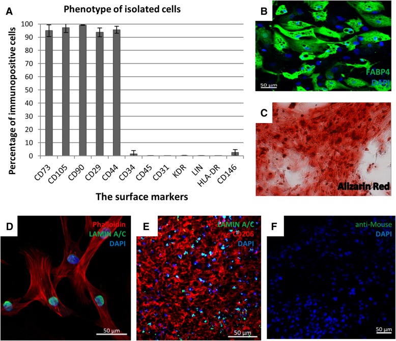
Background: Adipose-derived mesenchymal stromal cells (ADSCs) are multipotent stromal cells. The cells secrete a number of cytokines and growth factors and show immunoregulatory and proangiogenic properties. Their properties may be used to repair damaged tissues. The aim of our work is to explain the muscle damage repair mechanism with the utilization of the human adipose-derived mesenchymal stromal cells (hADSCs).

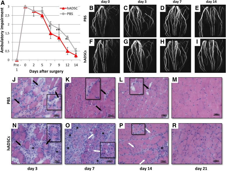
Methods: For the hADSCs isolation, we used the subcutaneous adipose tissue collected during the surgery. The murine hind limb ischemia was used as a model. The unilateral femoral artery ligation was performed on 10-12-week-old male C57BL/6NCrl and NOD SCID mice. The mice received PBS- (controls) or 1 × 10⁶ hADSCs. One, 3, 7, 14 and 21 days after the surgery, we collected the gastrocnemius muscles for the immunohistochemical analysis. The results were analyzed with relevant tests using the Statistica software.

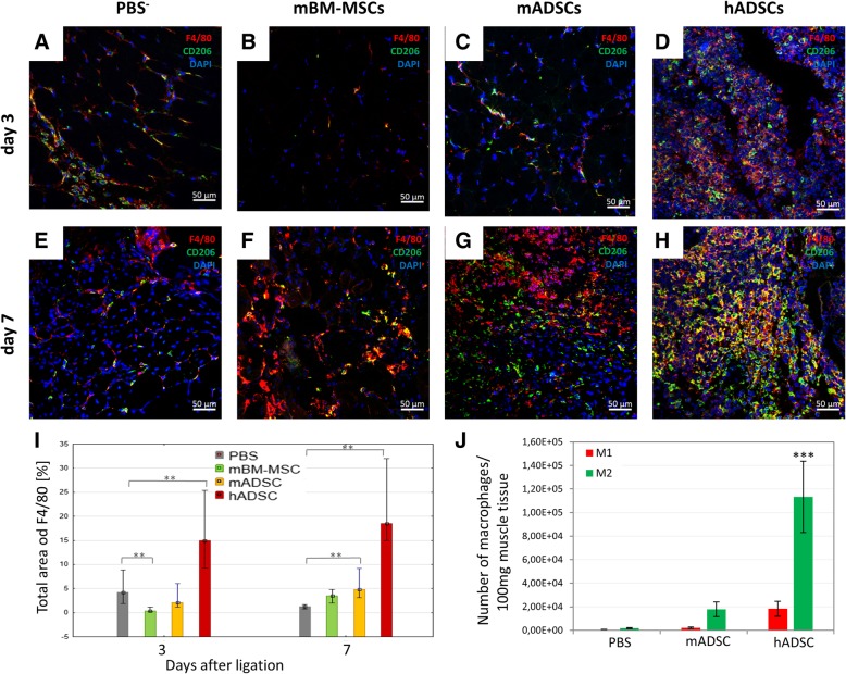
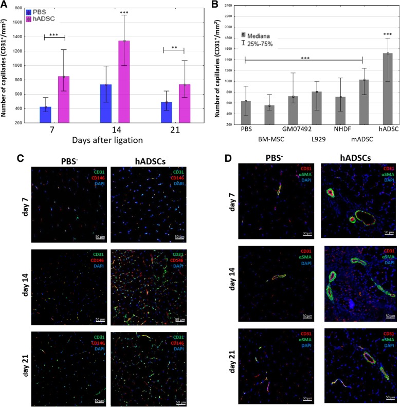
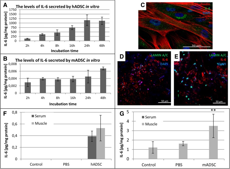
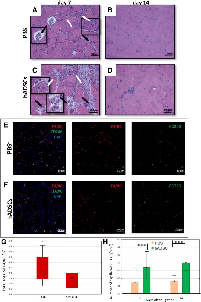
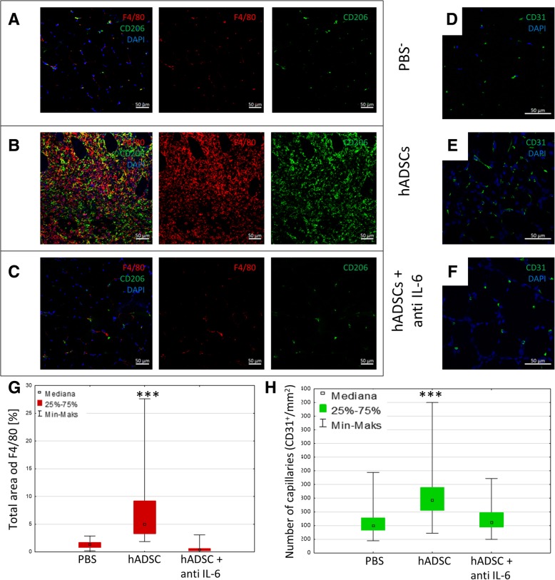
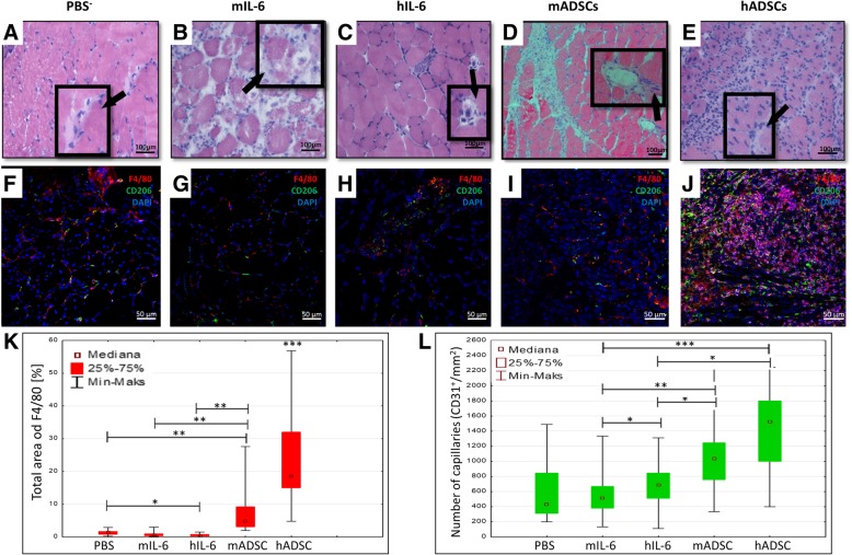
Results: The retention time of hADSCs in the limb lasted about 14 days. In the mice receiving hADSCs, the improvement in the functionality of the damaged limb occurred faster than in the control mice. More new blood vessels were formed in the limbs of the mice receiving hADSCs than in limbs of the control mice. hADSCs also increased the infiltration of the macrophages with the M2 phenotype (7-AAD-/CD45+/F4/80+/CD206+) into the ischemic limbs. hADSCs introduced into the limb of mice secreted interleukin-6. This cytokine stimulates the emergence of the proangiogenic M2 macrophages, involved, among others, in the repair of a damaged tissue. Both macrophage depletion and IL-6 blockage suppressed the therapeutic effect of hADSCs. In the mice treated with hADSCs and liposomes with clodronate (macrophages depletion), the number of capillaries formed was lower than in the mice treated with hADSCs alone. Administration of hADSCs to the mice that received siltuximab (human IL-6 blocker) did not cause an influx of the M2 macrophages, and the number of capillaries formed was at the level of the control group, as in contrast to the mice that received only the hADSCs.

Conclusions: The proposed mechanism for the repair of the damaged muscle using hADSCs is based on the activity of IL-6. In our opinion, the cytokine, secreted by the hADSCs, stimulates the M2 macrophages responsible for repairing damaged muscle and forming new blood vessels.

Keywords: Blood vessels; IL-6; M2 macrophages; Repairing damaged muscle; Xenograft; hADSCs.


Conflict of interest statement

Ethics approval and consent to participate

The experiments with animals were performed in accordance with the Declaration of Helsinki and with the approval of the Local Ethics Committee for Animal Experiments in Katowice (Permit Number: KB430–17/14).

Researches using human adipose tissue has been accepted by the Committee on the Bioethics Maria Sklodowska-Curie Institute - Oncology Center, Gliwice Branch (Permit Number: KB / 430–35 / 14).

Consent for publication

Not applicable.

Competing interests

The authors declare that they have no competing interests.

Publisher’s Note

Springer Nature remains neutral with regard to jurisdictional claims in published maps and institutional affiliations.

Figures

Fig. 1
Fig. 1
The hADSCs characteristics in vitro and their retention time after the administration to the murine muscle tissue. a Phenotype of hADSC (passage #2; n = 12 Flow cytometry). The phenotype fulfills all the accepted criteria for MSCs identification; b adipogenic differentiation—formation of lipid vesicles stained with FABP4 antibody (green, passage #2, n = 5); c osteogenic differentiation—calcium deposits stained with Alizarin Red (red, passage #2, n = 7, magn × 20); d immunofluorescence phalloidin staining of actin filaments (red) and Lamin A/C (green) in hADSCs in vitro. e The representative image of double immunofluorescent staining of human specific Lamin A/C (green) and macrophages (CD206, red) on day 7. Image shows that injected hADSC localize in clusters, presumably at the injection site. f Image shows control immunofluorescence staining with secondary Biotinylated Anti-Mouse and Fluorescein Avidin antibodies alone. Nuclei were stained with DAPI (blue), scale bars = 50 μm
Fig. 2
Fig. 2
MR imaging of vascular system and muscle regeneration in mouse gastrocnemius muscle after the hindlimb ischemia. The mice given hADSC demonstrated improved functional outcomes as compared to the control mice (PBS) (n = 10)*p < 0.05, **p < 0.01 by the U Mann-Whitney test (a). Effectiveness of femoral artery ligation and consecutive restoration of the blood flow in the ischemic hindlimbs were visually evaluated using magnetic resonance angiography. Representative magnetic resonance angiography images (maximum intensity projections) of mouse hindlimbs were acquired immediately after the femoral artery ligation (b—control mouse, f—hADSC treated mouse), on day 3 (c—control mouse, g—hADSC treated mouse), on day 7 (d—control mouse, h—hADSC-treated mouse) and on day 14 (e—control mouse, i—hADSC-treated mouse). The representative images of the transverse sections of the gastrocnemius muscle tissues by H&E staining after PBS (j, k, l, m) and hADSC (n, o, p, r) injection at 3, 7, 14, and 21 days after surgery. Necrotic muscle fibers (black arrows) with pale cytoplasm were observed at 3 (j), 7 (k), and 14 (l) days after the PBS administration. Regenerative small, basophilic muscle fibers with one or more centrally located nuclei (white arrows) were observed in all hADSC groups. Scale bar: 100 μm (× 20 magnification)
Fig. 3
Fig. 3
hADSCs injection in the ischemic muscles increased the infiltration of the proangiogenic M2 macrophages. Immunofluorescence analyzes in collected muscles 3 (a, b, c, d) and 7 (e, f, g, h) days after the ligation of the artery and the administration of hADSC (d, h), mADSC (c, g), mBM-MSC (b, f), and control (PBS) (a, e). The representative images of M2 macrophages (F4/80+ (red) / CD206+ (green)). The images show that after the hADSC injection into ischemic muscle the infiltration of the proangiogenic macrophages M2 was observed on the 3rd day after the surgery and it increased at day 7. After the administration of mBM-MSC and PBS, the significant M2 macrophage infiltration was not observed. However, after the administration of mADSC an increase in macrophage infiltration was observed on day 7. The graph of the total area of F4/80 macrophages in collected muscles on the 3rd and 7th day after the surgery (I). The total area occupied by F4/80 macrophages was calculated using ImageJ software in 10 fields of view at a magnification of 40 × (n = 5; 5 muscles per group were analyzed; 10 pictures of each muscle were taken). Using the flow cytometry 7 days after the artery ligation and administration of the hADSCs, mADSCs, and PBS in collected muscles the number of M1 (defined as 7-AAD/CD45+/F4/80+/ CD86+) and M2 (defined as 7-AAD/CD45+/F4/80+/CD206+) macrophages was evaluated (j). In all researched groups we observed a greater number of M2 macrophages in comparison to M1 macrophages. The largest number of M2 macrophages was found in muscles after the administration of hADSCs: six times more than mADSCs group and sixty times more than PBS group. The ratio of M2 to M1 macrophages was 686 in hADSCs group, 824 in mADSCs group and 364 in PBS group (n = 5). **p < 0.001 evaluated with Kruskal-Wallis one-way analysis of variance and multiple comparison of mean ranks for all groups. ***p < 0.0005 by the ANOVA followed by the Tukey’s post hoc test. Nuclei stained with DAPI (blue), scale bars = 50 μm
Fig. 4
Fig. 4
The administration of hADSCs increases the angiogenesis in mouse model of the hindlimb ischemia. Capillaries (CD31 positives cells) count in collected muscles 7, 14, and 21 days after the artery ligation and the administration of hADSC or PBS (a). The Number of capillaries in collected muscles 14 days after the artery ligation and the administration of hADSC, mADSC, mBM-MSC, L929, GM07492, NHDF, and PBS (b). The number of the vessels (CD31+) in each group was determined in 10 fields of view at a magnification of 40x and calculated per 1mm2 of tissue (n = 5; 5 muscles per group were analyzed; 5 (for total area of F4/80) or 10 (for number of capillaries) pictures of each muscle were taken). **p < 0.05, ***p < 0.001 evaluated with Kruskal-Wallis one-way analysis of variance and multiple comparison of mean ranks for all groups (hADSCs statistically significant with each group except the mADSC group, p = 0.235). Nuclei stained with DAPI (blue), scale bars = 50 μm. The immunostaining of pericytes CD146 (red)/endothelial cells (green) with nuclei staining (blue) (c). The immunostaining of αSMA (green)/endothelial cells (red) with nuclei staining (blue) (d)
Fig. 5
Fig. 5
hADSCs secrete interleukin 6. The levels of IL-6 secreted by hADSCs (a) and mADSCs (b) in vitro were determined by ELISA. The amounts of secreted IL-6 were calculated for 1 mg of total protein content. The immunofluorescence staining of interleukin 6 (green) and hADSCs (red) (c). The representative images of the double immunofluorescence staining of human specific Lamin A/C (green) and interleukin 6 (red) (d, e). The images show that the injected hADSC localized in clusters, presumably at the injection site, and interleukin 6 was located around hADSCs on the third (d) and the seventh (e) day after the cell injection in the ischemic limb muscles. The amounts of the secreted IL-6 by human ADSCs (f) and murine ADSCs (g) in the murine muscle and serum was determined by ELISA test on the 3rd after artery ligation (n = 5). **p < 0.01 evaluated with U Mann-Whitney test. Nuclei were stained with DAPI (blue), scale bars = 50 μm
Fig. 6
Fig. 6
Depletion of macrophages using liposomes with clodronate abolishes the therapeutic effect induced by hADSCs. The immunofluorescence analyzes of blood vessels and macrophages 7 days after the artery ligation and hADSCs and clodronate liposomes administration (hADSCs+clodronate liposomes) and control group (hADSCs+PBS liposomes). The representative images of M2 macrophages (F4/80+ (red)/CD206+ (green)) (a, b, c) and blood vessels CD31+ (green) (d, e, f). The graph of the total area of F4/80 macrophages (g) and capillaries (CD31) (h) in collected muscles. The images and the graph show that liposomes containing clodronate effectively deplete F4/80+ and F4/80+CD206+ cells in mouse gastrocnemius muscles in vivo (c), (g). After the depletion of macrophages an increase in the number of blood vessels was not observed in muscles where hADSC were administrated (f), (h) (n = 10; 10 muscles per group were analyzed; 5 (for the total area of F4/80) or 10 (for the number of capillaries) pictures of each muscle were taken). *p < 0.05, ***p < 0.001 evaluated with Kruskal-Wallis one-way analysis of variance and multiple comparison of mean ranks for all groups. Nuclei stained with DAPI (blue), scale bars = 50 μm
Fig. 7
Fig. 7
The administration of the hADSCs into the ischemic muscle of immunodeficient NOD SCID mice increases the number of new blood vessels but does not increase the macrophages infiltration. The representative images of H&E staining of the transverse sections of gastrocnemius muscle after PBS (a, b) and hADSCs (c, d) injection on days 7 and 14 post surgery. Necrotic muscle fibers (black arrows) with pale cytoplasm and regenerative small, basophilic muscle fibers with one or more centrally located nuclei (white arrows) were observed at 7 days after PBS and hADSCs administration (a, c). The representative images of M2 macrophages (F4/80+ (red)/CD206+ (green)) after the PBS (e) and hADSCs (f) injection 7 days after surgery. Seven days after the hADSCs injection into ischemic muscle of immunodeficient NOD SCID mice, the infiltration of macrophages was not observed. The graph of the total area of F4/80 macrophages shows that there is no significant difference in the macrophages presence in the muscles between PBS and hADSCs groups (g). The number of capillaries (CD31+) in collected muscles 7 and 14 days after hADSCs and PBS administration (h). n = 5; 5 muscles per group were analyzed; 5 (for the total area of F4/80) or 10 (for the number of capillaries) pictures of each muscle were taken. *p < 0.05, ***p < 0.001 evaluated with U Mann-Whitney test. Nuclei stained with DAPI (blue), scale bars = 50 μm
Fig. 8
Fig. 8
IL-6 produced by hADSCs causes an infiltration of the proangiogenic and immunosuppressive M2 macrophages and increases the number of blood vessels. The immunofluorescence analysis of the muscles collected 7 days after the artery ligation and the administration of hADSCs with human IL-6 blocking antibody (hADSCs + anti IL-6) and the control group (PBS). The representative images of M2 macrophages (F4/80+ (red)/CD206+ (green)) (a, b, c) and blood vessels CD31+ (green) (d, e, f). After the administration of IL-6 producing hADSCs the increased infiltration of M2 macrophages (b) and the increase of blood vessels (e) were observed. After blocking of the IL-6 secreted by hADSCs in vivo no macrophage infiltration or increase in the number of blood vessels was observed (c, f). The number of capillaries (CD31+) (h) and the total area of F4/80 macrophages (g) counted in collected muscles 7 days after the administration of hADSC, hADSCs with siltuximab and PBS. n = 5; 5 muscles per group were analyzed; 5 (for the total area of F4/80) or 10 (for the number of capillaries) pictures of each muscle were taken. ***p < 0.001 evaluated with Kruskal-Wallis one-way analysis of variance and multiple comparison of mean ranks for all groups. Nuclei stained with DAPI (blue), scale bars = 50 μm
Fig. 9
Fig. 9
The administration of interleukin 6 into ischemic muscle increases the number of new blood vessels but not the macrophages infiltration. The representative images of H&E staining of the transverse sections of the gastrocnemius muscle after PBS (a), mouse recombinant interleukin 6 (b), human recombinant interleukin 6 (c), mADSC (d), and hADSC (e) injection on day 7 post surgery. Several necrotic muscle fibers (black arrows) were observed in muscles after the administration of PBS (a) human recombinant interleukin 6 (c) and mADSC were observed? (d). Many necrotic muscle fibers with pale cytoplasm and the irregular internal architecture were observed in muscles after the administration of mouse recombinant interleukin 6 (b). Regenerative small muscle fibers with one or more centrally located nuclei (white arrows) were observed only in the muscles after hADSCs administration (d). The representative images of M2 macrophages (F4/80+ (red)/CD206+ (green)) after PBS, mIL-6, hIL-6, mADSC, and hADSC injection on the seventh day after the surgery (f, g, h, i, j). The number of capillaries (CD31+) (l) and the total area of F4/80 macrophages (k) counted in collected muscles 7 days after the administration of hADSC, mouse and human recombinant interleukin 6, and PBS. n = 5; 5 muscles per group were analyzed; 5 (for the total area of F4/80) or 10 (for the number of capillaries) pictures of each muscle were taken. *p < 0.05, ***p < 0.001 evaluated with Kruskal-Wallis one-way analysis of variance and multiple comparison of mean ranks for all groups. Nuclei stained with DAPI (blue), scale bars = 50 μm

Similar articles

Cited by

References

    1. Liew A, O’Brien T. Therapeutic potential for mesenchymal stem cell transplantation in critical limb ischemia. Stem Cell Res Ther. 2012;3(4):28. - PMC - PubMed
    1. Henning RJ. Therapeutic angiogenesis: angiogenic growth factors for ischemic heart disease. Futur Cardiol. 2016;12(5):585–599. - PubMed
    1. Zhao L, Johnson T, Liu D. Therapeutic angiogenesis of adipose-derived stem cells for ischemic diseases. Stem Cell Res Ther. 2017;8(1):125. - PMC - PubMed
    1. Moon MH, Kim SY, Kim YJ, Kim SJ, Lee JB, Bae YC, Sung SM, Jung JS. Human adipose tissue-derived mesenchymal stem cells improve postnatal neovascularization in a mouse model of hindlimb ischemia. Cell Physiol Biochem. 2006;17(5–6):279–290. - PubMed
    1. Frese L, Petra E, Dijkman PE, Hoerstrup SP. Adipose tissue-derived stem cells in regenerative medicine. Transfus Med Hemother. 2016;43(4):268–274. - PMC - PubMed

Publication types