. 2019 Mar 13;7(1):40.
doi: 10.1186/s40478-019-0684-8.

Postnatal development and maturation of layer 1 in the lateral prefrontal cortex and its disruption in autism

Affiliations

Postnatal development and maturation of layer 1 in the lateral prefrontal cortex and its disruption in autism

Iris Margalit Trutzer et al. Acta Neuropathol Commun. .

Abstract

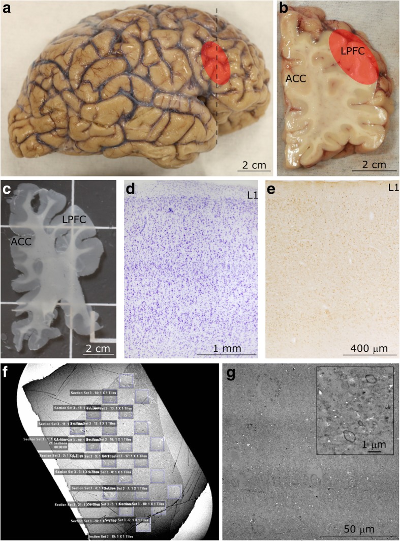
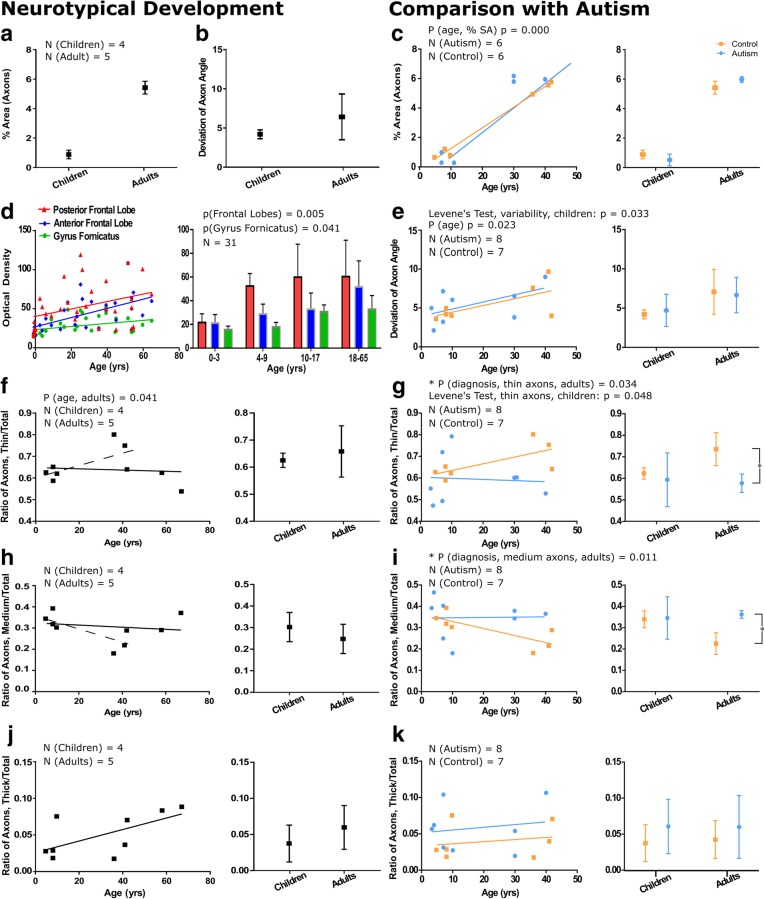
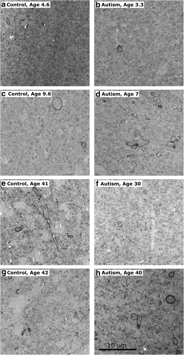
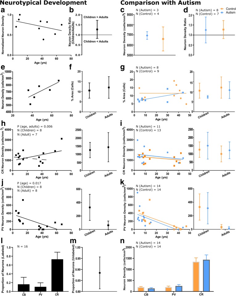
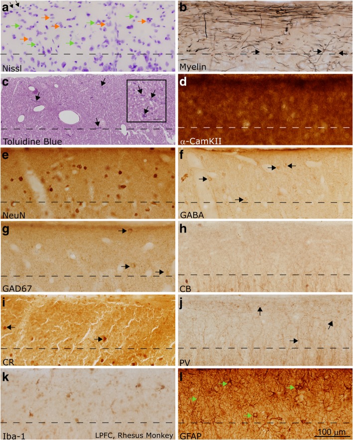
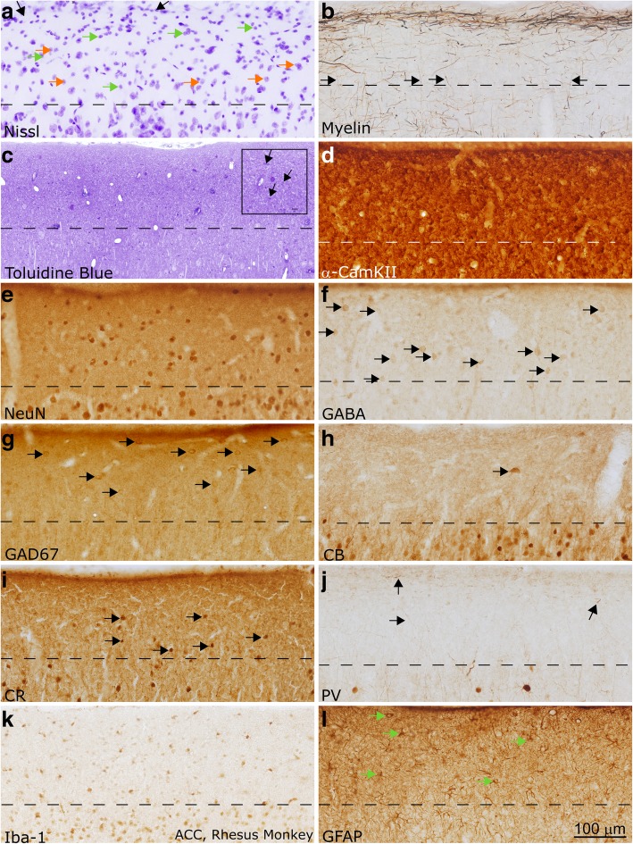
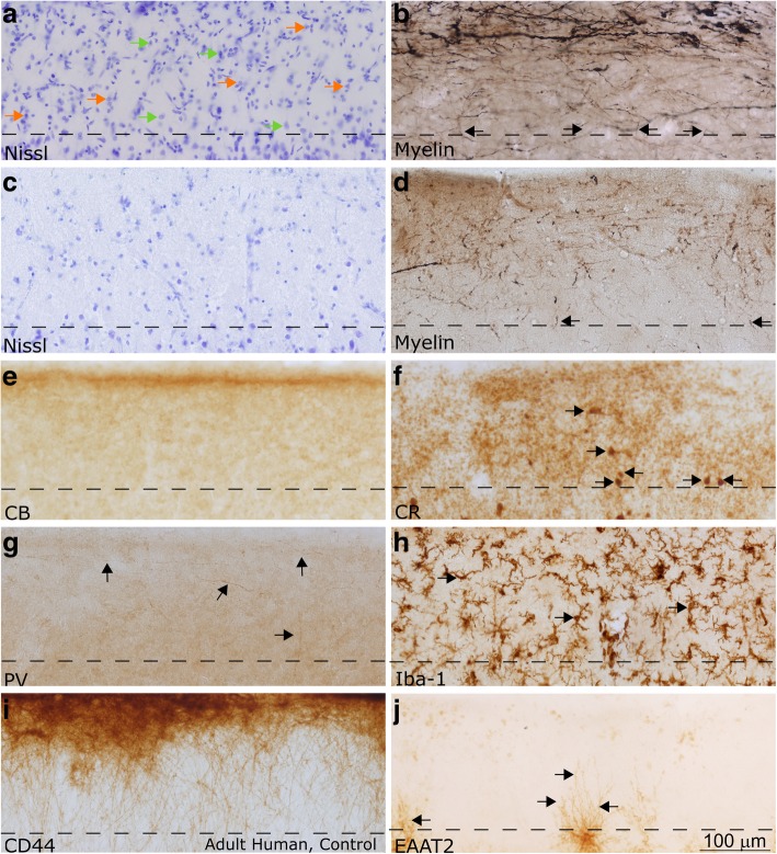
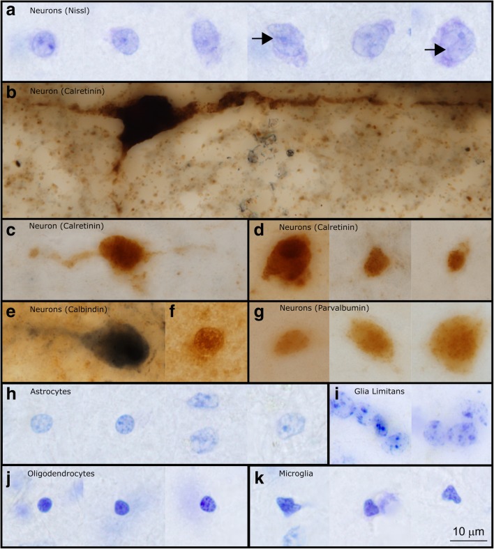
Autism is a neurodevelopmental connectivity disorder characterized by cortical network disorganization and imbalance in excitation/inhibition. However, little is known about the development of autism pathology and the disruption of laminar-specific excitatory and inhibitory cortical circuits. To begin to address these issues, we examined layer 1 of the lateral prefrontal cortex (LPFC), an area with prolonged development and maturation that is affected in autism. We focused on layer 1 because it contains a distinctive, diverse population of interneurons and glia, receives input from feedback and neuromodulatory pathways, and plays a critical role in the development, maturation, and function of the cortex. We used unbiased quantitative methods at high resolution to study the morphology, neurochemistry, distribution, and density of neurons and myelinated axons in post-mortem brain tissue from children and adults with and without autism. We cross-validated our findings through comparisons with neighboring anterior cingulate cortices and optimally-fixed non-human primate tissue. In neurotypical controls we found an increase in the density of myelinated axons from childhood to adulthood. Neuron density overall declined with age, paralleled by decreased density of inhibitory interneurons labeled by calretinin (CR), calbindin (CB), and parvalbumin (PV). Importantly, we found PV neurons in layer 1 of typically developing children, previously detected only perinatally. In autism there was disorganization of cortical networks within layer 1: children with autism had increased variability in the trajectories and thickness of myelinated axons in layer 1, while adults with autism had a reduction in the relative proportion of thin axons. Neurotypical postnatal changes in layer 1 of LPFC likely underlie refinement of cortical activity during maturation of cortical networks involved in cognition. Our findings suggest that disruption of the maturation of feedback pathways, rather than interneurons in layer 1, has a key role in the development of imbalance between excitation and inhibition in autism.

Keywords: Anterior cingulate cortex; Autism neuropathology; Feedback pathways; Inhibitory neuron; Laminar architecture; Postnatal axon myelination.


Conflict of interest statement

Ethics approval and consent to participate

The use of human post-mortem tissue for this study was approved by the Institutional Review Board (IRB) of Boston University. Experiments and procedures with animals were designed to minimize animal suffering and reduce the number of animals used. Detailed protocols of the procedures were approved by the Institutional Animal Care and Use Committee (IACUC) at Harvard Medical School and Boston University School of Medicine in accordance with NIH guidelines (DHEW Publication no. [NIH] 80–22, revised 1996, Office of Science and Health Reports, DRR/NIH, Bethesda, Maryland, United States).

Consent for publication

Not applicable.

Competing interests

The authors declare that they have no competing interests.

Publisher’s Note

Springer Nature remains neutral with regard to jurisdictional claims in published maps and institutional affiliations.

Figures

Fig. 1
Fig. 1
Experimental design. a Lateral view of the right hemisphere of the adult human brain. The region of the lateral prefrontal cortex (LPFC) analyzed in this study is shown in red. b Coronal tissue slab taken from the frontal cortex at the level marked by the dotted line in a. The red overlay highlights the LPFC at the level of Brodmann’s area 46. c Representative free-floating tissue section, cut at 50 μm, used for staining. d-e Representative cortical columnar regions of interest processed for Nissl d and calretinin e in the LPFC. Layer 1 (L1) is labeled in both columns. f Representative image of a 50 nm-thick section from LPFC gray matter on a pioloform-coated slot grid to show the unbiased systematic sampling scheme used to analyze axons at the EM level. This scheme also resembles the sampling used in quantitative analysis of cell and axon populations at the light microscope. g Representative high-resolution electron micrograph of an ultrathin section (50 nm), sampled from f, and acquired using a scanning-transmission electron microscope (STEM) system. Myelinated axon profiles can be identified by the darkly stained, electron dense ring of myelin surrounding the axolemma (shown at high magnification in inset). This representative image was taken from layer 5 of LPFC
Fig. 2
Fig. 2
Myelinated axons in layer 1 of prefrontal cortex show significant changes in development and in autism. a The percent surface area occupied by axons in children is low (0.89 ± 0.29%), but increases significantly to 5.4 ± 0.44% in adults. b Axon trajectories are less variable in children than in adults in neurotypical development, suggesting increased myelination of diverse pathways in adulthood. c The percent surface area occupied by myelinated axons was similar in autism and neurotypical groups. The percent surface area occupied by axons increased significantly in adults from both groups (p = 0.000), shown on a case-by-case basis (left graph) and average by age group (right graph). d We analyzed images of layer 1 from the atlas of Kaes [62] to cross-validate our findings. Trends of myelin development in layer 1 of the anterior and posterior frontal lobes, which are similar to anterior and lateral prefrontal cortices, and Gyrus Fornicatus, which includes the anterior cingulate cortex, showed increases in myelination with age. In the posterior frontal lobe, layer 1 in children aged 0–3 was less myelinated than older children and adults. The anterior frontal lobe and anterior cingulate cortex had a more prolonged period of myelination and overall had a lower level of myelination compared with the posterior frontal lobe. e In children with autism there was a significant increase in the variability of axon trajectory heterogeneity when compared to neurotypical children (p = 0.033). In adults, this variability was similar between groups. f The relative proportion of thin axons remained approximately stable through neurotypical development. The solid line shows the overall stable trend, the dotted line shows the increase in the relative proportion of thin axons when comparing children and young adults. There was a trend towards a decline in the proportion of thin axons with age in adulthood. g The proportion of thin axons in neurotypical children was on average 64%; in children with autism, there was significant variation in the percentage of axons that were thin (range: 47–75%). Neurotypical adults had a higher average proportion of thin axons than adults with autism (mean control = 73.6 ± 7.7%, mean autism = 57.8 ± 4.1%, p = 0.034). h The relative proportion of medium-caliber axons remained approximately stable through neurotypical development. The solid line shows the overall stable trend, the dotted line shows the decrease in the relative proportion of medium-caliber axons when comparing children and young adults. There was a trend towards an increase in the proportion of medium-caliber axons with age in adulthood. i There was significant variability in the proportion of medium-caliber axons in children with autism. In adulthood, there was a significant increase in the relative proportion of medium-caliber axons in adults with autism compared with neurotypical adults (mean control = 22.5 ± 5.1%, mean autism = 36.2 ± 1.8%, p = 0.011). j There was a trend towards an increase in the relative proportion of thick axons in neurotypical development. k Developmental trends in the proportion of thick axons were not different between autism and control groups. Left panels in e-k present developmental trends on a case-by-case basis, and right panels show mean ± SD for children and adults with and without an autism diagnosis. Autism cases 4021, 4029, AN 03221, 5144, 1182, B-5173, B-6232, B-6677, and control cases 451, 4203, 4337, 3835, B-6004, B-5353, and B-4981 were used for electron microscopy comparisons. Control adults HAW and HAY were included in the study of neurotypical development and aging
Fig. 3
Fig. 3
Myelinated axons in LPFC layer 1 analyzed at the electron microscope level in young children [cases 451 (a), 4021 (b)], older children [cases M3835 M (c), AN 03321 (d)], and adults [cases 5353 (e), 5173 (f), 4981(g), 6232 (h)] illustrate described trends. In younger children, who overall had fewer myelinated axons, there is still visible reduction in density of myelinated axons in individuals with autism (b). In older children there was a clear increase in the density of myelinated axons in autistic children (d), representing the variability inherent in autism, and multiple branching axons can be seen within the field. In adults, there was a decrease in proportion of small axons in individuals with autism with no visible change in overall axon density (p = 0.390) (f, h). Scale bar in (h) applies to all panels
Fig. 4
Fig. 4
Neuronal populations in LPFC layer 1 change in parallel between children and adults with and without autism. a There was a trend towards a reduction in neuron density between neurotypical children and adults. Neuron density values were normalized such that the highest density has a value of 1. Cases used in this analysis: 451, 4337, M3835 M, 6004, 4981, HAW, HAY, HCD, HCF. b There was a reduction in neuron density between typical children and adults, with a ratio of mean neuron density in children/adults of approximately 1.27. c Mean neuron density in adults with and without autism (mean control = 6602 ± 1186 cells/mm3, mean autism = 6939 ± 217 neurons/mm3) was not statistically different. d The decrease in neuron density from childhood to adulthood was not different between groups. e There was a trend towards an increase in neuron density with age between young and older adults. f The area fraction containing cells increased slightly in neurotypical development, likely representing simultaneous dilution of neurons within the neuropil and expansion of glial cell populations with age. g There was no difference between the percent surface area occupied by cells between autistic and neurotypical groups. h CR neuron density increased between children and adults. In this sample, this trend was primarily driven by high density of CR neurons in older adults. The solid line shows the overall trend towards an increase in CR neuron density with age, the dotted line shows the decrease in CR neuron density when comparing children and young adults i The trend towards a decrease in CR neuron density in layer 1 was similar in neurotypical individuals and individuals with autism. j PV neurons were present in layer 1 in neurotypical children; their density decreased significantly in adults, and in many adult cases these neurons were not found. k PV neurons were prominent in layer 1 of non-autistic (mean = 329 ± 69 neurons/mm3) and autistic children (mean = 312 ± 231 neurons/mm3), and their density decreased significantly in adults (mean control = 33 ± 39 cells/mm3, mean autism =15 ± 26 neurons/mm3, p = 0.006). l We calculated the relative proportions of the three inhibitory interneuron subtypes in layer 1. CR was the most prominent inhibitory interneuron subtype, labeling 73.07 ± 16.07% of labeled neurons. CB was expressed by 16.32 ± 15.81% of labeled inhibitory neurons, while PV was the least prominent interneuron subtype labeling only 10.60 ± 9.10% of labeled interneurons in layer 1. m 88.44 ± 7.24% of neurons were not labeled with calcium binding proteins in layer 1. n We calculated mean inhibitory neuron densities, including cases of all ages. CR-immunoreactive neurons were the densest neuron population (mean control = 1385 ± 696 neurons/mm3, mean autism = 1436 ± 733 neurons/mm3), followed by CB- (mean control = 194 ± 127 neurons/mm3, mean autism = 138 ± 124 neurons/mm3) and PV- neurons (mean control = 220 ± 215 neurons/mm3, mean autism = 248 ± 239 neurons/mm3). There were no significant differences between groups. Autism cases AN 03345, 1182, AN 04682, and B-6677 and control case HCF were not included in CR quantitative analyses due to inconsistent staining. Autism case B-6677 was not included in PV quantitative analyses due to inconsistent staining. Left panels in g-k present developmental trends on a case-by-case basis, and right panels show mean ± SD for children and adults
Fig. 5
Fig. 5
Layer 1 in the LPFC of the adult non-human primate (rhesus macaque). a Section labeled with Nissl showed a moderate density of neurons and glia, mainly astrocytes, in layer 1 (examples of neurons are marked with orange arrows, glial cells are marked with green arrows). Superficially, the glia limitans was visible as a dense band of astrocytes (shown with black arrows). b Myelin (Gallyas) stain showed the dense band of myelinated axons within superficial layer 1. Myelinated axons were seen penetrating layer 1 with diverse trajectories prior to joining the myelinated superficial plexus (black arrows). These axons came from long-range pathways or local interneurons. c Toluidine blue labeling of a 1 μm thick section from osmicated tissue revealed Nissl-stained cells and their processes, including profiles of myelinated axons (shown with black arrows). There was a higher density of thin axon profiles in superficial layer 1, in line with myelin stain shown in (b). Scale bar in inset measures 10 μm. d α-CamKII is a marker of synaptic turnover, and is used to identify areas of high plasticity. It labeled a dense population of processes in layer 1. e NeuN labels most neuronal nuclei, and in our material all layer 1 neurons that were labeled with Nissl were also labeled with NeuN (not shown). NeuN labeling clearly showed that the density of labeled neurons in layer 1 was low, and the majority of the cellular density in layer 1, as seen in (a), was not due to neurons but instead could be attributed to glia. f-g GABA (f) and GAD67 (g) label inhibitory neurons in the cortex (labeled with black arrows). There was a low density of labeling in both sections. Comparison with sections stained with NeuN (c) suggested that many neurons in layer 1 did not express GABA and its synthesizing enzymes strongly. h-j CB, CR, and PV labeled subpopulations of inhibitory interneurons. CB (h) and PV (j) did not label cell bodies in layer 1 in the adult non-human primate, while CR (i) labeled few cell bodies (shown with black arrows). PV (j) labeled a population of axons which joined the superficial plexus, and may represent either thalamocortical axons or axons of local inhibitory interneurons (shown with black arrows). CB (h) and CR (i) also labeled few axons in layer 1, but were not as visible as the PV-labeled processes. k Microglia with various morphologies, labeled with Iba-1, could be seen in layer 1. l GFAP labeled the cell bodies and dense processes of astrocytes within layer 1 (shown with green arrows). Images were acquired such that the top edge of the images underlie the pia. Dotted lines indicate the border with layer 2 in all panels. Calibration bar in (l) applies to all panels
Fig. 6
Fig. 6
Layer 1 in the ACC of the adult non-human primate (rhesus macaque). a Section labeled with Nissl showed a moderate density of neurons and glia, mainly astrocytes, in layer 1 (examples of neurons are marked with orange arrows, glial cells are marked with green arrows). Superficially, the glia limitans was visible as a dense band of astrocytes (shown with black arrows). b Myelin (Gallyas) stain showed the relatively light band of myelinated axons within superficial layer 1 in ACC. This band was lighter than that seen in LPFC, following overall trends in myelination between those areas. Black arrows show axons entering layer 1 from layer 2. c Toluidine blue labeling of a 1 μm thick section from osmicated tissue revealed Nissl-stained cells and their processes, including profiles of myelinated axons (shown with black arrows in inset). There was a low density of axon profiles in superficial layer 1, in line with the light myelin stain shown in (b). Scale bar in inset measures 10 μm. d α-CamKII is a marker of synaptic plasticity. It labeled a dense population of processes in layer 1. e NeuN labeled all layer 1 neurons that were labeled with Nissl (not shown). NeuN labeling clearly showed that the density of labeled neurons in layer 1 was low, and the majority of the cellular density in layer 1, as seen in (a), was not due to neurons but instead could be attributed to glia. f-g GABA (f) and GAD67 (g) labeled inhibitory neurons in the cortex (labeled with black arrows). There was a moderate density of labeling in both sections, comparable to what was seen in the NeuN stain (c). h-j CB, CR, and PV labeled subpopulations of inhibitory interneurons. CB (h) and PV (j) labeled primarily neuron processes in layer 1 in the adult non-human primate, while CR (i) labeled few cell bodies (shown with black arrows). Black arrows in (j) show PV-labeled axons. k Microglia with various morphologies, labeled with Iba-1, could be seen in layer 1. l GFAP labeled the cell bodies and dense processes of astrocytes within layer 1 (shown with green arrows). Images were acquired such that the top edge of the images underlie the pia. Dotted lines indicate the border with layer 2 in all panels. Calibration bar in (l) applies to all panels
Fig. 7
Fig. 7
Layer 1 in the LPFC and ACC of the adult human. All images in this figure were acquired from neurotypical adult cases (HCD, HCF, HAW, HAY). a-d Nissl (a) and myelin (b) in the LPFC showed stark differences from ACC (c-d). ACC had a thicker layer 1, and reduced density and thickness of myelinated axons in layer 1. Permeating blood vessels and endothelial cells were also visible in the Nissl-labeled section from LPFC layer 1 in (a). Examples of neurons in (a) are marked with orange arrows, glial cells are marked with green arrows. Myelin (Gallyas) stained tissue (b, d) showed a dense plexus of myelinated axons in superficial layer 1, consistent with observations in the non-human primate. While the majority of axons were horizontal, some also had diagonal trajectories, consistent with axons from incoming pathways (black arrows). e-g CB, CR, and PV labeled inhibitory interneuron classes. CB (e) labeled scant processes in layer 1 (labeled with black arrows), while CR (f) labeled a low density of neurons and few processes in layer 1 (labeled with black arrows). PV (g) labeled processes that joined the plexus of axons in superficial layer 1 (labeled with black arrows). h Iba-1 labeled microglia within the cortex, including layer 1 (examples of microglia are labeled with black arrows). Superficial microglia extended processes mostly parallel to the pial surface, while microglia deeper in layer 1 had processes oriented in multiple directions. i CD44 labeled interlaminar astrocytes, which sat on the pial surface and sent processes towards layer 2. These astrocytes are exclusive to higher-order primates. j Astrocytes labeled with excitatory amino acid transporter (EAAT2) were not present in layer 1; however, the processes of labeled astrocytes from layer 2 penetrated into the deep part of layer 1 (labeled with black arrows). Images were acquired such that the top edge of the images underlie the pia. Dotted lines indicate the border with layer 2 in all panels. Calibration bar in (j) applies to all panels
Fig. 8
Fig. 8
High magnification images of representative neurons and glia in layer 1 of the LPFC in the human brain. a Neurons in layer 1 had a broad range of sizes. In Nissl preparations it was possible to see the nucleus, nucleolus, and a rim of cytoplasm around labeled neurons. In larger neurons it was possible to observe folding of the nucleus (shown with black arrows in figure) (Case HCD). b This large, subpial neuron labeled with CR is likely a Cajal-Retzius cell. This image shows its large cell body and horizontal extensions, along with a descending process which is typical of Cajal-Retzius cells (Case AN4722). c-d CR labeled neurons of multiple sizes and shapes were found in layer 1 of the human cerebral cortex. CR-labeled neurons in layer 1 had multiple orientations within layer 1, including horizontally oriented cells (c) and cells with more typical interneuron morphology (d). e-f CB-labeled neurons were also diverse in shape and size. These neurons may also be oriented horizontally (e) (Case 4722, Case 3835). g Representative PV-labeled neurons in layer 1 (Case 4337). h-i Astrocytes in layer 1 had multiple morphologies. The astrocytes of the glial limitans were compact and formed tight groups (i) (Case HCD). j Three oligodendrocytes in human layer 1 (Case HCD). Oligodendrocytes in human tissue had varied staining characteristics: they could be darkly labeled, as seen in the first two images, or lightly labeled, as shown in the third image. k Microglia in layer 1 had irregularly shaped nuclei, distinguishing them from darkly stained oligodendrocytes (Case HCD). Calibration bar in (k) applies to all panels

Similar articles

Cited by

References

    1. Adorjan I, Ahmed B, Feher V, Torso M, Krug K, Esiri M, Chance SA, Szele FG. Calretinin interneuron density in the caudate nucleus is lower in autism spectrum disorder. Brain. 2017;140:2028–2040. doi: 10.1093/brain/awx131. - DOI - PMC - PubMed
    1. Bachevalier J, Loveland KA. The orbitofrontal-amygdala circuit and self-regulation of social-emotional behavior in autism. Neurosci Biobehav Rev. 2006;30:97–117. doi: 10.1016/j.neubiorev.2005.07.002. - DOI - PubMed
    1. Bailey A, Luthert P, Dean A, Harding B, Janota I, Montgomery M, Rutter M, Lantos P. A clinicopathological study of autism. Brain. 1998;121(Pt 5):889–905. doi: 10.1093/brain/121.5.889. - DOI - PubMed
    1. Barbaria EM, Kohl B, Buhren BA, Hasenpusch-Theil K, Kruse F, Kury P, Martini R, Muller HW. The alpha-chemokine CXCL14 is up-regulated in the sciatic nerve of a mouse model of Charcot-Marie-tooth disease type 1A and alters myelin gene expression in cultured Schwann cells. Neurobiol Dis. 2009;33:448–458. doi: 10.1016/j.nbd.2008.11.014. - DOI - PubMed
    1. Barbas H. Pattern in the laminar origin of corticocortical connections. J Comp Neurol. 1986;252:415–422. doi: 10.1002/cne.902520310. - DOI - PubMed

Publication types

LinkOut - more resources