. 2019 Mar 26;116(13):6162-6171.
doi: 10.1073/pnas.1814139116. Epub 2019 Mar 13.

Injured liver-released miRNA-122 elicits acute pulmonary inflammation via activating alveolar macrophage TLR7 signaling pathway

Affiliations

Injured liver-released miRNA-122 elicits acute pulmonary inflammation via activating alveolar macrophage TLR7 signaling pathway

Yanbo Wang et al. Proc Natl Acad Sci U S A. .

Abstract

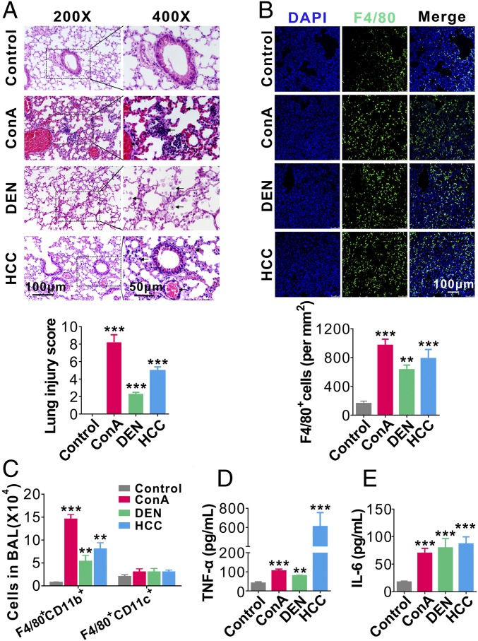
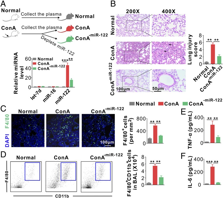
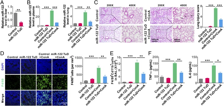
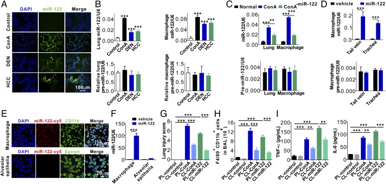
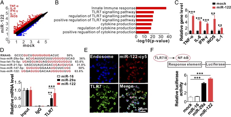
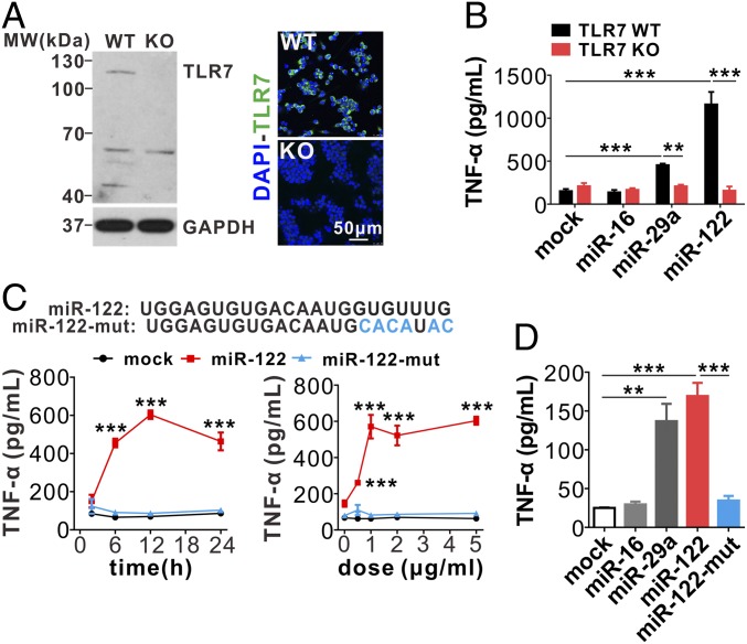
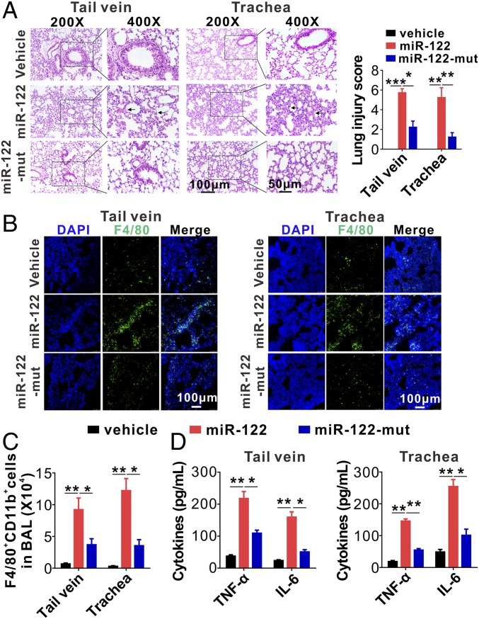
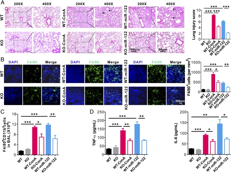
Hepatic injury is often accompanied by pulmonary inflammation and tissue damage, but the underlying mechanism is not fully elucidated. Here we identify hepatic miR-122 as a mediator of pulmonary inflammation induced by various liver injuries. Analyses of acute and chronic liver injury mouse models confirm that liver dysfunction can cause pulmonary inflammation and tissue damage. Injured livers release large amounts of miR-122 in an exosome-independent manner into the circulation compared with normal livers. Circulating miR-122 is then preferentially transported to mouse lungs and taken up by alveolar macrophages, in which it binds Toll-like receptor 7 (TLR7) and activates inflammatory responses. Depleting miR-122 in mouse liver or plasma largely abolishes liver injury-induced pulmonary inflammation and tissue damage. Furthermore, alveolar macrophage activation by miR-122 is blocked by mutating the TLR7-binding GU-rich sequence on miR-122 or knocking out macrophage TLR7. Our findings reveal a causative role of hepatic miR-122 in liver injury-induced pulmonary dysfunction.

Keywords: TLR7/8; circulating miR-122; liver injury; macrophage; pulmonary inflammatory.


Conflict of interest statement

The authors declare no conflict of interest.

Figures

Fig. 1.
Fig. 1.
Lung inflammation and tissue damage caused by liver injuries. (A, Upper) Representative H&E staining of lung tissue sections in mice with or without ongoing ConA-induced acute hepatitis, DEN-induced chronic hepatitis, or orthotopic transplanted HCC. (Lower) Mouse lung injury scores analyzed by a double-blind examination for the following parameters: pulmonary edema, inflammatory infiltration, hemorrhage, atelectasis, and hyaline membrane formation (six mice per group, three to five images per mouse). The scores were assigned as follows: 0, no injury; 1, <25% injury; 2, 25–50% injury; 3, 50–75% injury; and 4 >75% injury. Eight to 10 high-magnitude fields from each slide were analyzed. (B, Upper) Representative images of F4/80+ cell infiltration into mouse lungs from mice with or without liver injuries. (Lower) Analysis of alveolar infiltration of F4/80+ macrophages (six mice oer group, three to five images per mouse). (C) FACS analysis of alveolar macrophage subpopulations (F4/80+CD11b+ or F4/80+CD11c+ cells) in BAL from mice with or without various liver injuries (six mice per group, n = 3). (D and E) Levels of TNF-α (D) and IL-6 (E) in BALF from mice with or without various liver injuries (six mice per group, n = 3). Data are show as means ± SEM. **P < 0.01, ***P < 0.001. One-way ANOVA followed by Bonferroni’s multiple comparisons test.
Fig. 2.
Fig. 2.
Hepatic miR-122 induces acute pulmonary inflammation and tissue damage. (A, Upper) Schematic of experimental design: 200 μL plasma samples from normal mice and ConA-treated mice with or without miR-122 depletion were separately injected into WT mice (six mice per group) every 6 h six times. (Lower) miR-122 was depleted from mouse plasma by miR-122 antisense oligonucleotide-conjugated Sepharose beads. (B) Mouse lung tissue damage analyzed by H&E staining (Left) and injury score (Right). (C) Representative images (Left) and analysis (Right) of F4/80+ macrophage infiltration into mouse lungs. Note that the infiltration of F4/80+ macrophages was significantly enhanced by plasma from ConA-treated mice, while depleting circulating miR-122 in ConA-treated mouse plasma markedly attenuated the infiltration of F4/80+ cells into alveoli (n = 6 mice per group, three to five images per mouse). (D) FACS analysis of F4/80+CD11b+ macrophages in BAL harvested from the recipient mice (six mice per group, n = 3). (E and F) Levels of TNF-α (E) and IL-6 (F) in BALF from the recipient mice (six mice per group, n = 3). Data are presented as mean ± SEM. **P < 0.01, ***P < 0.001. One-way ANOVA followed by Bonferroni’s multiple comparisons test.
Fig. 3.
Fig. 3.
Depleting mouse liver miR-122 greatly attenuates lung inflammation and tissue damage induced by injured liver. (A) Relative level of miR-122 in mouse liver following miR-122 TuD AAV8 administration. (B) Relative levels of miR-122 in mouse plasma (Left) and lungs (Right) in control mice (Control), miR-122 TuD mice (miR-122 TuD), control mice treated with ConA (Control+ConA), and miR-122 TuD mice treated with ConA (miR-122 TuD+ConA). (C) Mouse lung tissue damage analyzed by H&E staining (Left) and injury score (Right). (D) Representative images (Left) and analysis (Right) of F4/80+ macrophage infiltration into mouse lungs. (E) FACS analysis of F4/80+CD11b+ macrophages in BAL harvested from the recipient mice (six mice per group, n = 3). (F) Levels of TNF-α (Left) and IL-6 (Right) in BALF from the four group mice (six mice per group, n = 3). Data are presented as mean ± SEM. *P < 0.05, **P < 0.01, ***P < 0.001. Student’s two-tailed, unpaired t-test (A), or one-way ANOVA followed by Bonferroni’s multiple comparisons test (BF).
Fig. 4.
Fig. 4.
Uptake of hepatic miR-122 by mouse alveolar macrophages. (A) In situ hybridization of miR-122 in mouse lungs following various liver injuries. (B) Levels of miR-122 or premiR-122 in mouse lungs and alveolar macrophages isolated from mice with various liver injuries. (C) Levels of miR-122 (Upper) and premiR-122 (Lower) in lungs and alveolar macrophages isolated from mice after adoptive transfer of ConA-treated mouse plasma with or without miR-122 depletion. (D) Levels of miR-122 (Upper) and premiR-122 (Lower) in alveolar macrophages isolated from mice with or without direct injection of miR-122 via tail vein or respiratory trachea. (E and F) Uptake of synthetic miR-122-cy5 by mouse primary macrophages and mouse alveolar epithelial cells assayed by immunofluorescence (E) and qRT-PCR (F). (G) Mouse lung tissue damage analyzed by injury score in mice of control group (PL-control), macrophage-depletion group (CL-control), control mice injected with ConA group (PL-ConA), control mice injected with synthetic miR-122 group (PL–miR-122), macrophage-depletion mice injected with ConA group (CL-ConA) and macrophage depletion mice injected with synthetic miR-122 group (CL–miR-122). (H) FACS analysis of F4/80+CD11b+ macrophages in BAL harvested from the recipient mice (six mice per group, n = 3). (I) Levels of TNF-α (Left) and IL-6 (Right) in BALF from the six group mice (six mice per group, n = 3). Data are presented as mean ± SEM. **P < 0.01, ***P < 0.001. One-way ANOVA followed by Bonferroni’s multiple comparisons test (B, C, and GI) or student’s two-tailed, unpaired t test (D and F).
Fig. 5.
Fig. 5.
MiR-122 specifically binds to mouse macrophage endosomal TLR7. (AC) Macrophage activation by internalized synthetic miR-122 detected by microarray gene-expression profiling (A and B) and qRT-PCR (C). (D, Upper) Comparison of GU enrichment in miR-122 with other TLR7/8-binding miRNAs; (Lower) Detection of miR-122 and miR-29a but not miR-16 in anti-TLR7 antibody-immunoprecipitated complex by qRT-PCR. (E) Colocalization of miR-122-Cy5 and TLR7 in endosomal structures of mouse macrophages. (F, Upper) schematic of NF-κB luciferase reporter assay of miR-122 binding human TLR8 in HEK293T cells. (Lower) Enhancement of NF-κB activity in HEK293T cells by miR-122 and miR-29a via specific binding to human TLR8. Data are presented as mean ± SEM (n = 3). **P < 0.01, ***P < 0.001. Student’s two-tailed, unpaired t test (C) or one-way ANOVA followed by Bonferroni’s multiple comparisons test (D and F).
Fig. 6.
Fig. 6.
MiR-122 elicits mouse macrophages inflammatory responses via activating Tlr7 signal pathway. (A) Tlr7 knockout in mouse macrophages by the CRISPR-cas9 technique. (B) Tlr7 KO abolishes the effect of miR-122 on mouse macrophage inflammatory activation. (C, Upper) Mutation of GU-rich sequences in miR-122. (Lower) miR-122 but not miR-122 mutant stimulates RAW246.1 cells to secrete TNF-α in a time- and dose-dependent manner. (D) TNF-α secretion by mouse alveolar macrophages treated with various miRNAs, including miR-16, miR-29a, miR-122, and miR-122 mutant. Data are presented as mean ± SEM. **P < 0.01, ***P < 0.001. Two-way ANOVA followed by Bonferroni’s multiple comparisons test (B), or student’s two-tailed, unpaired t test (C), or one-way ANOVA followed by Bonferroni’s multiple comparisons test (D).
Fig. 7.
Fig. 7.
MiR-122-mut fails to elicit mouse acute lung inflammation and tissue damage. (A) Mouse pulmonary inflammation and lung tissue damage quantitated by H&E staining (Left) and lung injury scoring (Right) (six mice per group, three to four images for each mouse). (B) Alveolar infiltration of F4/80+ macrophages in mice with injection of vehicle control, miR-122, or miR-122 mutant via tail vein or respiratory trachea, respectively. Macrophage infiltration was detected by immunofluorescence. (C) FACS analysis of F4/80+CD11b+ macrophages in BALF harvested from the mice injected with miR-122 or miR-122 mutant (six mice per group, n = 3). (D) Levels of inflammatory cytokines in BALF harvested from recipient mice following injection of miR-122 or miR-122 mutant via tail vein (Left) or respiratory trachea (Right) (six mice per group, n = 3). Data are presented as mean ± SEM. *P < 0.05, **P < 0.01, ***P < 0.001. One-way ANOVA followed by Bonferroni’s multiple comparisons test.
Fig. 8.
Fig. 8.
Lung inflammation and tissue damage induced by miR-122 is abolished by deleting Tlr7. (A) Mouse lung tissue damage analyzed by H&E staining (Left) and lung injury scoring (Right) in mice of control group (WT), Tlr7 depletion group (KO), control mice injected with ConA group (WT-ConA), control mice injected with synthetic miR-122 group (WT–miR-122), Tlr7-depletion mice injected with ConA group (KO-ConA), and Tlr7-depletion mice injected with synthetic miR-122 (KO–miR-122) (six mice per group, three to four images for each mouse). (B) Alveolar infiltration of F4/80+ macrophages in six group mice. Macrophage infiltration was detected by immunofluorescence. (C) FACS analysis of F4/80+CD11b+ macrophages in BAL harvested from the six group mice (six mice per group, n = 3). (D) Levels of TNF-α (Left) and IL-6 (Right) in BALF from the six group mice (six mice per group, n = 3). Data are presented as mean ± SEM. *P < 0.05, **P < 0.01, ***P < 0.001. One-way ANOVA followed by Bonferroni’s multiple comparisons test.

Similar articles

Cited by

References

    1. Huffmyer JL, Nemergut EC. Respiratory dysfunction and pulmonary disease in cirrhosis and other hepatic disorders. Respir Care. 2007;52:1030–1036. - PubMed
    1. Fallon MB, Abrams GA. Pulmonary dysfunction in chronic liver disease. Hepatology. 2000;32:859–865. - PubMed
    1. Rodriguez-Roisin R, Krowka MJ. Hepatopulmonary syndrome—A liver-induced lung vascular disorder. N Engl J Med. 2008;358:2378–2387. - PubMed
    1. Lange PA, Stoller JK. The hepatopulmonary syndrome. Ann Intern Med. 1995;122:521–529. - PubMed
    1. Lin HI, et al. Reperfusion liver injury induces down-regulation of eNOS and up-regulation of iNOS in lung tissues. Transplant Proc. 2006;38:2203–2206. - PubMed

Publication types

LinkOut - more resources