. 2019 Apr 16;116(16):7805-7812.
doi: 10.1073/pnas.1822148116. Epub 2019 Mar 13.

Diverse fate of ubiquitin chain moieties: The proximal is degraded with the target, and the distal protects the proximal from removal and recycles

Affiliations

Diverse fate of ubiquitin chain moieties: The proximal is degraded with the target, and the distal protects the proximal from removal and recycles

Hao Sun et al. Proc Natl Acad Sci U S A. .

Abstract

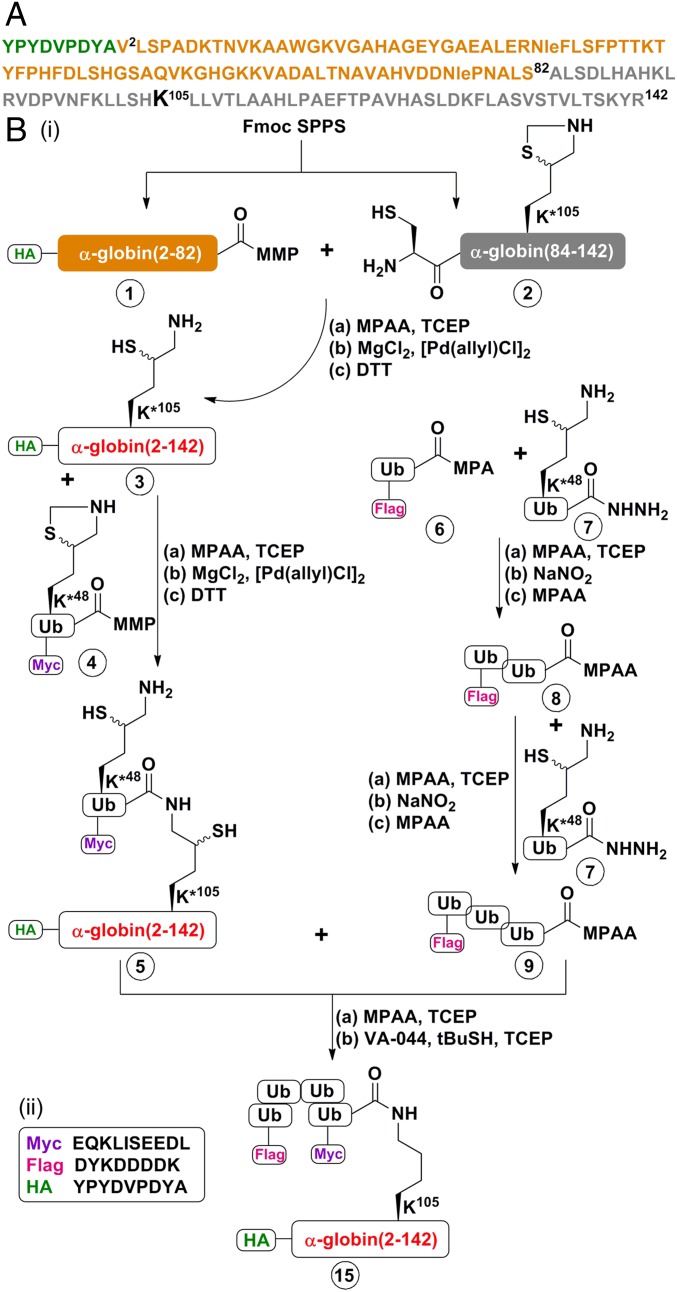
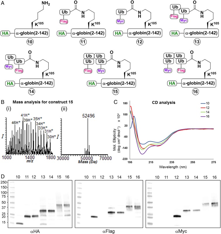
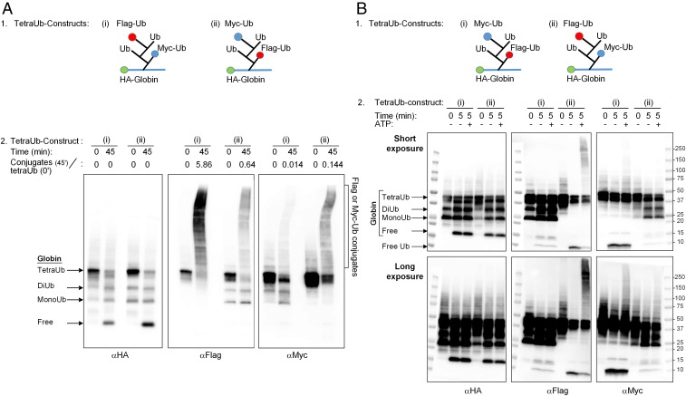
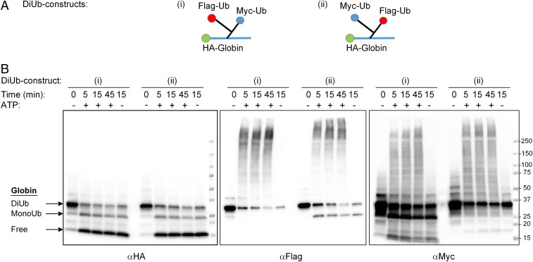
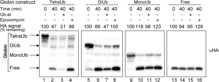
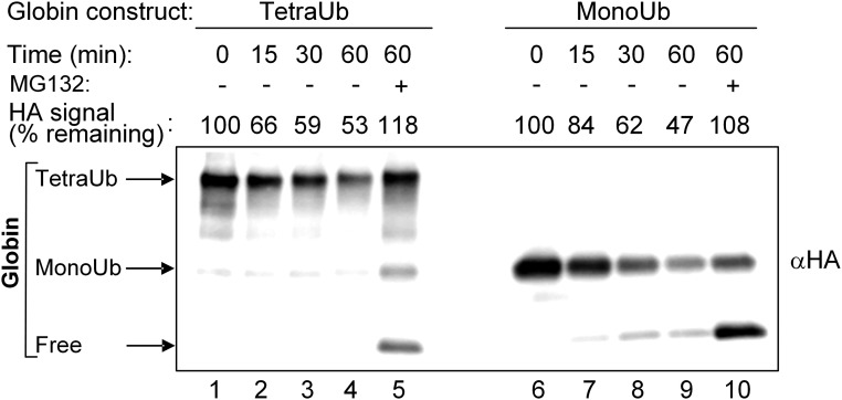
One of the enigmas in the ubiquitin (Ub) field is the requirement for a poly-Ub chain as a proteasomal targeting signal. The canonical chain appears to be longer than the distance between the two Ub-binding proteasomal receptors. Furthermore, genetic manipulation has shown that one receptor subunit is sufficient, which suggests that a single Ub can serve as a degradation signal. To shed light on this mystery, we chemically synthesized tetra-Ub, di-Ub (K48-based), and mono-Ub adducts of HA-α-globin, where the distal or proximal Ub moieties were tagged differentially with either Myc or Flag. When incubated in a crude cell extract, the distal Ub moiety in the tetra-Ub adduct was mostly removed by deubiquitinating enzymes (DUBs) and reconjugated to other substrates in the extract. In contrast, the proximal moiety was most likely degraded with the substrate. The efficacy of degradation was proportionate to the chain length; while tetra-Ub globin was an efficient substrate, with mono-Ub globin, we observed rapid removal of the Ub moiety with almost no degradation of the free globin. Taken together, these findings suggest that the proximal moieties are necessary for securing the association of the substrate with the proteasome along the proteolytic process, whereas the distal moieties are important in protecting the proximal moieties from premature deubiquitination. Interestingly, when the same experiment was carried out using purified 26S proteasome, mono- and tetra-Ub globin were similarly degraded, highlighting the roles of the entire repertoire of cellular DUBs in regulating the degradation of proteasomal substrates.

Keywords: 26S proteasome; chemical synthesis; deubiquitination; protein degradation; ubiquitination.


Conflict of interest statement

The authors declare no conflict of interest.

Figures

Fig. 1.
Fig. 1.
Representative scheme of the strategy used for chemical synthesis of differentially tagged tetra-Ub HA-α-globin. A detailed description of the complete synthesis of the building blocks and intermediates for the generation of HA-α-globin constructs is provided in SI Appendix, Figs. S1–S23. (A) Sequence of human HA-α-globin. The HA tag sequence is in green. The fragment 1 sequence (residues 2–82) is in light brown, and the fragment 2 sequence (residues Cys83-84–142) is in gray. Note that Ala83 in the native α-globin was substituted with Cys to enable chemical ligation and later desulfurized back to Ala. (B, i) Chemical synthesis steps of tetra-Ub HA-α-globin (construct 15). HA-α-globin K*105 (intermediate 3; K*105 denotes the presence of δ-mercaptolysine at this position) was generated from the ligation of fragments 1 and 2 of α-globin. Note that native α-globin contains a Cys residue at position 105. The ubiquitination sites of globins have not been identified; a previous study reported successful ubiquitination at position 105, generating a thiolester bond on the native Cys residue (15). In the present study, to generate an isopeptide bond, the native Cys residue was replaced with δ-mercaptolysine. Intermediate 3 was next ligated with (intermediate 4Myc-Ub(K*48)-MMP; K*48 denotes the presence of δ-mercaptolysine at this position) to obtain intermediate 5, which contains the whole HA-α-globin to which one Myc-Ub moiety is bound. In parallel, Flag-tri-Ub-MPAA (intermediate 9) was assembled from Ub monomers. Intermediates 5 and 9 were then ligated and desulfurized to obtain the final Flag-Ub-Ub-Ub-Myc-Ub-(tetra-Ub)-HA-α-globin (construct 15). (a), (b), and (c) denote the reagents used for each reaction. Details of the materials used and the synthetic steps are provided in SI Appendix, Materials and Methods). (B, ii) Sequences of Myc, Flag, and HA tags.
Fig. 2.
Fig. 2.
Characterization of the chemically synthesized HA-α-globin and its ubiquitinated derivatives. (A) Sketches of the seven different synthetic HA-α-globin constructs used in the study (constructs 10–16): 10, HA-α-globin; 11, Flag-Ub-HA-α-globin; 12, Myc-Ub-HA-α-globin; 13, Flag-Ub-Myc-Ub-HA-α-globin; 14, Myc-Ub-Flag-Ub-HA-α-globin; 15, Flag-Ub-Ub-Ub-Myc-Ub-HA-α-globin; 16, Myc-Ub-Ub-Ub-Flag-Ub-HA-α-globin. The synthesis of all intermediates and final constructs is described in SI Appendix, Figs. S1–S23). (B, i and ii) Representative mass analysis of tetra-Ub HA-α-globin (construct 15). (C) Circular dichroism analysis of HA-α-globin constructs 10, 12, 14, and 16. (D) Western blot analysis for HA-α-globin constructs 1016 with αHA, αFlag, and αMyc antibodies.
Fig. 3.
Fig. 3.
Fates of proximal and distal Ub moieties in a tetra-Ub chain. (A, 1) Sketches of differentially tagged tetra-Ub HA-α-globin: (i) tetra-Ub HA-α-globin with distal Flag-Ub and proximal Myc-Ub and (ii) tetra-Ub HA-α-globin with distal Myc-Ub and proximal Flag-Ub (also see Fig. 2A, constructs 15 and 16, respectively). (A, 2) Differentially tagged tetra-Ub HA-α-globin constructs were incubated for the indicated times in the presence of crude fraction II and ATP. Following SDS/PAGE, reactions were analyzed by Western blot analysis with αHA (Left), αFlag (Middle), and αMyc (Right). Quantification of the Flag-Ub- and Myc-Ub conjugates is presented as the ratio of the Flag or Myc signal in the generated conjugates at 45 min (minus the signal at time 0) relative to the Flag or Myc signal in the tetra-Ub HA-α-globin at time 0. (B, 1) Sketches of differentially tagged tetra-Ub HA-α-globins: (i) tetra-Ub HA-α-globin with distal Myc-Ub and proximal Flag-Ub and (ii) tetra-Ub HA-α-globin with distal Flag-Ub and proximal Myc-Ub (also see Fig. 2A, constructs 16 and 15, respectively). (B, 2) Differentially tagged tetra-Ub HA-α-globin constructs were incubated for the indicated times in the presence of fraction II with and without ATP. Following SDS/PAGE, reactions were analyzed by Western blot analysis as described in A, 2. The same image is shown with short (Upper) and long (Lower) exposures.
Fig. 4.
Fig. 4.
Similar fates of distal and proximal Ub moieties from di-Ub HA-α-globin. (A) Sketches of differentially tagged di-Ub HA-α-globins: (i) di-Ub HA-α-globin with distal Flag-Ub and proximal Myc-Ub and (ii) di-Ub HA-α-globin with distal Myc-Ub and proximal Flag-Ub (also seem Fig. 2A, constructs 13 and 14, respectively). (B) Differentially tagged di-Ub HA-α-globin constructs were incubated for the indicated times in the presence of crude fraction II with or without ATP. Following SDS/PAGE, reactions were analyzed by Western blot analysis with αHA (Left), αFlag (Middle), and αMyc (Right).
Fig. 5.
Fig. 5.
Ub-al inhibits the release of free α-globin from Ub-globin adducts and accelerates the degradation of tetra-Ub and di-Ub HA-α-globin. Tetra-Ub HA-α-globin with distal Flag-Ub and proximal Myc-Ub, di-Ub HA-α-globin with distal Flag-Ub and proximal Myc-Ub, mono-Ub HA-α-globin with Flag-Ub, and free HA-α-globin (Fig. 2A, constructs 15, 13, 11, and 10, respectively) were incubated for the indicated times in the presence of crude fraction II and ATP. Ub-al (1 μM) and epoxomicin (40 μM) were added to the reaction mixtures as indicated. Following SDS/PAGE, reactions were analyzed by Western blot analysis using αHA. Quantification of degradation was calculated as the HA signal along the lane at 40 min minus the background signal at time 0 and expressed as percentage of the total HA signal in the HA-α-globin construct at time 0.
Fig. 6.
Fig. 6.
Similar degradation of tetra-Ub HA-α-globin and mono-Ub HA-α-globin by purified 26S proteasome. Tetra-Ub HA-α-globin with distal Flag-Ub and proximal Myc-Ub and mono-Ub HA-α-globin with Flag-Ub (Fig. 2A, constructs 15 and 11, respectively) were incubated for the indicated times in the presence of purified human 26S proteasome (1.5 μg). MG132 (100 μM) was added as indicated. Following SDS/PAGE, reactions were analyzed by Western blot analysis using αHA. Quantification of degradation at the different time points was calculated as described in Fig. 5.

Comment in

References

    1. Mali SM, Singh SK, Eid E, Brik A. Ubiquitin signaling: Chemistry comes to the rescue. J Am Chem Soc. 2017;139:4971–4986. - PubMed
    1. Komander D, Rape M. The ubiquitin code. Annu Rev Biochem. 2012;81:203–229. - PubMed
    1. Kulathu Y, Komander D. Atypical ubiquitylation—The unexplored world of polyubiquitin beyond Lys48 and Lys63 linkages. Nat Rev Mol Cell Biol. 2012;13:508–523. - PubMed
    1. Finley D, Chen X, Walters KJ. Gates, channels, and switches: Elements of the proteasome machine. Trends Biochem Sci. 2016;41:77–93. - PMC - PubMed
    1. Budenholzer L, Cheng CL, Li Y, Hochstrasser M. Proteasome structure and assembly. J Mol Biol. 2017;429:3500–3524. - PMC - PubMed

Publication types

LinkOut - more resources