. 2019 May 15;93(11):e02230-18.
doi: 10.1128/JVI.02230-18. Print 2019 Jun 1.

Cellular DNA Topoisomerases Are Required for the Synthesis of Hepatitis B Virus Covalently Closed Circular DNA

Affiliations

Cellular DNA Topoisomerases Are Required for the Synthesis of Hepatitis B Virus Covalently Closed Circular DNA

Muhammad Sheraz et al. J Virol. .

Abstract

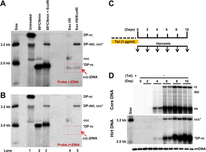
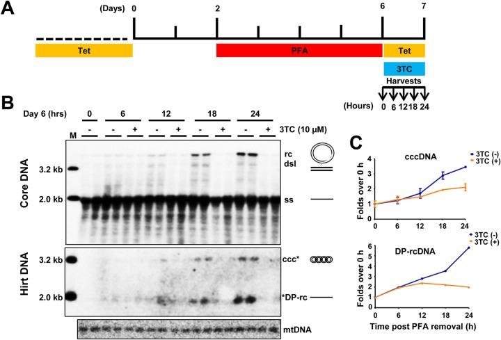
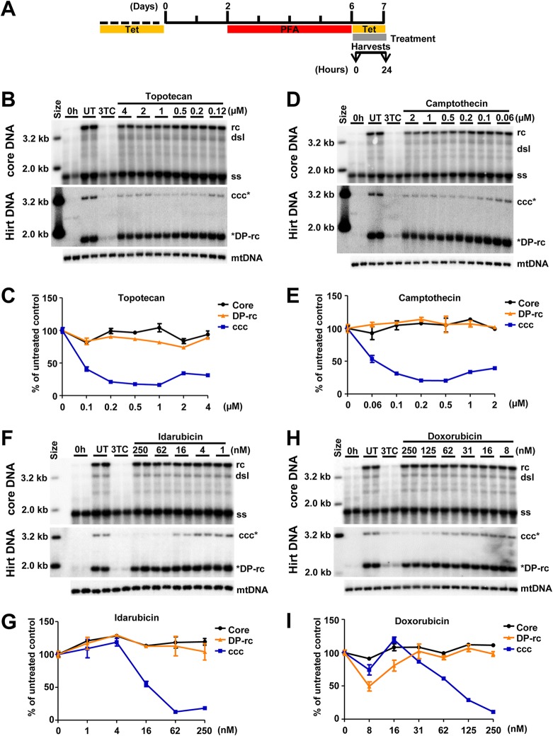
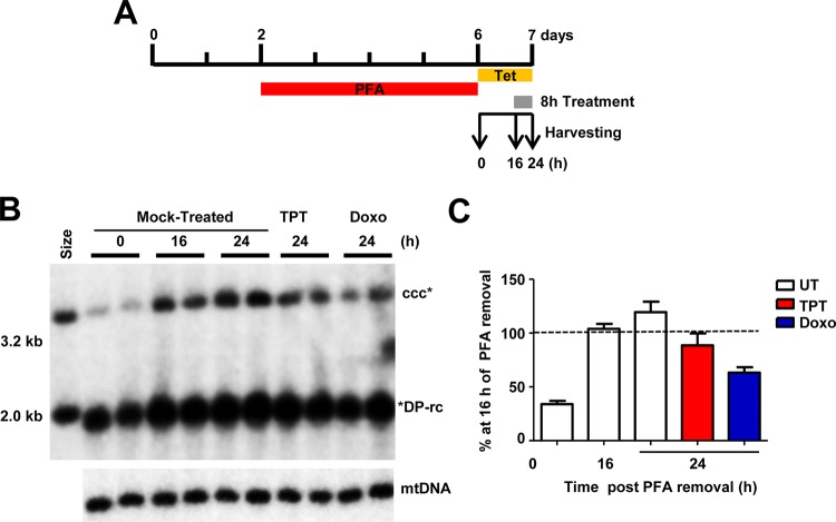
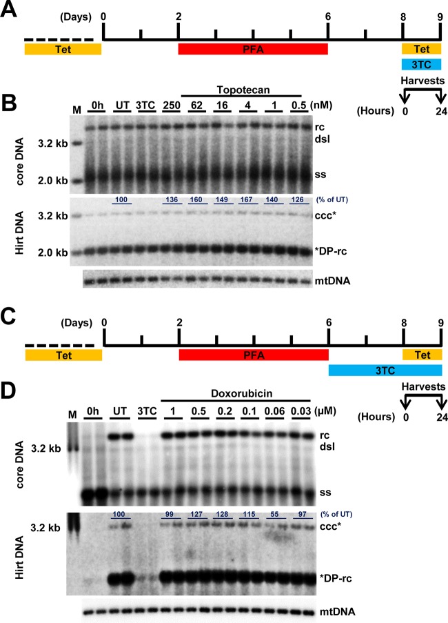
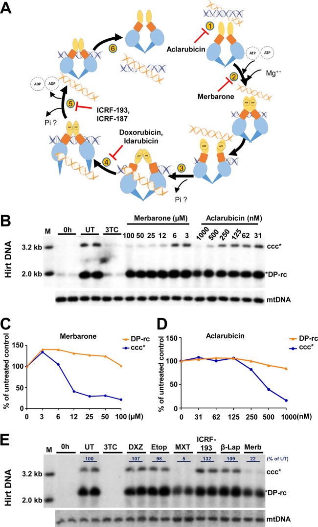
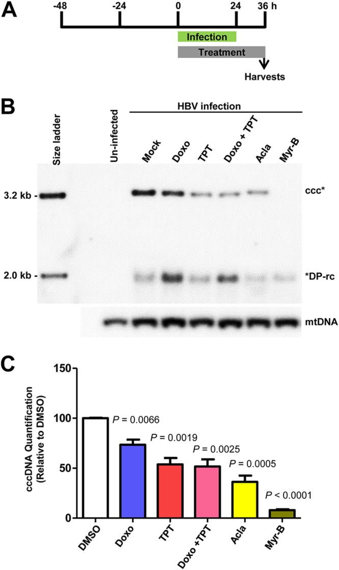
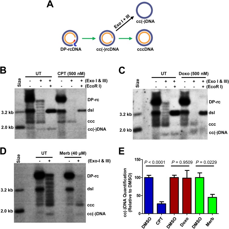
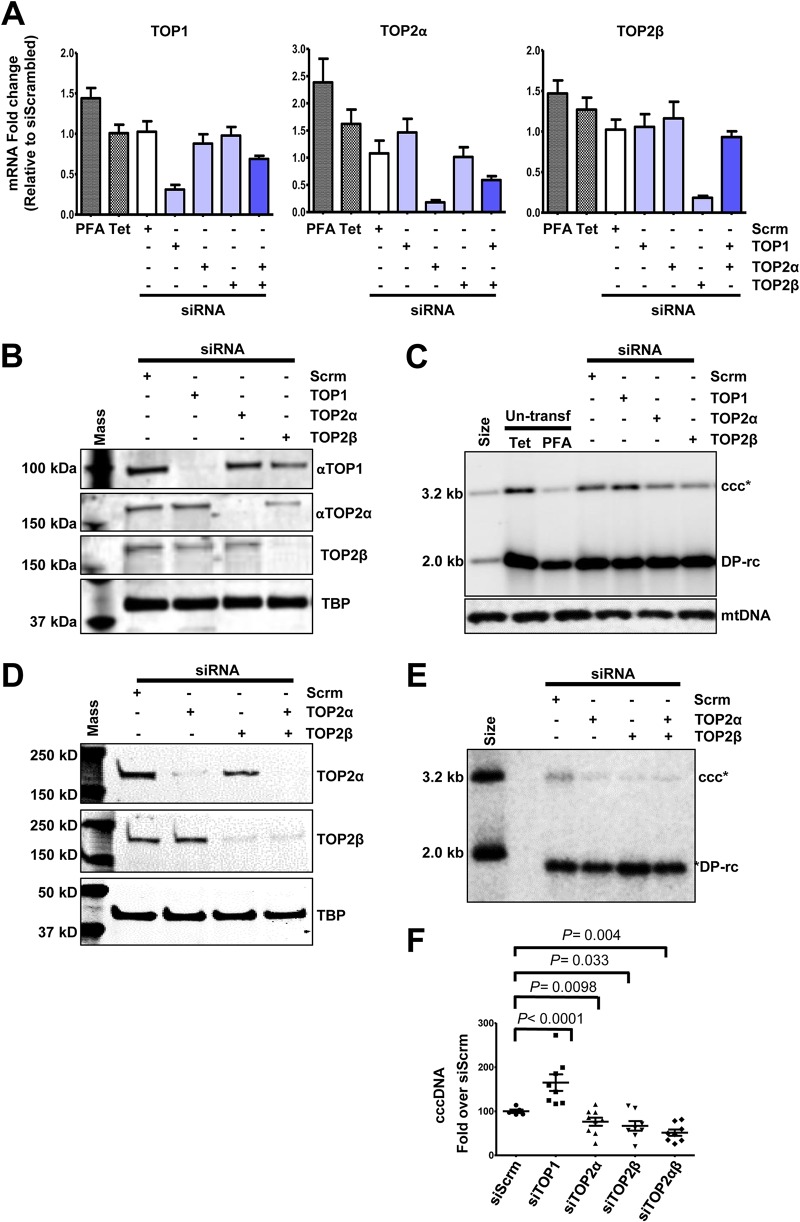
In order to identify host cellular DNA metabolic enzymes that are involved in the biosynthesis of hepatitis B virus (HBV) covalently closed circular (ccc) DNA, we developed a cell-based assay supporting synchronized and rapid cccDNA synthesis from intracellular progeny nucleocapsid DNA. This was achieved by arresting HBV DNA replication in HepAD38 cells with phosphonoformic acid (PFA), a reversible HBV DNA polymerase inhibitor, at the stage of single-stranded DNA and was followed by removal of PFA to allow the synchronized synthesis of relaxed circular DNA (rcDNA) and subsequent conversion into cccDNA within 12 to 24 h. This cccDNA formation assay allows systematic screening of the effects of small molecular inhibitors of DNA metabolic enzymes on cccDNA synthesis but avoids cytotoxic effects upon long-term treatment. Using this assay, we found that all the tested topoisomerase I and II (TOP1 and TOP2, respectively) poisons as well as topoisomerase II DNA binding and ATPase inhibitors significantly reduced the levels of cccDNA. It was further demonstrated that these inhibitors also disrupted cccDNA synthesis during de novo HBV infection of HepG2 cells expressing sodium taurocholate cotransporting polypeptide (NTCP). Mechanistic analyses indicate that whereas TOP1 inhibitor treatment prevented the production of covalently closed negative-strand rcDNA, TOP2 inhibitors reduced the production of this cccDNA synthesis intermediate to a lesser extent. Moreover, small interfering RNA (siRNA) knockdown of topoisomerase II significantly reduced cccDNA amplification. Taking these observations together, our study demonstrates that topoisomerase I and II may catalyze distinct steps of HBV cccDNA synthesis and that pharmacologic targeting of these cellular enzymes may facilitate the cure of chronic hepatitis B.IMPORTANCE Persistent HBV infection relies on stable maintenance and proper functioning of a nuclear episomal form of the viral genome called cccDNA, the most stable HBV replication intermediate. One of the major reasons for the failure of currently available antiviral therapeutics to cure chronic HBV infection is their inability to eradicate or inactivate cccDNA. We report here a chemical genetics approach to identify host cellular factors essential for the biosynthesis and maintenance of cccDNA and reveal that cellular DNA topoisomerases are required for both de novo synthesis and intracellular amplification of cccDNA. This approach is suitable for systematic screening of compounds targeting cellular DNA metabolic enzymes and chromatin remodelers for their ability to disrupt cccDNA biosynthesis and function. Identification of key host factors required for cccDNA metabolism and function will reveal molecular targets for developing curative therapeutics of chronic HBV infection.

Keywords: DNA topoisomerase; antiviral agents; cccDNA; hepatitis B virus.


Figures

FIG 1
FIG 1
Characterization of protein-free HBV DNA species in HepAD38 cells. (A and B) HepAD38 cells cultured for 12 days in the absence of Tet. Hirt DNA was extracted and detected by Southern blot hybridization. The size markers of HBV DNA (in length) are indicated. Hirt DNA without prior treatment (lane 1), after heat denaturalization without (lane 2) or with subsequent EcoRI digestion (lane 3), or with Exo I and Exo III treatment without (lane 4) or with subsequent EcoRI digestion (lane 5) were resolved by agarose gel electrophoresis and transferred on nylon membranes. The membranes were hybridized with a full-length riboprobe specifically hybridizing to the negative strand (A) and positive strand (B) of HBV DNA. The red arrow indicates covalently closed circular negative-strand HBV DNA. (C) Schematic presentation of the experimental schedule. HepAD38 cells were maintained in culture in the presence of Tet and harvested at the indicated time postremoval of Tet. (D) HBV core DNA (upper panel) and Hirt DNA after heat denaturalization and EcoRI digestion (lower panel) were resolved by agarose gel electrophoresis and detected by Southern blot hybridization with a full-length riboprobe specifically hybridizing to negative-strand HBV DNA. Mitochondrial DNA (mtDNA) served as a loading control for Hirt DNA. Biological duplicate samples were obtained and analyzed for each of the indicated time points. rc, relaxed circular DNA; DP-rc, deproteinized rcDNA; *DP-rc, denatured deproteinized rcDNA; dsl, double-stranded linear DNA; ss, single-stranded DNA; ccc, covalently closed circular DNA; ccc*, EcoRI-linearized cccDNA.
FIG 2
FIG 2
A synchronized and rapid cccDNA synthesis assay in HepAD38 cells. (A) Schematic presentation of the experimental schedule. HepAD38 cells were cultured in the absence of Tet, and 2 mM PFA was added in the culture medium 2 days after Tet removal to arrest viral DNA synthesis. Four days later, while PFA was withdrawn, Tet was added back to culture medium to stop viral pgRNA transcription from the transgene. Cells were harvested at the indicated time points. (B) HBV core DNA and Hirt DNA after heat denaturalization at 88°C for 8 min and EcoRI digestion were resolved by agarose gel electrophoresis, and HBV DNA species, indicated on the right, were detected by Southern blot hybridization with a riboprobe specifically hybridizing to negative-strand DNA. Lane M, molecular size marker. (C) The amounts of HBV rcDNA and cccDNA were quantified by phosphorimager and plotted as fold change relative to the amount of the corresponding DNA species at time zero of PFA removal. Means and standard deviations from two biological duplicates are presented.
FIG 3
FIG 3
TOP1 and TOP2 inhibitors reduce the level of cccDNA. (A) Schematic presentation of the experimental schedule. HepAD38 cells were cultured in the absence of Tet, and HBV DNA replication was arrested by PFA treatment between days 3 and 6 after Tet removal. The cells were immediately harvested (0 h) or cultured in the presence of Tet and absence of PFA and mock treated (untreated, UT) or treated with 10 μM 3TC or the indicated concentrations of DNA TOP poisons for 24 h. (B, D, F, and H) Intracellular HBV core DNA and Hirt DNA after heat denaturalization at 88°C for 8 min and EcoRI digestion were resolved by agarose gel electrophoresis, and HBV DNA species were detected by Southern blot hybridization with a riboprobe specifically hybridizing to negative-strand DNA. mtDNA served as a loading control of Hirt DNA analysis. (C, E, G, and I) Core DNA (including ssDNA and rcDNA), DP-rcDNA, and cccDNA were quantified by phosphorimager and normalized to the amount of mtDNA. The average level from two biological duplicates under compound treatment were plotted as the percentage of that in the mock-treated cells at 24 h post-PFA removal.
FIG 4
FIG 4
TOP1 and TOP2 inhibitors block cccDNA synthesis. (A) Schematic presentation of the experimental schedule. HepAD38 cells were cultured in the absence of Tet, and 2 mM PFA was added to the culture medium 2 days after Tet removal to arrest viral DNA synthesis. Four days later, while PFA was withdrawn, Tet was added back to the culture medium to stop viral pgRNA transcription from the transgene. Cells were left untreated or treated with topotecan (TPT; 1 μM) or doxorubicin (Doxo; 1 μM) at 16 h after PFA removal and harvested at the indicated time points. (B) Hirt DNA after heat denaturalization at 88°C for 8 min and EcoRI digestion was resolved by agarose gel electrophoresis and HBV DNA species were detected by Southern blot hybridization with a riboprobe specifically hybridizing to negative-strand DNA. mtDNA served as a loading control. (C) The amounts of cccDNA were quantified by phosphorimager, normalized to the amount of mtDNA, and plotted as the percentage of that in the mock-treated (UT) cells harvested at 16 h post-PFA removal. Means and standard deviations (n = 4) are presented. *DP-rc, denatured deproteinized rc DNA; ccc*, EcoRI-linearized cccDNA.
FIG 5
FIG 5
TOP1 and TOP2 inhibitors do not alter the level of preexisting cccDNA. (A and C) Schematic representation of the experimental schedules. (B and D) Intracellular HBV core DNA and Hirt DNA after heat denaturalization at 88°C for 8 min and EcoRI digestion were resolved by agarose gel electrophoresis, and HBV DNA species were detected by Southern blot hybridization with a riboprobe specifically hybridizing to negative-strand DNA. mtDNA served as a loading control of Hirt DNA analysis. For the data shown in panel D, treatment with 3TC alone was started at day 6, and the cells were harvested at day 9, whereas mock treatment or treatment with the indicated concentrations of doxorubicin was started at day 8, and the cells were harvested at day 9, as illustrated in panel C. The amounts of cccDNA were quantified by phosphorimager, normalized to the amount of the mtDNA. The average levels of cccDNA in compound-treated cells were denoted as the percentage of that in the mock-treated (UT) cells.
FIG 6
FIG 6
TOP1 and TOP2 inhibitors did not induce prominent cytokines. HepAD38 cells were mock treated (UT) or treated with 250 nM camptothecin (CTP) or 250 nM doxorubicin (Doxo) for 6 h or 24 h. Intracellular IFN-β, interleukin-6 (IL-6), IL-29, IL-28A, IL-28B, and tumor necrosis factor alpha (TNF-α) transcripts were quantified by qRT-PCR assays and normalized to the level of β-actin mRNA. Means and standard deviations (n = 3) are presented.
FIG 7
FIG 7
Effects of distinct TOP1 and TOP2 inhibitors on HBV cccDNA synthesis. (A) Illustration of DNA TOP2 catalytic cycle. Briefly, TOP2 enzyme binds to the DNA molecule (step 1). In the presence of Mg++, two ATP molecules bind to the ATPase domain which results in its dimerization and cleavage of one double-stranded DNA (blue) (step 2). The second DNA molecule (orange) is transported through the break (step 3). Upon transport of the DNA segment through the break, one molecule of the ATP is hydrolyzed (step 4), followed by the religation of the cleaved DNA segment along with hydrolysis of another ATP molecule (step 5) and release of a DNA fragment (step 6). Compounds that inhibit each of these steps are indicated. (B to E) HepAD38 cells were cultured in the absence of Tet, and HBV DNA replication was arrested by PFA treatment between days 3 and 6 after Tet removal. The cells were immediately harvested (0 h) or cultured in the presence of Tet and absence of PFA and mock treated (UT) or treated with 10 μM 3TC or the indicated concentrations of aclarubicin and merbarone (B) or 500 nM ICRF-187, 500 nM etoposide (Etop), 500 nM mitoxantrone (MXT), 500 nM ICRF-193, 500 nM β-lapachone (β-Lap), and 50 μM merbarone (Merb) (E) for 24 h. Hirt DNA was resolved by agarose gel electrophoresis after heat denaturalization at 88°C for 8 min and EcoRI digestion. HBV DNA species were detected by Southern blot hybridization with a riboprobe specifically hybridizing to negative-strand DNA. mtDNA served as a loading control of Hirt DNA analysis. The amounts of cccDNA were quantified by phosphorimager and normalized to the amount of mtDNA. The levels of cccDNA in compound-treated cells were plotted (C and D) or represented as the percentage of that in the mock-treated cells (E).
FIG 8
FIG 8
TOP1 and TOP2 inhibitors inhibited HBV cccDNA synthesis in de novo infection. (A) Schematic representation of the experimental schedule. C3AhNTCP cells were infected with HBV at an MOI of 250 genome equivalents for 24 h. The cells were mock treated or treated with 200 nM doxorubicin (Doxo), 200 nM topotecan (TPT), 200 nM doxorubicin and 200 nM topotecan (Doxo + TPT), 200 nM aclarubicin (Acla), or 1 μg/ml myrcludex B (Myr-B) starting from HBV infection for a total of 36 h. (B) Hirt DNA was resolved by agarose gel electrophoresis after heat denaturalization at 88°C for 8 min and EcoRI digestion. HBV DNA species were detected by Southern blot hybridization with a riboprobe specifically hybridizing to negative-strand DNA. mtDNA served as a loading control of Hirt DNA analysis. (C) cccDNAs were quantified by a phosphorimager. The data from three independent experiments are presented. P values calculated by Student's t test are presented.
FIG 9
FIG 9
TOP1 and TOP2 inhibitors disrupted distinct steps of HBV cccDNA synthesis. (A) Schematic illustration of cccDNA synthesis from DP-rcDNA via an intermediate, cc(−)rcDNA, and production of cc(−)DNA by exonuclease I and III digestion of cc(−)rcDNA. (B to E) HepAD38 cells were cultured in the absence of Tet, and HBV DNA replication was arrested by PFA treatment between days 3 and 6 after Tet removal. The cells were then cultured in the presence of Tet and absence of PFA and mock-treated (UT) or treated for 24 h with 500 nM camptothecin (CPT) (B), 500 nM doxorubicin (Doxo) (C), or 40 μM merbarone (Merb) (D). Hirt DNA without prior treatment, digested with Exo I and Exo III without or with subsequent EcoRI restriction, was resolved by agarose gel electrophoresis. HBV DNA species were detected by Southern blot hybridization with a riboprobe specifically hybridizing to negative-strand DNA. (E) The amounts of cc(−)DNA were quantified by a phosphorimager and normalized to the amount in mock (DMSO)-treated cells. Mean and standard derivations from six (camptothecin and doxorubicin) or three (merbarone) independent experiments are presented. P values calculated by Student's t test are presented.
FIG 10
FIG 10
Knockdown of TOP2 mRNA reduced cccDNA formation. HepAD38 cells were mock transfected or transfected with the indicated siRNA and cultured in the absence of Tet and presence of PFA for 2 days. The cells were then cultured in the presence of Tet and absence of PFA for an additional 24 h. (A) Total cellular RNAs were extracted, and TOP mRNAs were quantified by qRT-PCR assay, normalized to the level of β-actin mRNA, and expressed as the ratio to the level of the respective TOP mRNA in cells transfected with a scrambled siRNA. (B and D) The levels of TOP proteins were determined by Western blot assays. TATA-binding protein (TBP) served as a loading control. (C and E) Hirt DNA was resolved by agarose gel electrophoresis after heat denaturalization at 88°C for 8 min and EcoRI digestion. HBV DNA species were detected by Southern blot hybridization with a riboprobe specifically hybridizing to negative-strand DNA. mtDNA served as a loading control of Hirt DNA analysis. (F) cccDNA was quantified by a phosphorimager. Means and standard deviations are presented (n = 8). P values calculated by Student's t test are presented.

Similar articles

Cited by

References

    1. Ott JJ, Stevens GA, Groeger J, Wiersma ST. 2012. Global epidemiology of hepatitis B virus infection: new estimates of age-specific HBsAg seroprevalence and endemicity. Vaccine 30:2212–2219. doi:10.1016/j.vaccine.2011.12.116. - DOI - PubMed
    1. GBD 2013 Mortality and Causes of Death Collaborators. 2015. Global, regional, and national age-sex specific all-cause and cause-specific mortality for 240 causes of death, 1990–2013: a systematic analysis for the Global Burden of Disease Study 2013. Lancet 385:117–171. doi:10.1016/S0140-6736(14)61682-2. - DOI - PMC - PubMed
    1. Block TM, Guo H, Guo JT. 2007. Molecular virology of hepatitis B virus for clinicians. Clin Liver Dis 11:685–706. vii. doi:10.1016/j.cld.2007.08.002. - DOI - PMC - PubMed
    1. Dienstag JL. 2009. Benefits and risks of nucleoside analog therapy for hepatitis B. Hepatology 49:S112–S121. doi:10.1002/hep.22920. - DOI - PubMed
    1. Perrillo R. 2009. Benefits and risks of interferon therapy for hepatitis B. Hepatology 49:S103–S111. doi:10.1002/hep.22956. - DOI - PubMed

Publication types