. 2019 Mar 13;10(1):1198.
doi: 10.1038/s41467-019-09037-9.

Duodenal bacterial proteolytic activity determines sensitivity to dietary antigen through protease-activated receptor-2

Affiliations

Duodenal bacterial proteolytic activity determines sensitivity to dietary antigen through protease-activated receptor-2

Alberto Caminero et al. Nat Commun. .

Abstract

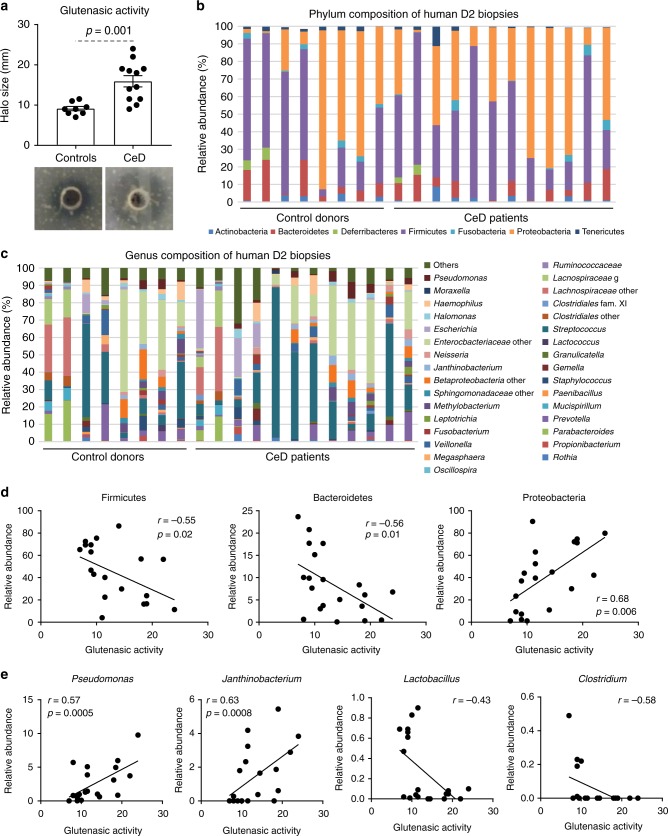
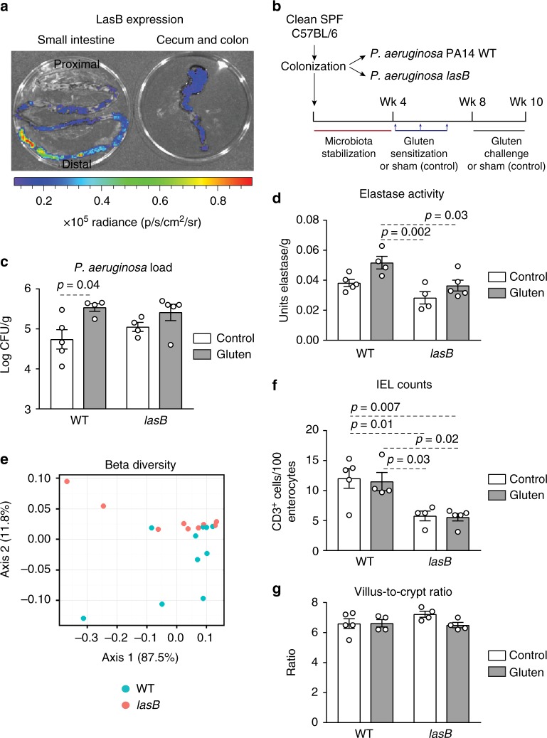
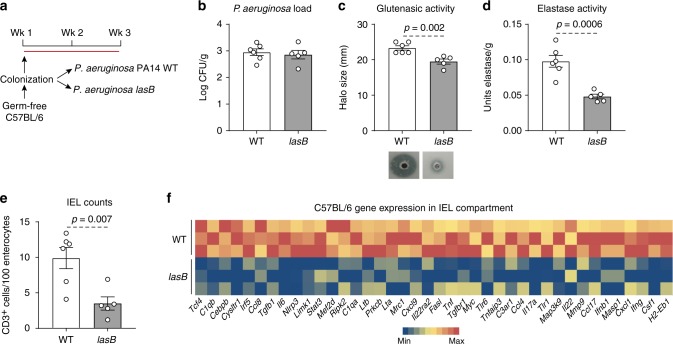
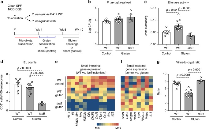
Microbe-host interactions are generally homeostatic, but when dysfunctional, they can incite food sensitivities and chronic diseases. Celiac disease (CeD) is a food sensitivity characterized by a breakdown of oral tolerance to gluten proteins in genetically predisposed individuals, although the underlying mechanisms are incompletely understood. Here we show that duodenal biopsies from patients with active CeD have increased proteolytic activity against gluten substrates that correlates with increased Proteobacteria abundance, including Pseudomonas. Using Pseudomonas aeruginosa producing elastase as a model, we show gluten-independent, PAR-2 mediated upregulation of inflammatory pathways in C57BL/6 mice without villus blunting. In mice expressing CeD risk genes, P. aeruginosa elastase synergizes with gluten to induce more severe inflammation that is associated with moderate villus blunting. These results demonstrate that proteases expressed by opportunistic pathogens impact host immune responses that are relevant to the development of food sensitivities, independently of the trigger antigen.


Conflict of interest statement

The authors declare no competing interests.

Figures

Fig. 1
Fig. 1
High proteolytic activity in celiac disease (CeD) duodenum correlates with microbiota changes. a Glutenasic activity measured in duodenal biopsies from non-celiac disease donors (controls; n = 8) and CeD patients (n = 12). Data presented as mean ± s.e.m. where each dot represents an individual human donor. Displayed P value was calculated by Student’s t-test. Representative bioassays are shown. b, c Relative abundance of the microbial composition at the phylum level (b) and genus level (c) of duodenal biopsies from controls (n = 8) and CeD patients (n = 12). d, e Correlation between microbial relative abundance at the phylum level (d) or genus level (e) and glutenasic activity in duodenal biopsies from controls (n = 8) and CeD (n = 12) human donors. Displayed P values survived 10% false discovery rate (FDR) correction. Each dot represents individual human donor. Correlation based on Spearman’s index
Fig. 2
Fig. 2
LasB induces intraepithelial lymphocytes (IELs) and microbiota shifts. a Visual expression of lasB in the small intestine (left panel) and large intestine (right panel) of clean specific pathogen free (SPF) mice 2 weeks following colonization with a luminescent producing P. aeruginosa mutant linked to the lasB promoter (n = 3). One representative image is shown. b Protocol for gluten sensitization and challenge (gluten treatment) in clean SPF C57BL/6 mice colonized with P. aeruginosa PA14 WT (wild-type (WT) gluten) or the lasB mutant (lasB gluten). Sham controls consisted of non-sensitized and non-gluten challenged clean SPF C57BL/6 mice colonized with P. aeruginosa PA14 WT (WT control) or the lasB mutant (lasB control). c P. aeruginosa bacterial load in the small intestine of clean SPF C57BL/6 colonized with P. aeruginosa PA14 WT or lasB mutant with gluten treatment (gray bars; n = 4 WT, n = 5 lasB) or without gluten treatment (white bars; n = 5 WT, n = 4 lasB). d Elastase activity measured in the small intestine of clean SPF C57BL/6 mice colonized with P. aeruginosa PA14 WT or the lasB mutant with gluten treatment (gray bars; n = 4 WT, n = 5 lasB) or without gluten treatment (white bars; n = 5 WT, n = 4 lasB). e β-Diversity Bray–Curtis dissimilarity principal coordinates analysis (PCoA) plot of microbiota profiles from clean SPF C57BL/6 mice colonized with P. aeruginosa PA14 WT (n = 10) or the lasB mutant (n = 9). Each dot represents an individual mouse. Differences between bacterial communities were tested by permutational multivariate analysis of variance (PERMANOVA) using Qiime. f Quantitative measurement of IELs/100 enterocytes in small intestinal villi tips of clean SPF C57BL/6 mice colonized with P. aeruginosa PA14 WT or the lasB mutant, with gluten treatment (gray bars; n = 4 WT, n = 5 lasB) or without gluten treatment (white bars; n = 5 WT, n = 4 lasB). g Small intestinal villus-to-crypt ratios of clean SPF C57BL/6 mice colonized with P. aeruginosa PA14 WT or the lasB mutant, with gluten treatment (gray bars; n = 4 WT, n = 5 lasB) or without gluten treatment (white bars; n = 5 WT, n = 4 lasB). Data for c, d, f, g are presented as mean ± s.e.m. where each dot represents an individual mouse. Displayed P values were calculated by a one-way analysis of variance (ANOVA) with Tukey's post-hoc test
Fig. 3
Fig. 3
LasB induces a pro-inflammatory response in the absence of microbiota. a Protocol for monocolonization of C57BL/6 mice with P. aeruginosa PA14 wild-type (WT) or the lasB mutant. b P. aeruginosa bacterial load recovered in the small intestine of WT (n = 6) and lasB mutant (n = 5) colonized mice 3 weeks post colonization. c Luminal small intestinal glutenasic activity from mice monocolonized with P. aeruginosa PA14 WT (n = 6) or the lasB mutant (n = 5). Representative bioassays are shown. d Luminal small intestinal elastase activity from mice monocolonized with P. aeruginosa PA14 WT (n = 6) or the lasB mutant (n = 5). e Quantitative measurement of intraepithelial lymphocytes (IELs)/100 enterocytes in small intestinal villi tips of P. aeruginosa PA14 WT (n = 6) or lasB mutant (n = 5) monocolonized mice. f Heat map of gene expression in the small intestinal IEL compartment of mice colonized with P. aeruginosa PA14 WT (n = 3) or the lasB mutant (n = 3) assessed by NanoString nCounter gene expression. Only statistically different genes generated by nSolver 2.5 using Student’s t-test and based on bacterial colonization are shown. Data for be are presented as mean ± s.e.m. where each dot represents an individual mouse. Displayed P values were calculated by Student’s t-test
Fig. 4
Fig. 4
LasB induces a pro-inflammatory response through protease-activated receptor- 2 (PAR-2). a In vitro cleavage of the external domain of PAR-2 by P. aeruginosa PA14 wild-type (WT) (n = 3 biological replicates) and the lasB mutant (n = 3 biological replicates). Data presented as mean ± s.e.m where each dot represents one biological replicate. b Protocol for monocolonization of germ-free C57BL/6 mice and protease-resistant PAR-2 mutant mice (PAR38E-PAR2) with P. aeruginosa PA14 WT. c P. aeruginosa bacterial load recovered in the small intestine of monocolonized C57BL/6 mice (n = 4) and PAR38E-PAR2 mice (n = 4). d Luminal small intestinal glutenasic activity from C57BL/6 mice (n = 4) and PAR38E-PAR2 mice (n = 4) monocolonized with P. aeruginosa PA14 WT. Representative bioassays are shown. e Luminal small intestinal elastase activity from C57BL/6 mice (n = 4) and PAR38E-PAR2 mice (n = 4) monocolonized with P. aeruginosa PA14 WT. f Quantitative measure of intraepithelial lymphocytes (IELs)/100 enterocytes in small intestinal villi tips of C57BL/6 mice (n = 4) and R38E-PAR2 mice (n = 4) monocolonized with P. aeruginosa PA14 WT. g Heat map of gene expression in the IEL compartment of the small intestine of C57BL/6 mice (n = 4) and PAR38E-PAR2 mice (n = 4) monocolonized with P. aeruginosa PA14 WT assessed by NanoString nCounter gene expression. Only statistically different genes generated by nSolver 2.5 using Student’s t-test and based on bacterial colonization are shown. Data for ce are presented as mean ± s.e.m. where each dot represents an individual mouse. Displayed P values were calculated by Student’s t-test
Fig. 5
Fig. 5
LasB enhances gluten sensitivity in genetically predisposed mice. a Protocol for gluten sensitization and challenge (gluten treatment) of clean specific pathogen free (SPF) NOD/DQ8 mice colonized with P. aeruginosa PA14 WT (wild-type (WT) gluten) or the lasB mutant (lasB gluten). Sham controls consisted of non-sensitized and non-gluten challenged clean SPF NOD/DQ8 mice colonized with P. aeruginosa PA14 WT (WT control). b P. aeruginosa bacterial load in the small intestine of clean SPF NOD/DQ8 colonized with P. aeruginosa PA14 WT or the lasB mutant, treated with gluten (gray bars; n = 10 WT, n = 4 lasB) or without gluten (white bars; n = 6). c Luminal small intestinal elastase activity of clean SPF NOD/DQ8 mice colonized with P. aeruginosa PA14 WT or the lasB mutant, treated with gluten (gray bars; n = 10 WT, n = 4 lasB) or without gluten (white bars; n = 6). d Quantitative measure of intraepithelial lymphocytes (IELs)/100 enterocytes in small intestinal villi tips in clean SPF NOD/DQ8 mice colonized with P. aeruginosa PA14 WT or the lasB mutant, treated with gluten (gray bars; n = 10 WT, n = 4 lasB) or without gluten (white bars; n = 7). e, f Heat map of significantly altered genes in whole small intestinal tissue of clean SPF NOD/DQ8 mice (n = 4/group), when comparisons were performed between P. aeruginosa PA14 WT and lasB mutant colonized mice (e) or when comparisons were performed between gluten treated and control mice (f). Gene expression was assessed by NanoString nCounter gene expression and only significantly altered genes generated by nSolver 2.5 using multiple t-tests are shown. g Small intestinal villus-to-crypt ratios in clean SPF NOD/DQ8 mice colonized with P. aeruginosa PA14 WT or the lasB mutant, treated with gluten (gray bars; n = 10 WT, n = 4 lasB) or without gluten (white bars; n = 8). Data for bd, g are presented as mean ± s.e.m. where each dot represents an individual mouse. Displayed P values were calculated by one-way analysis of variance (ANOVA) with Tukey's post-hoc test
Fig. 6
Fig. 6
Celiac duodenal microbiota induces proteolytic activity and intraepithelial lymphocytes (IELs). a Protocol for the colonization of germ-free C57BL/6 mice with human small intestinal aspirates from celiac disease (CeD) patients (n = 4) or controls (n = 5). b Beta-diversity of small intestinal microbiota profiles of recipient mice colonized with human aspirates of patients with CeD and without CeD (controls), using Bray–Curtis dissimilarity represented as non-metric multidimensional scaling (NMSD). Differences between bacterial communities were tested by permutational multivariate analysis of variance (PERMANOVA) using Qiime. Each donor was used to colonize 2–3 mice, and each dot represents an individual mouse. c Glutenasic activity per donor (left side) and pooled (right side), measured in the small intestine of mice colonized with small intestinal aspirates from control or CeD donors. Representative bioassays are shown. d Quantitative measurement of IELs/100 enterocytes in small intestinal villi tips of mice colonized with small intestinal aspirates from control or CeD donors. IEL counts per donor (left panel) and pooled counts (right panel) are shown. Data for c, d are presented as median with interquartile range and whiskers extending from minimum to maximum. Each dot represents an individual mouse. Each donor, as indicated by a different color, was used to colonize 2–3 mice. Displayed P values were calculated by Mann–Whitney test. Tissue from one recipient mouse receiving CeD donor 2 presented technical difficulties during embedding and processing and was dropped out. e Correlation between glutenasic activity and IEL counts in the small intestine of mice colonized with small intestinal aspirates (n = 23). Each dot represents an individual mouse. Correlation based on Spearman's index. f Correlation between relative abundance of Proteobacteria and Firmicutes and small intestinal glutenasic activity of mice colonized with small intestinal aspirates (n = 23). Each dot represents an individual mouse. Displayed P values from Proteobacteria and Firmicutes survived 10% false discovery rate (FDR) correction. Correlation based on Spearman's index

Similar articles

Cited by

References

    1. Vergnolle N. Protease inhibition as new therapeutic strategy for GI diseases. Gut. 2016;65:1215–1224. doi: 10.1136/gutjnl-2015-309147. - DOI - PMC - PubMed
    1. Antalis TM, Shea-Donohue T, Vogel SN, Sears C, Fasano A. Mechanisms of disease: protease functions in intestinal mucosal pathobiology. Nat. Clin. Pract. Gastroenterol. Hepatol. 2007;4:393–402. doi: 10.1038/ncpgasthep0846. - DOI - PMC - PubMed
    1. Cenac N, et al. Role for protease activity in visceral pain in irritable bowel syndrome. J. Clin. Investig. 2007;117:636–647. doi: 10.1172/JCI29255. - DOI - PMC - PubMed
    1. Gecse K, et al. Increased faecal serine protease activity in diarrhoeic IBS patients: a colonic lumenal factor impairing colonic permeability and sensitivity. Gut. 2008;57:591–599. doi: 10.1136/gut.2007.140210. - DOI - PubMed
    1. Bustos D, et al. Colonic proteinases: increased activity in patients with ulcerative colitis. Medicina. 1998;58:262–264. - PubMed

Publication types

MeSH terms