. 2019 Mar 13;10(1):1203.
doi: 10.1038/s41467-019-09174-1.

Poly(ADP-ribose) polymerase 1 accelerates vascular calcification by upregulating Runx2

Affiliations

Poly(ADP-ribose) polymerase 1 accelerates vascular calcification by upregulating Runx2

Cheng Wang et al. Nat Commun. .

Abstract

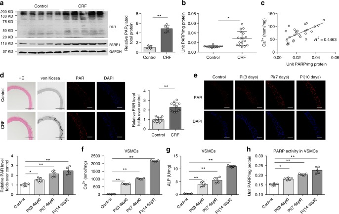
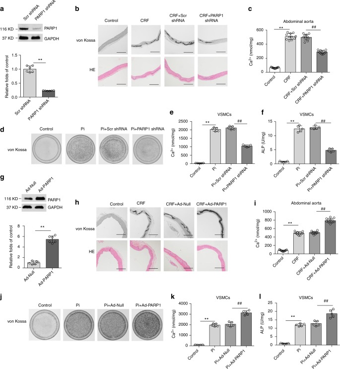
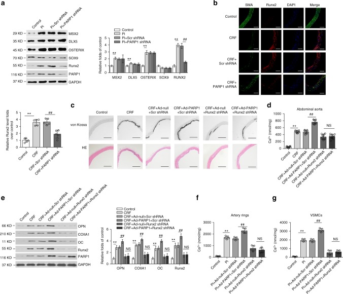
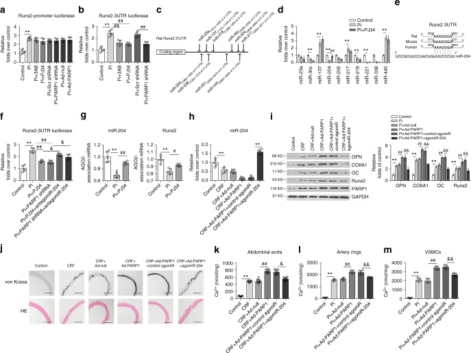
Vascular calcification is highly prevalent in end-stage renal diseases and is predictive of cardiovascular events and mortality. Poly(ADP-ribose) polymerase 1 (PARP1) inhibition or deletion is vasoprotective in several disease models. Here we show that PARP activity is increased in radial artery samples from patients with chronic renal failure, in arteries from uraemic rats, and in calcified vascular smooth muscle cells (VSMCs) in vitro. PARP1 deficiency blocks, whereas PARP1 overexpression exacerbates, the transdifferentiation of VSMCs from a contractile to an osteogenic phenotype, the expression of mineralization-regulating proteins, and calcium deposition. PARP1 promotes Runx2 expression, and Runx2 deficiency offsets the pro-calcifying effects of PARP1. Activated PARP1 suppresses miRNA-204 expression via the IL-6/STAT3 pathway and thus relieves the repression of its target, Runx2, resulting in increased Runx2 protein. Together, these results suggest that PARP1 counteracts vascular calcification and that therapeutic agents that influence PARP1 activity may be of benefit to treat vascular calcification.


Conflict of interest statement

The authors declare no competing interests.

Figures

Fig. 1
Fig. 1
PARP activity is increased in calcifying arteries and VSMCs. a Representative western blot analysis of PARP1 expression and PARP activity (Poly(ADP-ribosyl)ation) in radial arteries. Chronic renal failure (CRF): patients with CRF and uremia who underwent arterial venous fistular surgery (n = 5); Control (n = 5): patients who underwent amputation surgery due to arm trauma, without diagnosed complications of diabetes and chronic kidney disease. b PARP activity in radial arteries of control (n = 9) and CRF patients (n = 17) were assayed using the universal colorimetric PARP assay kit. c The calcium content and PARP activity were positively correlated in CRF arteries. R2 = 0.4463, P < 0.05. Statistical significance of correlations was determined by Pearson’s correlation coefficient analysis. d Wistar rats were fed an adenine diet or a chow diet for 6 weeks (n = 10–12 per group). Arteries were isolated and performed by hematoxylin/eosin (H&E) and von Kossa staining. The PAR level was determined by immunofluorescence staining. Scale bars: black, 200 μm; white, 100 μm. e Rat aortic rings were treated with high Pi (osteogenic medium containing 10 mM β-glycerophosphate) for indicated days (0, 3, 7, and 10 days), and PARP activity was then detected by immunofluorescence staining. (n = 5 per group). Scale bar, 100 μm. fh Rat VSMCs were treated with high Pi (10 mM β-glycerophosphate) for indicated days (0, 3, 7, and 14 days). The calcium content (f), ALP activity (g), and PARP activity (h) in calcified VSMCs were assayed. (n = 5 per group). Statistical significance was assessed using one-way ANOVA for multiple comparison and two-tailed t-tests for two groups and is presented as follows: *P < 0.05 and **P < 0.01. All values are means ± S.D.
Fig. 2
Fig. 2
Manipulation of PARP1 expression regulates vascular calcification. ac Rat abdominal aortas were inoculated with adenovirus encoding Scrambled (Scr shRNA) or PARP1 shRNA at three weeks after the adenine diet, and then fed for three weeks. PARP1 deficiency in arteries was identified by western blot (a) (n = 5 per group). Aortas were stained by H&E and von Kossa for mineralization (b), and the calcium deposition in arteries was quantified (c). (n = 10–12 per group). Scale bar, 100 μm. df Rat primary VSMCs were pre-infected with Scrambled or PARP1 shRNA adenovirus and then exposed to osteogenic medium for 14 days. VSMCs were stained for mineralization by Alizarin red S (d), and the quantitative analysis of calcium content (e) and ALP (f) were detected respectively. (n = 5 for each group). gi Rat abdominal aortas were inoculated with Ad-Null or Ad-PARP1 at three weeks after the adenine diet, and then fed for three weeks. PARP1 overexpression in arteries was evaluated via western blot (g) (n = 5 per group). Aortas were stained by von Kossa and H&E for mineral nodules (h) and the quantification of calcium deposition was calculated (i). (n = 10–12 per group). Scale bar, 100 μm. jl Rat primary VSMCs were pre-infected with Ad-Null or Ad-PARP1 adenovirus and then exposed to osteogenic medium for 14 days. Alizarin red S staining (j), calcium content (k), and ALP (l) in calcified VSMCs were then determined. (n = 5 per group). Statistical significance was assessed using one-way ANOVA for multiple comparison and two-tailed t-tests for two groups and is presented as follows: **P < 0.01 and ##P < 0.01. All values are means ± S.D.
Fig. 3
Fig. 3
PARP1 regulates VSMCs osteogenic transition, not Na/P co-transporter. a, b Quantitative RT-PCR (a) and western blot (b) analysis of PIT1 and PIT2 expression in VSMCs, which were infected with Scrambled (Scr shRNA) or PARP1 shRNA, and treated with high Pi. ce Rat VSMCs were infected with adenovirus carrying Scr shRNA, PARP1 shRNA, Null or PARP1 gene for 48 h and then incubated with osteogenic media for 14 days. PARP1 inhibitors 3AB (10 mM) and PJ34 (10 μM) were added every day. Western blot analysis and related quantification of osteogenic-related marker genes (OPN, ColIA1, and OC) and smooth muscle marker genes (SMA, SM22 and SMMHC) expression in VSMCs with PARP1 knockdown (c), PARP1 inhibitors 3AB and PJ34 (d), and PARP1 overexpression (e). (n = 5 for each group). Statistical significance was assessed using one-way ANOVA for multiple comparison and two-tailed t-tests for two groups and is presented as follows: NS: no significance, **P < 0.01 and ##P < 0.01. All values are means ± S.D.
Fig. 4
Fig. 4
Runx2 acts as a mediator of the pro-calcifying effects of PARP1. a Western blot analysis of several osteogenic factors expression in calcified VSMCs transfected with Scr or PARP1 shRNA. (n = 5 per group). b The expression levels of Runx2 and SMA in abdominal arteries of indicated groups were determined by immunofluorescence staining. (n = 5 per group). Scale bar, 100 μm. ce Rat abdominal aortas were inoculated with Ad-Scr shRNA, or Ad-Runx2 shRNA together with Ad-Null or Ad-PARP1 at three weeks after the adenine diet. Six weeks later, arteries were isolated and the calcification was analyzed by H&E, von Kossa staining (c) and the calcium quantification (d) (n = 10–12 per group), and the downstream osteogenic markers (OPN, ColIA1, OC, and Runx2) were analyzed by western blot (e). (n = 5 per group). Scale bar, 100 μm. f, g The aortic rings (f) or rVSMCs (g) were pre-infected with Ad-Scr shRNA, or Ad-Runx2 shRNA together with Ad-Null or Ad-PARP1, and exposed to the osteogenic medium for 14 days. The calcification was determined by the calcium assay respectively. (n = 5 for each group). Statistical significance was assessed using one-way ANOVA for multiple comparison and is presented as follows: NS: no significance, **P < 0.01 and ##P < 0.01. All values are means ± S.D. Source data are provided as a Source Data file
Fig. 5
Fig. 5
miR-204 mediates the regulation of PARP1 on Runx2. a A7r5 cells were transfected with the Runx2 promoter, along with 3AB, PJ34 or Scr shRNA and PARP1 shRNA treatments. The luciferase activity was analyzed. (n = 5 per group). b The Runx2-3′UTR plasmid was transiently transfected into A7r5 cells before stimulation as indicated. The luciferase reporter assay was analyzed. (n = 5 per group). c Seed regions of predicted Runx2-targeting miRNAs. d Selected miRNA expression levels in calcified VSMCs were analyzed by qRT-PCR assays. (n = 5 per group). e The putative miR-204 sequence present in Runx2 is conserved across different species. f A7r5 cells were co-transfected with the Runx2-3′UTR plasmid and miR-204 antagomir, and then treated with high Pi in the absence or presence of PARP inhibitor or PARP1 deficiency. The luciferase activity was analyzed. (n = 5 per group). g Rat VSMCs were treated with high Pi or high Pi + PJ34 for 3 days. The binding ability of miR-204 to Runx2 3′UTR in RISC complex was detected by RIP assay. hk Rat abdominal arteries were inoculated with Ad-Null or Ad-PARP1 at three weeks after the adenine diet, and miR-204 agomir was intravenously injected through the tail vein for three consecutive days. miR-204 expression was determined by qRT-PCR (h). (n = 5 per group). The expression of osteogenic genes (OPN, ColIA1, OC, and Runx2) was analyzed by western blot (i). (n = 5 per group). The vascular calcification was detected by von Kossa staining (j) and the calcium quantification (k). (n = 10–12 per group). Scale bar, 100 μm. l, m The aortic rings (l) or rVSMCs (m) were transfected with control or miR-204 agomir together with Ad-Null or Ad-PARP1, and exposed to the osteogenic medium for 14 days. The calcification was determined by the calcium assay respectively (n = 5 per group). Statistical significance was assessed using one-way ANOVA for multiple comparison and is presented as follows: **P < 0.01, #P < 0.05, ##P < 0.01, &P < 0.05 and &&P < 0.01. All values are means ± S.D. Source data are provided as a Source Data file
Fig. 6
Fig. 6
PARP1 regulates miR-204 expression through the IL-6/JAK2/STAT3 pathway. a Rat VSMCs was pre-infected with Scr shRNA or PARP1 shRNA, and then exposed to high Pi for 3 days. The phosphorylation of STAT3 (pSTAT3) and STAT3 were determined by western blot. (n = 5 per group). b–d, VSMCs were infected with Scr shRNA or STAT3 shRNA, along with Ad-Null or Ad-PARP1, and then incubated with osteogenic media for 14 days. MiR-204 expression was determined by qRT-PCR (b). The expression of pSTAT3/STAT3 and Runx2 was analyzed by western blot (c), and the calcium deposition was quantified by the calcium assay (d). (n = 5 per group). e The phosphorylation of JAK2 and SRC, and their total proteins in calcified VSMCs infected with Scr shRNA or PARP1 shRNA were determined by western blot. f The level of IL-6 in the supernatant of calcified VSMCs was detected by ELISA assay. g, h The mRNA (g) and protein (h) levels of IL-6 in the abdominal arteries of CRF rats inoculated with Scr shRNA or PARP1 shRNA were determined by qRT-PCR and western blot. (n = 5 per group). ik Rat VSMCs were pre-infected with Ad-Null or Ad-PARP1, and then treated with neutralizing antibodies against IL-6 in the osteogenic media for 14 days. The levels of pJAK2/JAK2, pSTAT3/STAT3 and osteogenic markers (OC and Runx2) were determined by western blot (i), and the calcification was detected by Alizarin red S staining (j) and calcium quantification (k). (n = 5 per group). Statistical significance was assessed using one-way ANOVA followed by for multiple comparison and is presented as follows: NS: no significance, **P < 0.01, #P < 0.05, ##P < 0.01, &P < 0.05 and &&P < 0.01. All values are means ± S.D. Source data are provided as a Source Data file

References

    1. Demer LL, Tintut Y. Vascular calcification: pathobiology of a multifaceted disease. Circulation. 2008;117:2938–2948. doi: 10.1161/CIRCULATIONAHA.107.743161. - DOI - PMC - PubMed
    1. Lanzer P, et al. Medial vascular calcification revisited: review and perspectives. Eur. Heart J. 2014;35:1515–1525. doi: 10.1093/eurheartj/ehu163. - DOI - PMC - PubMed
    1. Shroff R, Long DA, Shanahan C. Mechanistic insights into vascular calcification in CKD. J. Am. Soc. Nephrol. 2013;24:179–189. doi: 10.1681/ASN.2011121191. - DOI - PubMed
    1. Jablonski KL, Chonchol M. Vascular calcification in end-stage renal disease. Hemodial. Int. 2013;17(Suppl 1):S17–S21. doi: 10.1111/hdi.12084. - DOI - PMC - PubMed
    1. Steitz SA, et al. Smooth muscle cell phenotypic transition associated with calcification: upregulation of Cbfa1 and downregulation of smooth muscle lineage markers. Circ. Res. 2001;89:1147–1154. doi: 10.1161/hh2401.101070. - DOI - PubMed

Publication types

MeSH terms