

Deletion of hematopoietic Dectin-2 or CARD9 does not protect against atherosclerotic plaque formation in hyperlipidemic mice
- PMID: 30867470
- PMCID: PMC6416398
- DOI: 10.1038/s41598-019-40663-x
Deletion of hematopoietic Dectin-2 or CARD9 does not protect against atherosclerotic plaque formation in hyperlipidemic mice
Abstract
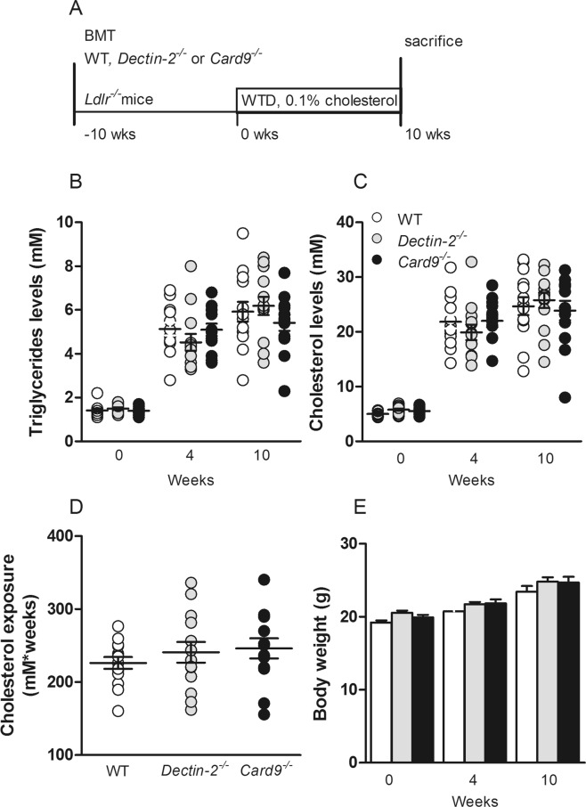
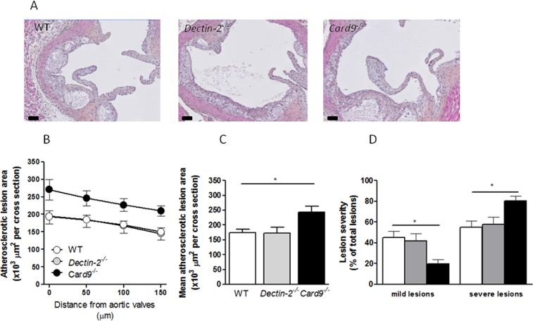
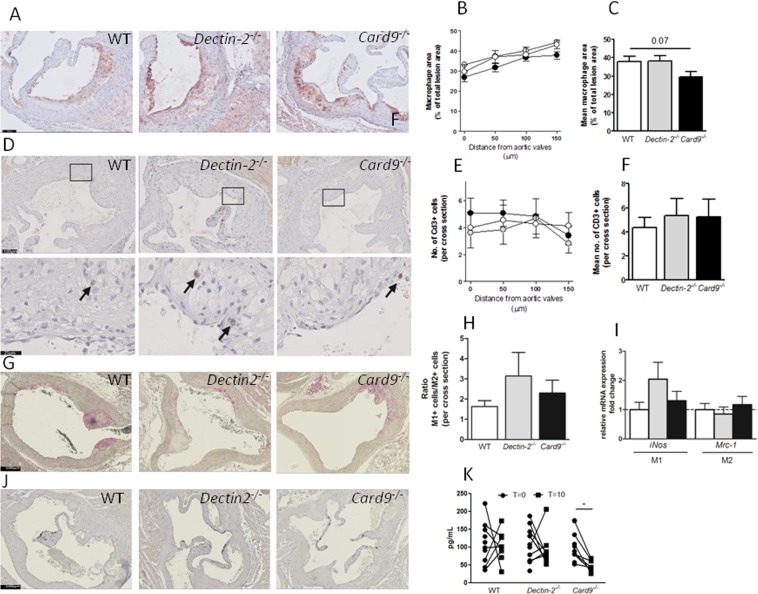
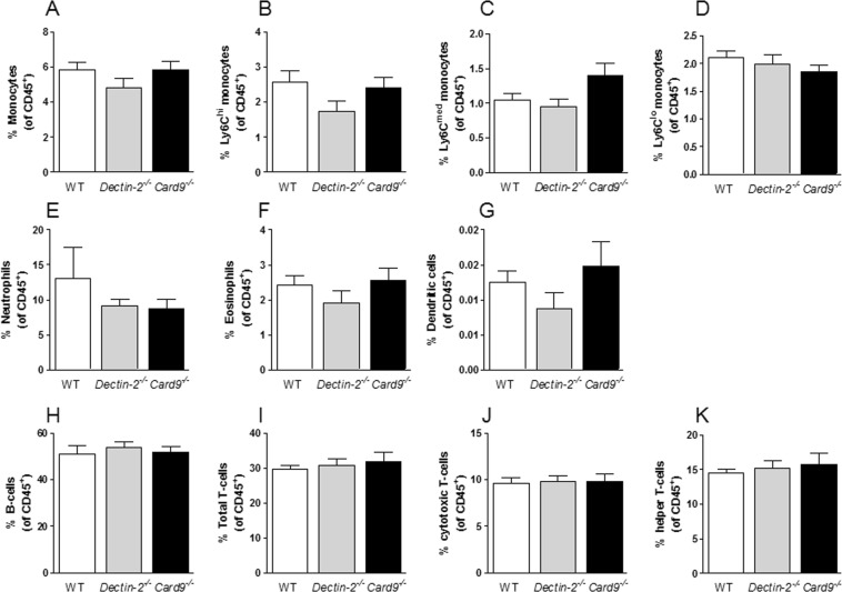
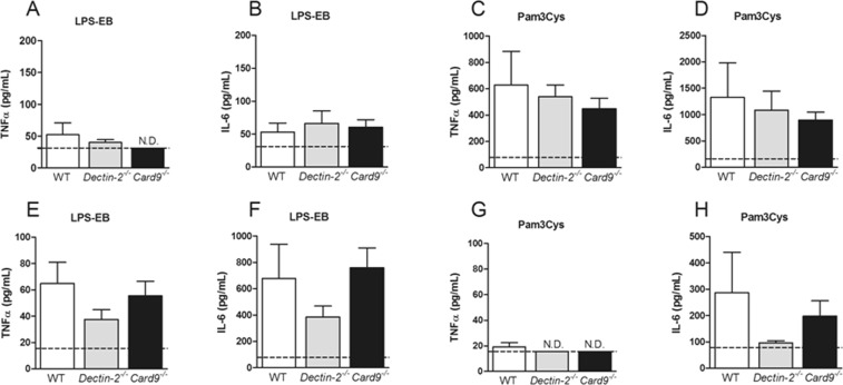
Inflammatory reactions activated by pattern recognition receptors (PRRs) on the membrane of innate immune cells play an important role in atherosclerosis. Whether the PRRs of the C-type lectin receptor (CLR) family including Dectin-2 may be involved in the pathogenesis of atherosclerosis remains largely unknown. Recently, the CLR-adaptor molecule caspase recruitment domain family member 9 (CARD9) has been suggested to play a role in cardiovascular pathologies as it provides the link between CLR activation and transcription of inflammatory cytokines as well as immune cell recruitment. We therefore evaluated whether hematopoietic deletion of Dectin-2 or CARD9 reduces inflammation and atherosclerosis development. Low-density lipoprotein receptor (Ldlr)-knockout mice were transplanted with bone marrow from wild-type, Dectin-2- or Card9-knockout mice and fed a Western-type diet containing 0.1% (w/w) cholesterol. After 10 weeks, lipid and inflammatory parameters were measured and atherosclerosis development was determined. Deletion of hematopoietic Dectin-2 or CARD9 did not influence plasma triglyceride and cholesterol levels. Deletion of hematopoietic Dectin-2 did not affect atherosclerotic lesion area, immune cell composition, ex vivo cytokine secretion by peritoneal cells or bone marrow derived macrophages. Unexpectedly, deletion of hematopoietic CARD9 increased atherosclerotic lesion formation and lesion severity. Deletion of hematopoietic CARD9 did also not influence circulating immune cell composition and peripheral cytokine secretion. Besides a tendency to a reduced macrophage content within these lesions, plasma MCP-1 levels decreased upon WTD feeding. Deletion of hematopoietic Dectin-2 did not influence atherosclerosis development in hyperlipidemic mice. The absence of CARD9 unexpectedly increased atherosclerotic lesion size and severity, suggesting that the presence of CARD9 may protect against initiation of atherosclerosis development.
Conflict of interest statement
The authors declare no competing interests.
Figures
Similar articles
-
Deletion of haematopoietic Dectin-2 or CARD9 does not protect from atherosclerosis development under hyperglycaemic conditions.Diab Vasc Dis Res. 2020 Jan-Feb;17(1):1479164119892140. doi: 10.1177/1479164119892140. Epub 2019 Dec 23. Diab Vasc Dis Res. 2020. PMID: 31868000 Free PMC article.
-
Differential use of CARD9 by dectin-1 in macrophages and dendritic cells.J Immunol. 2009 Jan 15;182(2):1146-54. doi: 10.4049/jimmunol.182.2.1146. J Immunol. 2009. PMID: 19124758 Free PMC article.
-
The CARD9-Associated C-Type Lectin, Mincle, Recognizes La Crosse Virus (LACV) but Plays a Limited Role in Early Antiviral Responses against LACV.Viruses. 2019 Mar 26;11(3):303. doi: 10.3390/v11030303. Viruses. 2019. PMID: 30917612 Free PMC article.
-
Mechanistic Insights into the Role of C-Type Lectin Receptor/CARD9 Signaling in Human Antifungal Immunity.Front Cell Infect Microbiol. 2016 Apr 5;6:39. doi: 10.3389/fcimb.2016.00039. eCollection 2016. Front Cell Infect Microbiol. 2016. PMID: 27092298 Free PMC article. Review.
-
Dectin-1-Syk-CARD9 Signaling Pathway in TB Immunity.Front Immunol. 2018 Feb 13;9:225. doi: 10.3389/fimmu.2018.00225. eCollection 2018. Front Immunol. 2018. PMID: 29487599 Free PMC article. Review.
Cited by
-
The Role of CARD9 in Metabolic Diseases.Curr Med Sci. 2020 Apr;40(2):199-205. doi: 10.1007/s11596-020-2166-4. Epub 2020 Apr 26. Curr Med Sci. 2020. PMID: 32337681 Review.
-
CARD9 Signaling, Inflammation, and Diseases.Front Immunol. 2022 Mar 30;13:880879. doi: 10.3389/fimmu.2022.880879. eCollection 2022. Front Immunol. 2022. PMID: 35432375 Free PMC article. Review.
-
Genetic inhibition of CARD9 accelerates the development of atherosclerosis in mice through CD36 dependent-defective autophagy.Nat Commun. 2023 Aug 1;14(1):4622. doi: 10.1038/s41467-023-40216-x. Nat Commun. 2023. PMID: 37528097 Free PMC article.
-
CARD9: key player or bystander in cardiac remodeling under hypertension?Hypertens Res. 2020 Dec;43(12):1454-1456. doi: 10.1038/s41440-020-00542-9. Epub 2020 Sep 2. Hypertens Res. 2020. PMID: 32873900 No abstract available.
-
Deletion of haematopoietic Dectin-2 or CARD9 does not protect from atherosclerosis development under hyperglycaemic conditions.Diab Vasc Dis Res. 2020 Jan-Feb;17(1):1479164119892140. doi: 10.1177/1479164119892140. Epub 2019 Dec 23. Diab Vasc Dis Res. 2020. PMID: 31868000 Free PMC article.
References
-
- Hoseini, Z. et al. NLRP3 inflammasome: Its regulation and involvement in atherosclerosis. J Cell Physiol, 10.1002/jcp.25930 (2017). - PubMed
Publication types
MeSH terms
Substances
Grants and funding
LinkOut - more resources
Full Text Sources
Molecular Biology Databases
Miscellaneous
