. 2019 Mar 13;9(1):4340.
doi: 10.1038/s41598-019-40983-y.

Identification of fibrinogen as a natural inhibitor of MMP-2

Affiliations

Identification of fibrinogen as a natural inhibitor of MMP-2

Hassan Sarker et al. Sci Rep. .

Abstract

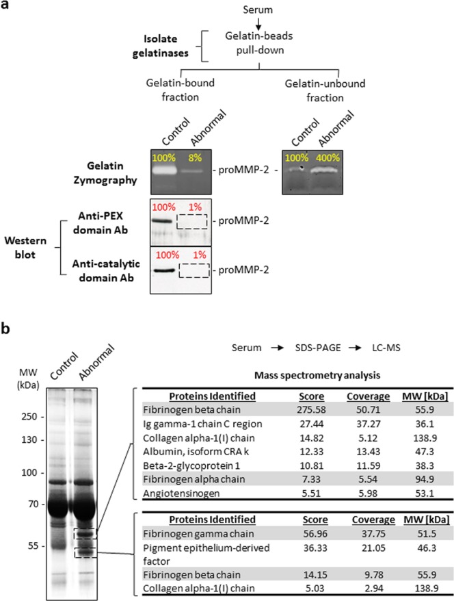
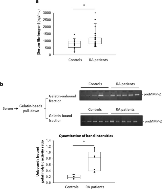
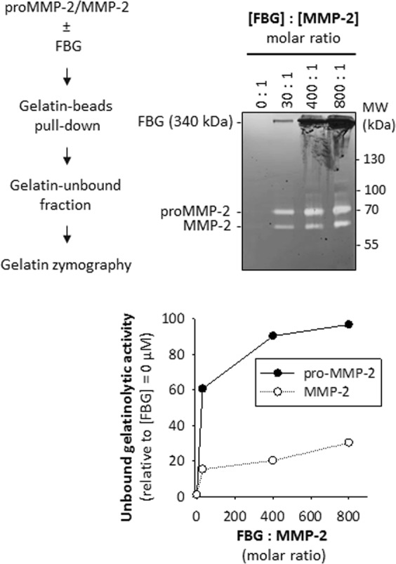
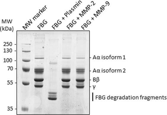
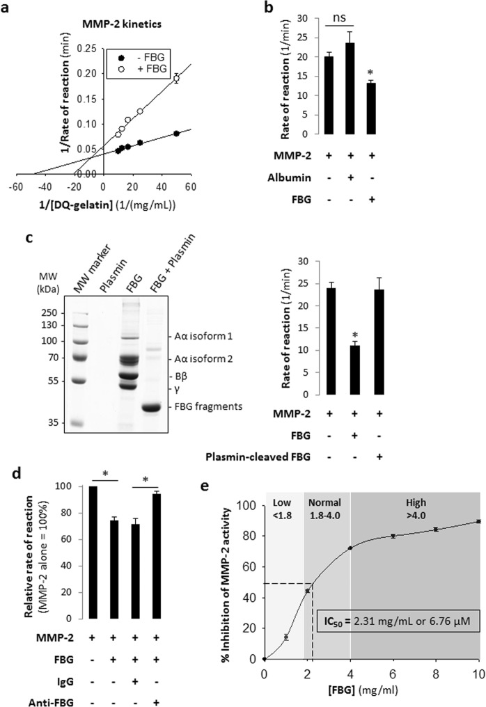
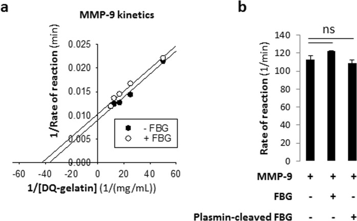
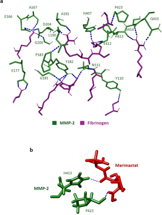
Non-genetic MMP-2 insufficiency is a relatively unexplored condition which could be induced by pathological overexpression of endogenous MMP-2 inhibitors such as TIMPs and/or the acute phase reactant alpha-2-macroglobulin. Here, we investigate the hypothesis that human fibrinogen (FBG) - an acute phase reactant - inhibits human MMP-2. Following an unexpected observation where sera from human donors including arthritis patients with increased levels of serum FBG exhibited reduced binding of serum proMMP-2 to gelatin, we found that human FBG (0 to 3.6 mg/mL i.e., 0 to 10.6 μM) concentration-dependently inhibited human proMMP-2 and MMP2 from binding to gelatin. Moreover, at normal physiological concentrations, FBG (5.29-11.8 μM) concentration-dependently inhibited (40-70% inhibition) the cleavage of fluorescein-conjugated gelatin by MMP-2, but not MMP-9. Indicative of a mixed-type (combination of competitive and non-competitive) inhibition mechanism, FBG reduced the Vmax (24.9 ± 0.7 min-1 to 17.7 ± 0.9 min-1, P < 0.05) and increased the Michaelis-Menten constant KM (204 ± 6 nM to 478 ± 50 nM, P < 0.05) for the reaction of MMP-2 cleavage of fluorescein-conjugated gelatin. In silico analyses and studies of FBG neutralization with anti-FBG antibodies implicated the domains D and E of FBG in the inhibition of MMP-2. In conclusion, FBG is a natural selective MMP-2 inhibitor, whose pathological elevation could lead to MMP-2 insufficiency in humans.


Conflict of interest statement

The authors declare no competing interests.

Figures

Figure 1
Figure 1
Identification of a blood donor with elevated serum fibrinogen exhibiting impaired binding of MMP-2 to gelatin. (a) Analysis of serum samples by gelatin zymography and Western blot to detect MMP-2 and/or to quantitate its gelatinolytic activity. Undiluted serum samples (10 μL) were incubated with an estimated 43 μg of immobilized gelatin at 4 °C for 1 hour. Gelatin-bound and gelatin-unbound proteins were subjected to gelatin zymography to quantitate MMP-2 activity. Gelatin-bound proteins were subjected to Western blot to detect MMP-2 using anti-PEX and anti-catalytic domain (MMP-2) antibodies. Top: Gelatin zymogram showing the majority of activity of serum MMP-2 of the abnormal sample remained in the gelatin-unbound fraction. Bottom: Western blots showing serum MMP-2 of the abnormal sample was undetected in the gelatin-bound fraction by both the anti-PEX domain and anti-catalytic domain antibodies. Band intensities are shown as a percentage relative to the band of the representative control serum sample. Uncropped gels and blots are presented in Supplementary Fig. S7. (b) SDS-PAGE coupled with LC-MS (ESI) was used to identify the compositions of two unique protein bands in the abnormal sample as indicated. Uncropped gel is presented in Supplementary Fig. S7. LC-MS, liquid chromatography – mass spectrometry; ESI, electrospray ionization; PEX, hemopexin-like; Ab, antibody; MMP-2, matrix metalloproteinase 2.
Figure 2
Figure 2
Reduced binding of serum proMMP-2 to gelatin in sera with higher fibrinogen concentrations. (a) Quantitation of serum fibrinogen concentrations of rheumatoid arthritis patients (n = 25) and healthy controls (n = 19) by ELISA. P = 0.019 controls vs RA patients, determined by Mann-Whitney Rank Sum Test. (b) Top: Gelatin binding assay of selected RA patient (high FBG) and control (low FBG) serum samples, followed by gelatin zymography shows the gelatinolytic activity of proMMP-2 in the gelatin-unbound fraction and the gelatin-bound fraction. Bottom: Quantitation and comparison of the intensities of the lysis bands in the gelatin zymograms above by densitometry. *P < 0.05 controls vs RA patients, determined by Mann-Whitney Rank Sum Test. Uncropped gels are presented in Supplementary Fig. S7. ELISA, enzyme-linked immunosorbent assay; RA, rheumatoid arthritis; FBG, fibrinogen.
Figure 3
Figure 3
Purified fibrinogen concentration-dependently reduces binding of recombinant MMP-2 to gelatin. Top left: Strategy for assessing a lack of binding of MMP-2 to gelatin in the presence or absence of FBG. Top right: Gelatin binding assay followed by gelatin zymography shows an increase in gelatinolytic activity of MMP-2 remaining in the gelatin-unbound fraction with increasing FBG concentration. Gelatin zymogram showing gelatinolytic activity of MMP-2 in the gelatin-unbound fraction for varying [FBG]: [MMP-2] ratios (0: 1 to 800: 1). Uncropped gel is presented in Supplementary Fig. S7. Bottom: Quantitation of the intensities of the lysis bands in the gelatin zymogram (left) by densitometry. Relative intensity for each band was calculated by dividing the absolute intensity of each band by the absolute intensity of the band at [FBG] = 0 μM ([FBG]: [MMP-2] = 0:1).
Figure 4
Figure 4
Human fibrinogen is not cleaved by recombinant human MMP-2 or human MMP-9. Proteolysis of human FBG by plasmin, MMP-2 and MMP-9. FBG (3 mg/mL) was incubated with near physiological concentrations of either plasmin (12 nM) or MMP-2 (12 nM) or MMP-9 (12 nM) for 24 hours at 37 °C. The reaction mixtures were analysed by SDS-PAGE.
Figure 5
Figure 5
Purified human fibrinogen inhibits gelatinolytic activity of recombinant human MMP-2. (a) Lineweaver-Burk plot of the proteolytic processing of DQ-gelatin by MMP-2 in the absence or presence of 3 mg/mL FBG; [MMP-2] = 0.001 mg/mL (13.9 nM) and [DQ-gelatin] = 0.02, 0.04, 0.06, 0.08 or 0.1 mg/mL. (b) Bar graphs showing the effect of intact fibrinogen (3 mg/mL) on the activity of MMP-2 (0.001 mg/mL); MMP-2 alone and human serum albumin (3 mg/mL) plus MMP-2 were used as a control. *P < 0.05 vs MMP-2 determined by student’s t-test. ns, not significant. Data are shown as mean of triplicates. (c) Left: SDS-PAGE confirming complete degradation of fibrinogen (6 mg/mL) when incubated with plasmin (0.001 mg/mL) for 12 hours at 37 °C. Right: Bar graphs showing that and plasmin-degraded fibrinogen fragments (3 mg/mL) vs intact fibrinogen (3 mg/mL) have no effect on the activity of MMP-2 (0.001 mg/mL). *P < 0.05 vs MMP-2 determined by student’s t-test. ns, not significant. (d) Bar graph showing restoration of MMP-2 activity when FBG is selectively removed from solution by an anti-FBG antibody. [MMP-2] = 0.001 mg/mL (13.9 nM); [DQ-gelatin] = 0.05 mg/mL; [FBG] = 2 mg/mL (5.88 µM); [IgG] and [anti-FBG] = 5.88 µM. *P < 0.05; determined by two-tailed student’s t-test (n = 4). (e) Plot showing the effect of increasing fibrinogen concentrations (0.0 to 10 mg/mL) on MMP-2 (0.001 mg/mL) activity. Note that high circulating FBG concentrations (as those found in RA patients (Fig. 2a)) effectively inhibit MMP-2 activity by more than 50%. Data are presented as mean ± standard error of mean. FBG, fibrinogen; RFU, relative fluorescence unit.
Figure 6
Figure 6
Gelatinolytic activity of recombinant human MMP-9 is not inhibited by purified human fibrinogen. (a) Lineweaver-Burk plot of the proteolytic processing of DQ-gelatin by MMP-9 in the absence or presence of 3 mg/mL FBG; [MMP-9] = 0.001 mg/mL (12.0 nM) and [DQ-gelatin] = 0.02, 0.04, 0.06, 0.08 or 0.1 mg/mL. (b) Bar graphs showing that intact fibrinogen (3 mg/mL) or plasmin-degraded fibrinogen fragments (3 mg/mL) has no effect on the activity of MMP-9 (0.001 mg/mL). ns, not significant. Data are presented as mean ± standard error of mean. FBG, fibrinogen; RFU, relative fluorescence unit.
Figure 7
Figure 7
Fibrinogen and Marimastat bind at a common region of the catalytic domain of MMP-2. (a) Molecular docking of MMP-2 to FBG. Labelled residues of MMP-2 (green) that form hydrogen bonds (blue dotted lines) with FBG residues (magenta) are presented. (b) Molecular docking of MMP-2 to Marimastat. Labelled residues of MMP-2 (green) that form hydrogen bonds (blue dotted lines) with Marimastat (red) are presented.

Similar articles

Cited by

References

    1. Parks WC, Wilson CL, Lopez-Boado YS. Matrix metalloproteinases as modulators of inflammation and innate immunity. Nat Rev Immunol. 2004;4:617–629. doi: 10.1038/nri1418nri1418. - DOI - PubMed
    1. Fernandez-Patron C, Kassiri Z, Leung D. Modulation of Systemic Metabolism by MMP-2: From MMP-2 Deficiency in Mice to MMP-2 Deficiency in Patients. Compr Physiol. 2016;6:1935–1949. doi: 10.1002/cphy.c160010. - DOI - PubMed
    1. Nissinen L, Kahari VM. Matrix metalloproteinases in inflammation. Biochim Biophys Acta. 2014;1840:2571–2580. doi: 10.1016/j.bbagen.2014.03.007. - DOI - PubMed
    1. McQuibban GA, et al. Matrix metalloproteinase activity inactivates the CXC chemokine stromal cell-derived factor-1. J Biol Chem. 2001;276:43503–43508. doi: 10.1074/jbc.M107736200. - DOI - PubMed
    1. McQuibban GA, et al. Matrix metalloproteinase processing of monocyte chemoattractant proteins generates CC chemokine receptor antagonists with anti-inflammatory properties in vivo. Blood. 2002;100:1160–1167. - PubMed

Publication types

MeSH terms