. 2019 Mar;17(3):3253-3260.
doi: 10.3892/ol.2019.9980. Epub 2019 Jan 28.

MicroRNA-1291 mediates cell proliferation and tumorigenesis by downregulating MED1 in prostate cancer

Affiliations

MicroRNA-1291 mediates cell proliferation and tumorigenesis by downregulating MED1 in prostate cancer

Qi Cai et al. Oncol Lett. 2019 Mar.

Abstract

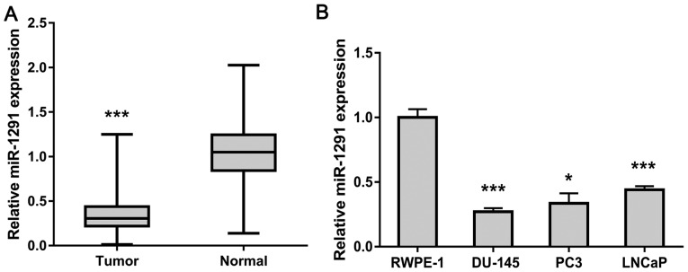
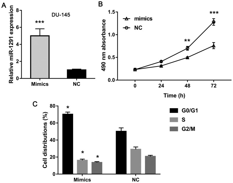
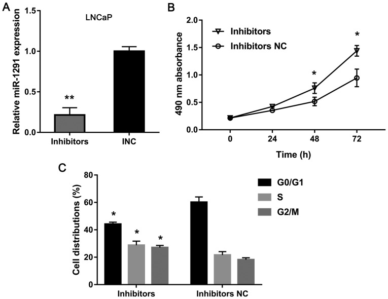
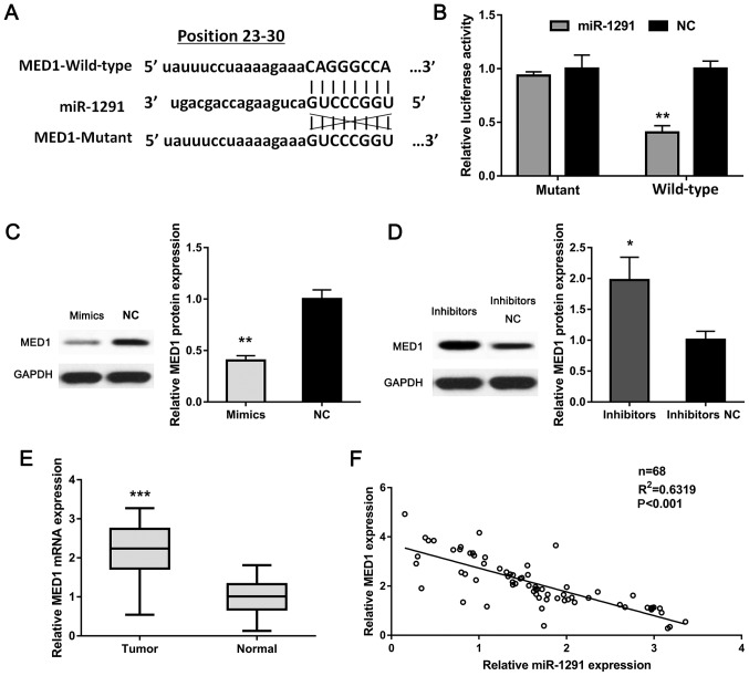
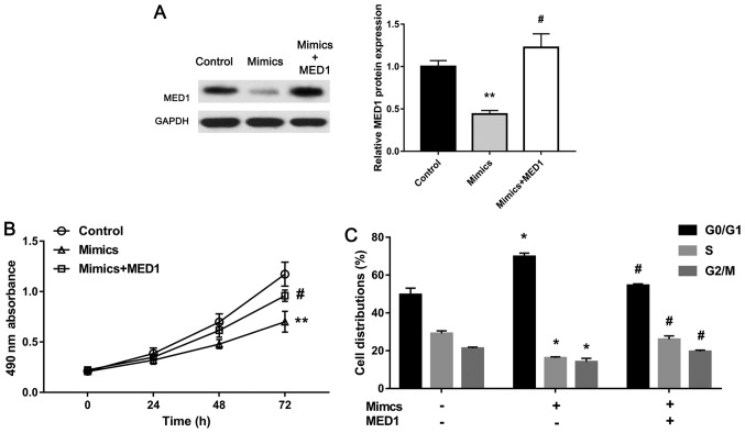
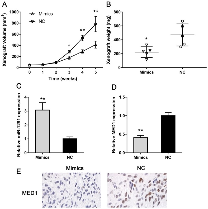
miRNAs are important factors involved in the regulation of tumor development. miR-1291 was found to have regulatory effects in many tumors, but its role in prostate cancer (PCa) still remains unclear. We explored the expression of miR-1291 in PCa to reveal its role in regulating the progression of PCa as well as its underlying mechanism. Reverse transcription-quantitative polymerase chain reaction (RT-qPCR) was used to detect the expression of miR-1291 in PCa tissues and cell lines compared to normal tissues and cell lines. miR-1291 mimics and inhibitors were applied to overexpress or inhibit the level of miR-1291 in PCa cells. The ability of cell proliferation was measured using MTT assay, and cell cycle distribution was determined by flow cytometry. The potential target of miR-1291 was identified via western blot analysis and luciferase assays. Then a xenograft model was established to explore the function of miR-1291 in PCa in vivo. The results revealed that the expression level of miR-1291 was significantly lower in the PCa tissues than that in the normal adjacent tissues. In PCa-derived cells, there was also a downregulated expression level of miR-1291. Overexpression of miR-1291 obviously inhibited DU-145 cell proliferation and induced cell cycle transition from G0/G1 to S phase. However, inhibition of miR-1291 promoted the growth of LNCaP cells, and promoted the cell cycle transition to S phase and G2/M phase. MED1 was proven to be a potential target gene of miR-1291, and miR-1291 significantly inhibited its expression. At the in vivo level, overexpression of miR-1291 inhibited the growth of xenograft tumors and significantly inhibited the expression of MED1 protein. Our study demonstrated that miR-1291 inhibits cell proliferation and tumorigenesis of PCa via MED1, which might provide a novel target for PCa diagnosis and biological therapy.

Keywords: MED1; miR-1291; proliferation; prostate cancer; tumorigenesis.


Figures

Figure 1.
Figure 1.
miR-1291 expression in PCa tissues and cell lines. (A) The expression levels of miR-1291 in PCa tissues as well as in the paired adjacent prostate tissues were analyzed by RT-qPCR. (B) miR-1291 expression in RWPE-1 cells and PCa cell lines (DU-145, PC3, LNCaP). Data are shown as mean ± SD of three separate experiments. *P<0.05 and ***P<0.001. PCa, prostate cancer.
Figure 2.
Figure 2.
Overexpression of miR-1291 inhibits proliferation and induced cell cycle of DU-145 cells. (A) DU-145 cells were transfected with miR-1291 mimics or NC. (B) Cell viability was detected by MTT assay. (C) After transfection for 48 h, DU-145 cells were stained with PI, and the distributions of cell cycle were determined using flow cytometric analysis. *P<0.05, **P<0.01 and ***P<0.001. NC, negative control; PI, propidium iodide.
Figure 3.
Figure 3.
Downregulation of miR-1291 promotes proliferation and cell cycle of LNCaP cells. (A) LNCaP cells were transfected with miR-1291 inhibitors or INC. (B) Cell viability was detected by MTT assay. (C) After transfection for 48 h, LNCaP cells were stained with PI, and the distribution of cell cycle was determined using flow cytometric analysis. *P<0.05 and **P<0.01. INC, inhibitor negative control; PI, propidium iodide.
Figure 4.
Figure 4.
miR-1291 targets MED1 in PCa. (A) The predicted binding site of miR-1291 at the MED1 mRNA 3′-UTR. (B) A luciferase reporter assay using pMIR-REPORT (vector), pMIR-REPORT MED1-3′-UTR-WT, or MED1-3′-UTR-Mut plasmid in DU-145 cells transfected with the miR-1291 mimics or NC. (C and D) Western blot analysis of MED1 protein in established DU-145 and LNCaP cells. (E) The expression of MED1 mRNA in PCa specimens and paired adjacent normal tissues was analyzed by RT-qPCR. (F) Correlation between miR-1291 expression and the MED1 mRNA level in PCa specimens (R2=0.6319, P<0.001). *P<0.05, **P<0.01 and ***P<0.001. PCa, prostate cancer; 3′-UTR, 3′-untranslated region; NC, negative control.
Figure 5.
Figure 5.
MED1 overexpression restores miR-1291-mediated inhibition of the growth of DU-145 cells. (A) Western blot analysis of MED1. GAPDH was used as an internal control. (B) Analysis of cell proliferation ability by MTT assay in control, mimics, or mimics + MED1 treated DU-145 cells. (C) The distributions of cell cycle were determined. Data are presented as mean ± SD of three replicates. *P<0.05, **P<0.01 and #P<0.05.
Figure 6.
Figure 6.
miR-1291 inhibits cell growth of PCa in vivo. (A) Tumor growth curves of xenografts. (B) The weight of xenografts was analyzed. (C) The expression of miR-1291 in xenografts was analyzed by RT-qPCR. (D) The expression of MED1 mRNA in xenografts was analyzed by RT-qPCR. (E) The expression of MED1 protein in xenografts was analyzed by IHC (magnification, ×400). *P<0.05 and **P<0.01. PCa, prostate cancer; IHC, immunohistochemistry.

Similar articles

Cited by

References

    1. Siegel RL, Miller KD, Jemal A. Cancer statistics, 2018. CA Cancer J Clin. 2018;68:7–30. doi: 10.3322/caac.21442. - DOI - PubMed
    1. Blumenthal-Barby JS, Lee D, Volk RJ. Toward ethically responsible choice architecture in prostate cancer treatment decision-making. CA Cancer J Clin. 2015;65:257–260. doi: 10.3322/caac.21283. - DOI - PMC - PubMed
    1. Chan JM, Stampfer MJ, Giovannucci EL. What causes prostate cancer? A brief summary of the epidemiology. Semin Cancer Biol. 1998;8:263–273. doi: 10.1006/scbi.1998.0075. - DOI - PubMed
    1. Levesque C, Nelson PS. Cellular constituents of the prostate stroma: Key contributors to prostate cancer progression and therapy resistance. Cold Spring Harb Perspect Med. 2018;8(pii):a030510. doi: 10.1101/cshperspect.a030510. - DOI - PMC - PubMed
    1. Garisto JD, Klotz L. Active surveillance for prostate cancer: How to do it right. Oncology (Williston Park) 2017;31:333–340, 345. - PubMed