Review
. 2019 Feb 12;9(5):1303-1322.
doi: 10.7150/thno.30309. eCollection 2019.

The Applications of Radiomics in Precision Diagnosis and Treatment of Oncology: Opportunities and Challenges

Affiliations
Review

The Applications of Radiomics in Precision Diagnosis and Treatment of Oncology: Opportunities and Challenges

Zhenyu Liu et al. Theranostics. .

Abstract

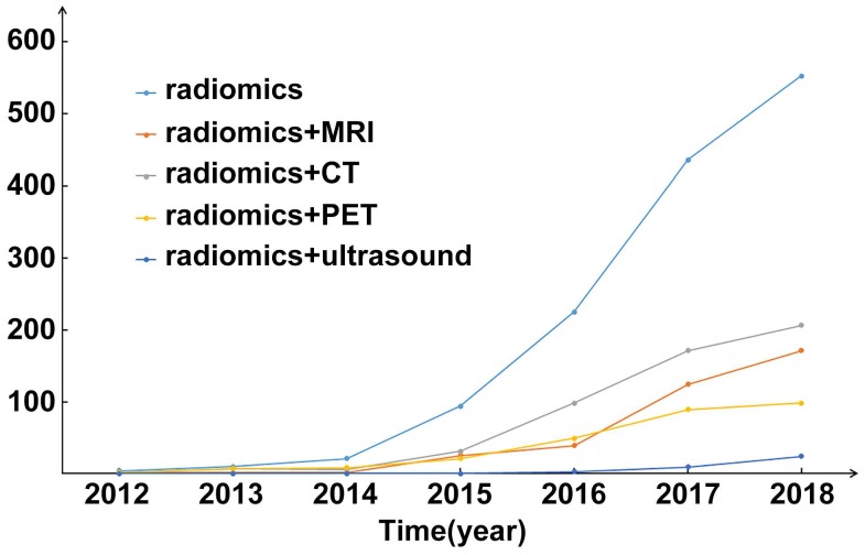
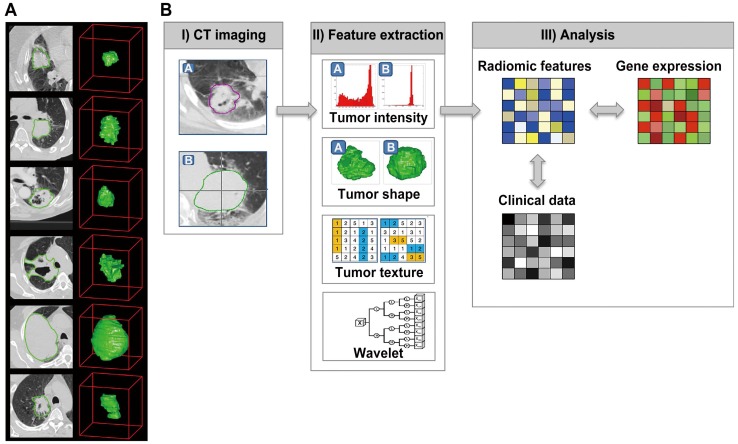
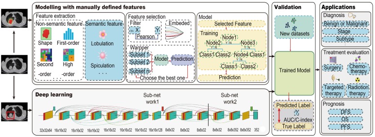
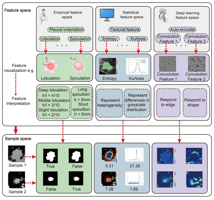
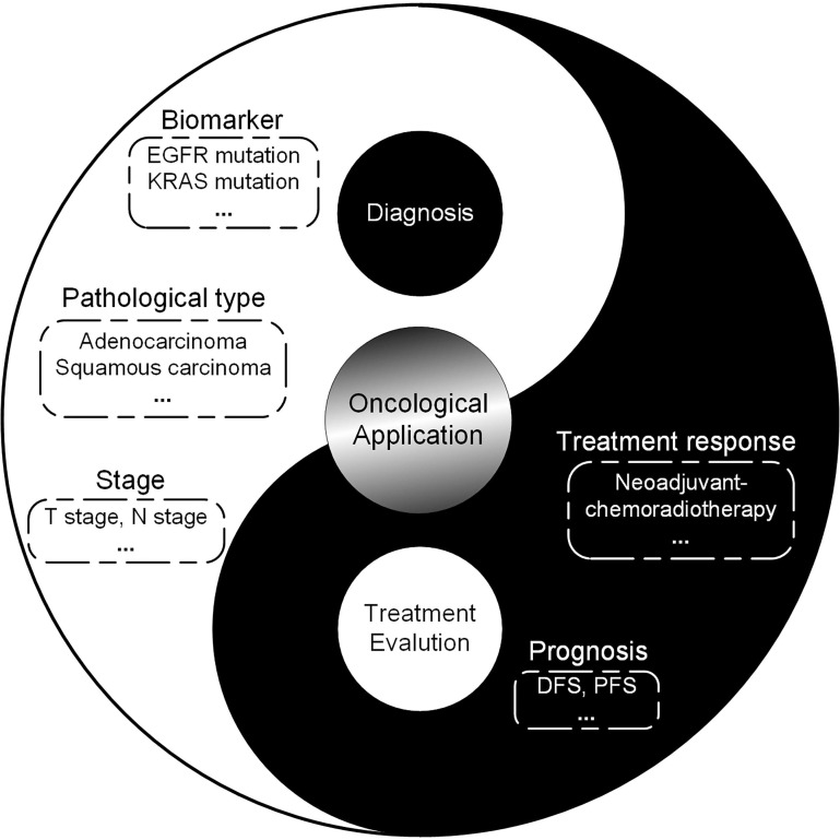
Medical imaging can assess the tumor and its environment in their entirety, which makes it suitable for monitoring the temporal and spatial characteristics of the tumor. Progress in computational methods, especially in artificial intelligence for medical image process and analysis, has converted these images into quantitative and minable data associated with clinical events in oncology management. This concept was first described as radiomics in 2012. Since then, computer scientists, radiologists, and oncologists have gravitated towards this new tool and exploited advanced methodologies to mine the information behind medical images. On the basis of a great quantity of radiographic images and novel computational technologies, researchers developed and validated radiomic models that may improve the accuracy of diagnoses and therapy response assessments. Here, we review the recent methodological developments in radiomics, including data acquisition, tumor segmentation, feature extraction, and modelling, as well as the rapidly developing deep learning technology. Moreover, we outline the main applications of radiomics in diagnosis, treatment planning and evaluations in the field of oncology with the aim of developing quantitative and personalized medicine. Finally, we discuss the challenges in the field of radiomics and the scope and clinical applicability of these methods.

Keywords: medical imaging; oncology; precision diagnosis and treatment; radiomics.


Conflict of interest statement

Competing Interests: The authors have declared that no competing interest exists.

Figures

Figure 1
Figure 1
Publication statistics of radiomics since 2012. The number of publications is going straight up. Abbreviations: CT, Computed Tomography; MRI, Magnetic Resonance Imaging; PET, Positron Emission Tomography
Figure 2
Figure 2
The initially radiomics pipeline with medical images. Reproduced with permission from . (a) Example CT images of patients with lung cancer. (b) Strategy of radiomic analysis.
Figure 3
Figure 3
The radiomics pipeline of Modelling with manually defined features and Deep learning. For Modelling with manually defined features, it includes the main steps: data acquisition and preprocessing, tumor segmentation, feature extraction and selection, and modeling. For deep learning, it is an end-to-end method without separate steps of feature extraction, feature selection and modelling. Trained model from both two methods should be validated with new dataset, and then could be applied. Abbreviations: AUC, area under the receiver operating characteristic curve; C-index, concordance index; DFS, disease-free survival; PFS, progression-free survival; OS, overall survival
Figure 4
Figure 4
The main features we used for radiomic analysis could be divided into three parts: Empirical features, Statistical features and Deep learning features. All these features could be visualized and interpreted with physical meanings. However, what we should do further is to unravel their physiological significance.
Figure 5
Figure 5
Scope of radiomics for diagnosis and treatment evaluation, suggesting the potential directions radiomics could be applied for. Abbreviations: EGFR: epidermal growth factor receptor; IDH: isocitrate dehydrogenase; KRAS: Kirsten rat sarcoma viral oncogene homolog; MGMT: O-6-methylguanine-DNA methyltransferase
Figure 6
Figure 6
Developed radiomics nomogram for prediction of pCR to NCRT in rectal cancer. Reproduced with permission from .
Figure 7
Figure 7
Flowchart depicting the workflow of radiomics and the application of the RQS. Reproduced with permission from . Abbreviations: RQS, radiomics quality score.

References

    1. Lambin P, Rios-Velazquez E, Leijenaar R, Carvalho S, van Stiphout RG, Granton P. et al. Radiomics: extracting more information from medical images using advanced feature analysis. Eur J Cancer. 2012;48:441–6. - PMC - PubMed
    1. Kumar V, Gu Y, Basu S, Berglund A, Eschrich SA, Schabath MB. et al. Radiomics: the process and the challenges. Magn Reson Imaging. 2012;30:1234–48. - PMC - PubMed
    1. Gatenby RA, Grove O, Gillies RJ. Quantitative Imaging in Cancer Evolution and Ecology. Radiology. 2013;269:8–15. - PMC - PubMed
    1. Aerts HJ, Velazquez ER, Leijenaar RT, Parmar C, Grossmann P, Carvalho S. et al. Decoding tumour phenotype by noninvasive imaging using a quantitative radiomics approach. Nat Commun. 2014;5:4006. - PMC - PubMed
    1. Aerts HJ. The potential of radiomic-based phenotyping in precision medicine: a review. JAMA Oncol. 2016;2:1636–42. - PubMed

Publication types

MeSH terms