. 2019 Mar 5:10:3.
doi: 10.1186/s13227-019-0116-7. eCollection 2019.

Modular co-option of cardiopharyngeal genes during non-embryonic myogenesis

Affiliations

Modular co-option of cardiopharyngeal genes during non-embryonic myogenesis

Maria Mandela Prünster et al. Evodevo. .

Abstract

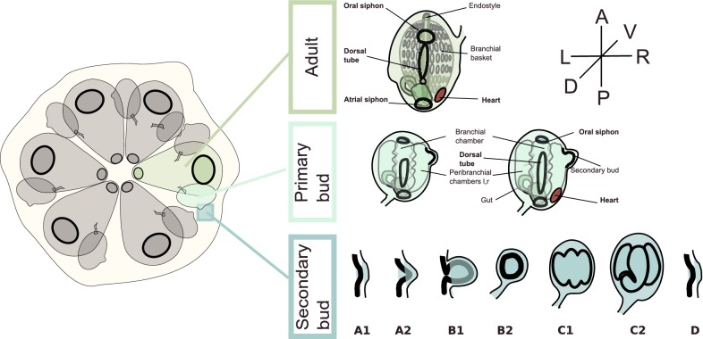
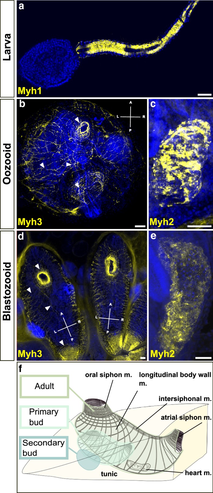
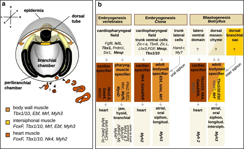
Background: In chordates, cardiac and body muscles arise from different embryonic origins. In addition, myogenesis can be triggered in adult organisms, during asexual development or regeneration. In non-vertebrate chordates like ascidians, muscles originate from embryonic precursors regulated by a conserved set of genes that orchestrate cell behavior and dynamics during development. In colonial ascidians, besides embryogenesis and metamorphosis, an adult can propagate asexually via blastogenesis, skipping embryo and larval stages, and form anew the adult body, including the complete body musculature.

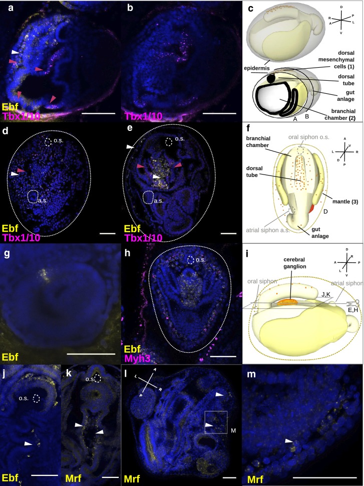
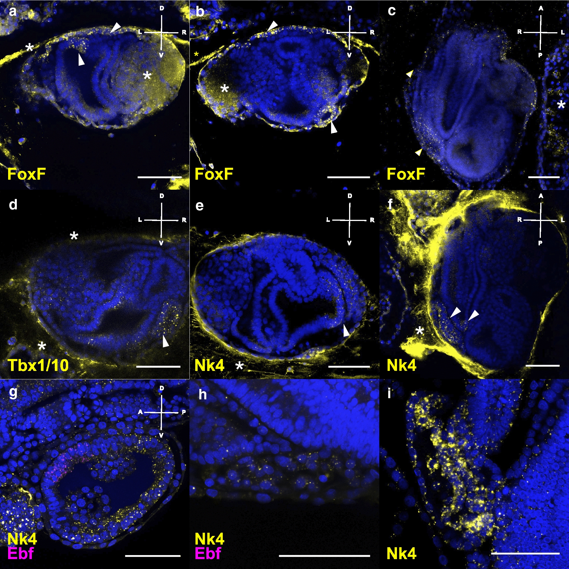
Results: To investigate the cellular origin and mechanisms that trigger non-embryonic myogenesis, we followed the expression of ascidian myogenic genes during Botryllus schlosseri blastogenesis and reconstructed the dynamics of muscle precursors. Based on the expression dynamics of Tbx1/10, Ebf, Mrf, Myh3 for body wall and of FoxF, Tbx1/10, Nk4, Myh2 for heart development, we show that the embryonic factors regulating myogenesis are only partially co-opted in blastogenesis, and that markers for muscle precursors are expressed in two separate domains: the dorsal tube and the ventral mesenchyma.

Conclusions: Regardless of the developmental pathway, non-embryonic myogenesis shares a similar molecular and anatomical setup as embryonic myogenesis, but implements a co-option and loss of molecular modules. We then propose that the cellular precursors contributing to heart and body muscles may have different origins and may be coordinated by different developmental pathways.

Keywords: Ascidians; Blastogenesis; Botryllus schlosseri; Budding; Muscle; Regeneration.


Figures

Fig. 1
Fig. 1
Development and staging of a Botryllus schlosseri colony. The staging of the animals was performed after Lauzon [63]. The secondary bud develops as thickening of the peribranchial epithelium and the epidermis (stages A1, A2), which evaginates and closes forming a double monolayered vesicle (stages B1, B2). The inner vesicle undergoes morphogenesis and is subdivided into three chambers (stages C1, C2). During “takeover” (stage D), the adult degenerate and get resorbed, the primary buds become adult, the secondary buds become the primary buds and start to bud again a new secondary bud, so a new blastogenetic cycle begins for the next secondary bud (A1) [32, 34]
Fig. 2
Fig. 2
Fluorescent in situ hybridization showing the expression of myosin heavy chain mRNAs at different stages of Botryllus schlosseri life cycle. a Expression of Myh1 (yellow) along the striated muscle in the larval tail. b Myh3 expression along muscle circular (oral and atrial siphons) and longitudinal muscle fibers in the oozooid body wall. c Expression of Myh2 in the heart of the adult oozooid. d Myh3 expression in the body wall of a blastozooid: circular and longitudinal fibers. bd Arrowheads indicating the circular muscles around the oral siphon, the longitudinal body muscles and the intersiphonal muscles. e Myh2 expression in the heart of an adult blastozooid. Gene names are indicated in the lower left corner of the pictures in yellow. Arrowheads point circular and longitudinal fibers. Blue nuclei: Hoechst staining. Scale bar: 50 μm. f Schematic drawing of the body musculature of a colony in stage C1. Adult, a primary bud, and a secondary bud are embedded in common tunic. At stage C1 adult and primary bud, the muscle fibers are developed and both are able to contract. The body wall musculature consists of circular muscles around the oral and atrial siphon, longitudinal muscles, and a band of intersiphonal muscles, connecting the two siphons. The heart lies to the ventral and right side of the zooid body
Fig. 3
Fig. 3
Fluorescent in situ hybridization (FISH) showing expression of body muscle myogenic TFs during blastogenetic development of Botryllus schlosseri. ae Double ISH showing the expression of Tbx1/10 (magenta) and Ebf (yellow). a, b FISH performed on the same animal in stage C2-D. a Magenta arrowheads point to the expression of Tbx1/10 in the dorsal mesenchyme, the dorsal region of the branchial basket, and the ventral region of the branchial basket lateral to the endostyle, and white arrowheads point to the expression of Ebf in the dorsal mesenchyme. b Posterior and transversal section of the same sample showing the expression of Tbx1/10 in the dorsal and ventral branchial basket. c Schematic representation of a developing Botryllus secondary bud in stage C2-D, showing the dorsal tube the branchial chamber, the gut, and the epidermis. de FISH performed in stage A1. d Magenta arrowheads point to the expression of Tbx1/10 and white arrowheads to the expression of Ebf in the mantle. O.s: oral siphon, a.s.: atrial siphon. e Magenta arrowheads point to the bilateral expression of Tbx1/10 in the intersiphonal region and white arrowheads to the expression of Ebf in the mantle and the forming cerebral ganglion. f Schematic representation of a developing Botryllus primary bud in stage A1. g Earliest expression of Ebf in few single cells of the vesicle-like secondary bud in stage B2, oriented to the primary bud and hence the future dorsal side. h Double ISH showing the expression of Ebf (yellow) and Myh3 (magenta), Ebf is expressed in mantle together with Myh3, in the cerebral ganglion and in the dorsal tube. i Schematic representation of a developing Botryllus primary bud in stage A, sagittal view. j Ebf expression in the region of the intersiphonal muscle. k Max projection showing the expression of Mrf in the region of the intersiphonal muscle. l, m Mrf expression in the mantle of the primary bud. Blue nuclei: Hoechst staining, dashed line: outline of primary bud, scale bar: 50 μm. Asterisk: unspecific staining in either tunic, gonads, or insufficiently bleached auto-fluorescent cells
Fig. 4
Fig. 4
Expression of TFs heart development-related revealed by FISH. ac Expression of FoxF in secondary bud at stage D: a in the dorsal branchial chamber and epidermis, b in the forming heart vesicle, and c FoxF expression in the heart and epidermis in a ventral view shown by a max projection of 13 slides, stage A2. d Tbx1/10 expression in stage D. e Asymmetric Nk4 expression in stage C2, over the entire right peribranchial chamber (white arrowhead) and the right side of the branchial chamber. f Nk4 expression in a ventral view of a primary bud in stage B1. g, h Double FISH with Nk4 and Ebf in the same B2-C1 colony g in the secondary bud and h the heart of the primary bud. Nk4 restricting to the myocardium in late primary bud. Probes are indicated in the lower left corner of the pictures in the same color of the expression. Blue nuclei: Hoechst staining, dashed line: outline of primary bud, scale bar: 50 μm. Asterisk: unspecific staining in either tunic, gonads, or insufficiently bleached auto-fluorescent cells
Fig. 5
Fig. 5
Proposed model showing a modular nature of muscle development. a Proposed model for Botryllus schlosseri muscle development. Three myogenic regions are depicted: the body wall muscles are derived from mesenchymal cells that evaginated from the dorsal tube, the intersiphonal muscles from the posterior branchial chamber, and the heart formed by either evagination or clustering of mesenchymal cells in the ventrolateral region. The myocardium is formed by invagination; hence, the two-layered tubular heart of the adult is put in place. b Comparison of the cardiopharyngeal muscle network in chordate development. The expression of genes has been assayed in multiple vertebrates [, –67], Ciona intestinalis [31, 40] and Botryllus schlosseri. A number of similarities characterize the inductive signals specifying the cardiopharyngeal field in vertebrate and ascidian embryogenesis, while in blastogenesis a common origin of body wall muscles and heart muscles is unlikely. The cardiac lineage expresses Nk2.5 and Gata4 in the forming vertebrate hearts as well as their orthologs in both ascidian species. For what concern Mef2 expression no information is currently available in ascidians [68]. An ortholog to vertebrate Mef2 is present in the transcriptomic dataset of blastogenesis. Pharyngeal muscle starts differentiation by activating the paralogues Myf5 and MyoD; only one ortholog, Mrf, is found in ascidians. To activate such in ascidians, Ebf is expressed in the body muscle lineage. Two myosin heavy chain forms characterize the vertebrate heart. Pharyngeal muscles do not express the same isoforms in all vertebrates: ruminants and rodents express the same myosins as in trunk and limb, namely, Myh1 and Myh2; in other animals Myh6 and Myh16 can be expressed in addition. In ascidians, Myh2 is expressed in the heart, and Myh3 in the body wall. Bold text indicates conserved expression within at least two species, gray only transcriptomic data, asterisk only in some species

Similar articles

Cited by

References

    1. Hooper SL. Invertebrate muscles: muscle specific genes and proteins. Physiol Rev. 2005;85:1001–1060. doi: 10.1152/physrev.00019.2004. - DOI - PubMed
    1. Bernadskaya Y, Christiaen L. Transcriptional control of developmental cell behaviors. Annu Rev Cell Dev Biol. 2016 doi: 10.1146/annurev-cellbio-111315-125218. - DOI - PubMed
    1. Buckingham M. Gene regulatory networks and cell lineages that underlie the formation of skeletal muscle. Proc Natl Acad Sci. 2017;114:5830–5837. doi: 10.1073/pnas.1610605114. - DOI - PMC - PubMed
    1. Diogo R, et al. A new heart for a new head in vertebrate cardiopharyngeal evolution. Nature. 2015;520:466–473. doi: 10.1038/nature14435. - DOI - PMC - PubMed
    1. Gopalakrishnan S, et al. A cranial mesoderm origin for esophagus striated muscles. Dev Cell. 2015;34:694–704. doi: 10.1016/j.devcel.2015.07.003. - DOI - PubMed

LinkOut - more resources