. 2019 Mar 4:9:22.
doi: 10.1186/s13578-019-0287-x. eCollection 2019.

The Y-linked proto-oncogene TSPY contributes to poor prognosis of the male hepatocellular carcinoma patients by promoting the pro-oncogenic and suppressing the anti-oncogenic gene expression

Affiliations

The Y-linked proto-oncogene TSPY contributes to poor prognosis of the male hepatocellular carcinoma patients by promoting the pro-oncogenic and suppressing the anti-oncogenic gene expression

Tatsuo Kido et al. Cell Biosci. .

Abstract

Background: Liver cancer is one of the major causes of cancer death worldwide, with significantly higher incidence and mortality among the male patients. Although sex hormones and their receptors could contribute to such sex differences, the story is incomplete. Genes on the male-specific region of the Y chromosome could play a role(s) in this cancer. TSPY is the putative gene for the gonadoblastoma locus on the Y chromosome (GBY) that is ectopically expressed in a subset of male hepatocellular carcinomas (HCCs). Although various studies showed that TSPY expression is associated with poor prognosis in the patients and its overexpression promotes cell proliferation of various cancer cell lines, it remains unclear how TSPY contributes to the clinical outcomes of the HCC patients. Identifying the downstream genes and pathways of TSPY actions would provide novel insights on its contribution(s) to male predominance in this deadly cancer.

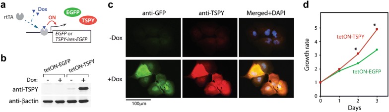
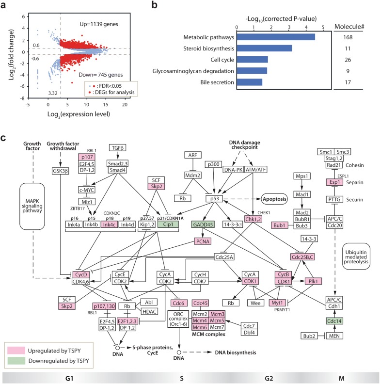
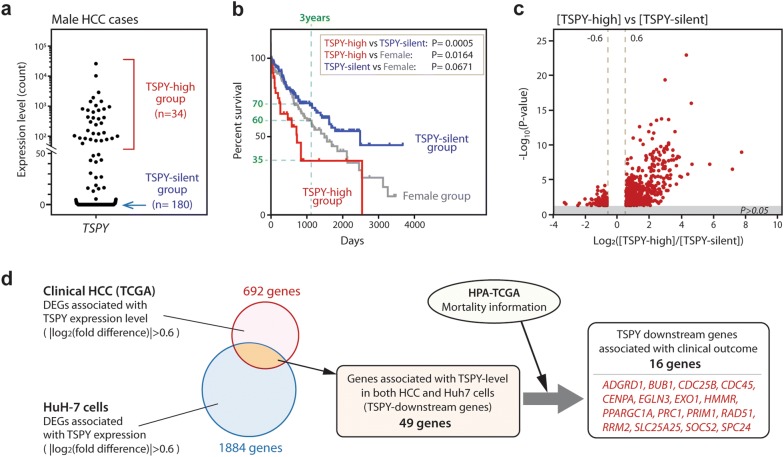
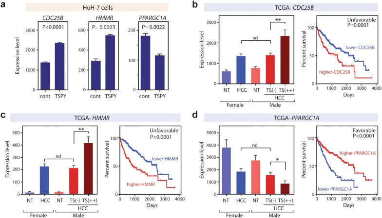
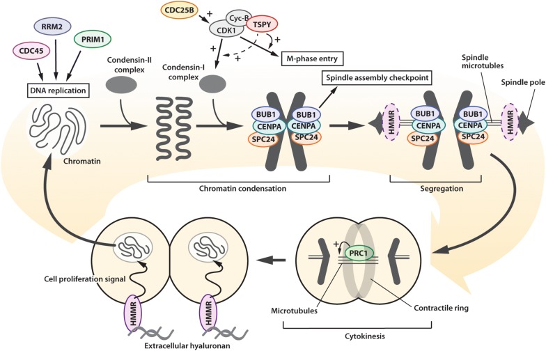
Results: To determine the effects of TSPY on HCC, a TSPY transgene was introduced to the HCC cell line, HuH-7, and studied with RNA-Seq transcriptome analysis. The results showed that TSPY upregulates various genes associated with cell-cycle and cell-viability, and suppresses cell-death related genes. To correlate the experimental observations with those of clinical specimens, transcriptomes of male HCCs with high TSPY expression were analyzed with reference to those with silent TSPY expression from the Cancer Genome Atlas (TCGA). The comparative analysis identified 49 genes, which showed parallel expression patterns between HuH-7 cells overexpressing TSPY and clinical specimens with high TSPY expression. Among these 49 genes, 16 likely downstream genes could be associated with survival rates in HCC patients. The major upregulated targets were cell-cycle related genes and growth factor receptor genes, including CDC25B and HMMR, whose expression levels are negatively correlated with the patient survival rates. In contrast, PPARGC1A, SLC25A25 and SOCS2 were downregulated with TSPY expression, and possess favorable prognoses for HCC patients.

Conclusion: We demonstrate that TSPY could exacerbate the oncogenesis of HCC by differentially upregulate the expression of pro-oncogenic genes and downregulate those of anti-oncogenic genes in male HCC patients, thereby contributing to the male predominance in this deadly cancer.

Keywords: Datamining; Hepatocellular carcinoma; Male predominance; TCGA dataset; TSPY; Transcriptome analysis; Y-chromosome.


Figures

Fig. 1
Fig. 1
Overexpression of TSPY in HuH-7 cells using the Tet-ON system. a Schematic diagram of the Tet-ON system, in which the expression levels of TSPY and EGFP are upregulated by doxycycline (Dox) treatment via recruitment of the rtTA transcription activator onto the Tet-responsive promoter. b Confirmation of doxycycline-induced TSPY expression by western-blot analysis. β-actin was tested as a reference. c Immunofluorescence analysis showed that TSPY (red) and co-expressed EGFP (green) were expressed in the HuH-7-tetON-TSPY cells cultured with and without Dox. DNA was visualized by DAPI staining (blue). d Cell proliferation assay showing HuH-7-tetON-TSPY cells (red line) proliferated faster than HuH-7-tetO-EGFP cells (green line) in the presence of 1 µg/mL Dox
Fig. 2
Fig. 2
Transcriptome and DAVID analyses of the HuH-7-tetOn-TSPY cells. a MA-plots showing the differentially expressed genes (DEGs) between HuH-7-tetOn-TSPY cells and HuH-7-tetOn-EGFP cells. Genes plotted within the areas of Log2[expression level] > 3.32 and |log2[fold change]| > 0.6 (red) were analyzed as differentially expressed genes (DEGs). b DAVID gene-annotation enrichment analysis identified top 5 KEGG pathways enriched in DEGs with FDR < 0.05; FDR = false discovery rate or corrected P-value. c Mapping of the 26 DEGs on the KEGG cell-cycle pathway [49]. Red indicates the upregulated genes and green indicates the downregulated genes in the Dox-treated HuH-7-tetON-TSPY cells
Fig. 3
Fig. 3
Identification of the TSPY downstream genes associated with clinical outcomes in primary HCC. a Based on the TSPY expression level, male HCC cases were classified into the TSPY-high group (n = 36, expression count > 50) and the TSPY-silent group (n = 180, expression count = 0). b Survival curves showing that the survival rates of the male TSPY-high group (red), male TSPY-silent group (blue), and female group (gray). Log-rank test P-values are indicated. c Volcano plot representing the DEGs between TSPY-high and TSPY-silent HCC groups (red plots). d The diagram shows the workflow to identify the downstream genes regulated by TSPY in HCC tissues. The genes whose expression levels consistently correlated with the TSPY expression level in both HuH-7 cells and clinical TSPY-high HCC tissue samples were selected as described in the body text. Sixteen genes (red), whose expression patterns correlated with that of TSPY, were identified to be associated with clinical outcomes.
Fig. 4
Fig. 4
Representative expression patterns and associated with survival rates of TSPY downstream genes in HCC. a TSPY stimulated the expression levels of CDC25B, HMMR, and PPARGC1A, in HuH-7 cells. The results of transcriptome analysis (n = 3) were presented with the P-values by Student t-tests. bd Left panels, expression levels of TSPY and selected downstream genes, e.g. CDC25B, HMMR, and PPARGC1A, among female non-tumor liver (NT), female HCC, male non-tumor liver (NT), male TSPY-silent HCC (TS(−)), and male TSPY-high HCC (TS(++)), in the TCGA transcriptome dataset. Y-axis indicates the expression level as RSEN normalized count. Right panels, survival analyses of the TCGA data, comparing the high-expresser HCC patients (red lines) and the low-expresser HCC patients (blue lines) for the respective genes. Both male and female cases were included in these analyses. Log-rank test P-values are indicated. Abbreviations; **t-test P-value < 0.005; *P-value < 0.05; nd, P-value > 0.05. See Additional file 5: Figure S2 and Additional file 6: Figure S3 for same analyses for all 16 potential TSPY downstream genes
Fig. 5
Fig. 5
Schematic diagram illustrating the functions of TSPY and TSPY-downstream genes in cell cycle events, particularly mitosis, in HCC. From top-left; CDC45, RRM2 and PRIM1 participate in DNA replication at S-phase; CDC25B activates cyclin-B/CDK1 complex and promotes M-phase entry. TSPY binds cyclin-B/CDK1 complex and enhances its activity. BUB1, CENPA, and SPC24 interact with the condensed chromatin and participate in spindle assembly checkpoint and chromatin segregation respectively. HMMR promotes spindle pole formation; PRC1 binds microtubules and modulates cytokinesis. HMMR also activates the signaling cascades of cell-proliferation via extracellular hyaluronan

Similar articles

Cited by

References

    1. Salo P, Kaariainen H, Petrovic V, Peltomaki P, Page DC, de la Chapelle A. Molecular mapping of the putative gonadoblastoma locus on the Y chromosome. Genes Chromosom Cancer. 1995;14(3):210–214. - PubMed
    1. Tsuchiya K, Reijo R, Page DC, Disteche CM. Gonadoblastoma: molecular definition of the susceptibility region on the Y chromosome. Am J Hum Genet. 1995;57(6):1400–1407. - PMC - PubMed
    1. Skaletsky H, Kuroda-Kawaguchi T, Minx PJ, Cordum HS, Hillier L, Brown LG, Repping S, Pyntikova T, Ali J, Bieri T, et al. The male-specific region of the human Y chromosome is a mosaic of discrete sequence classes. Nature. 2003;423(6942):825–837. - PubMed
    1. Schnieders F, Dork T, Arnemann J, Vogel T, Werner M, Schmidtke J. Testis-specific protein, Y-encoded (TSPY) expression in testicular tissues. Hum Mol Genet. 1996;5(11):1801–1807. - PubMed
    1. Zhang JS, Yang-Feng TL, Muller U, Mohandas TK, de Jong PJ, Lau YF. Molecular isolation and characterization of an expressed gene from the human Y chromosome. Hum Mol Genet. 1992;1(9):717–726. - PubMed