

Agreement between chest ultrasonography and chest X-ray in patients who have undergone thoracic surgery: preliminary results
- PMID: 30867907
- PMCID: PMC6398240
- DOI: 10.1186/s40248-019-0171-x
Agreement between chest ultrasonography and chest X-ray in patients who have undergone thoracic surgery: preliminary results
Abstract
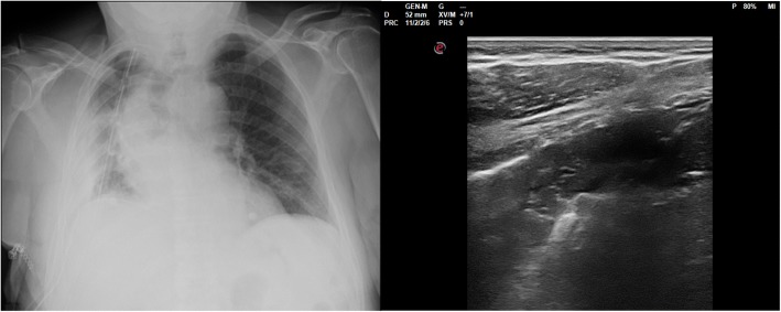
Background: Chest Ultrasonography (chest US) has shown good sensibility in detecting pneumothorax, pleural effusions and peripheral consolidations and it can be performed bedside.
Objectives: The aim of the study was to analyze agreement between chest US and chest X-ray in patients who have undergone thoracic surgery and discuss cases of discordance.
Methods: Patients undergoing thoracic surgery were retrospectively selected. Patients underwent routinely Chest X-ray (CXR) during the first 48 h after surgery. Chest US have been routinely performed in all selected patients in the same date of CXR. Chest US operators were blind to both reports and images of CXR. Ultrasonographic findings regarding pneumothorax (PNX), subcutaneous emphysema (SCE), lung consolidations (LC), pleural effusions (PE) and hemi-diaphragm position were collected and compared to corresponding CXR findings. Inter-rater agreement between two techniques was determined by Cohen's kappa-coefficient.
Results: Twenty-four patients were selected. Inter-rater agreement showed a moderate magnitude for PNX (Cohen's Kappa 0.5), a slight/fair magnitude for SCE (Cohen's Kappa 0.21), a fair magnitude for PE (Cohen's Kappa 0.39), no agreement for LCs (Cohen's Kappa 0.06), high levels of agreement for position of hemi-diaphragm (Cohen's Kappa 0.7).
Conclusion: Analysis of agreement between chest X-ray and chest US showed that ultrasonography is able to detect important findings for surgeons. Limitations and advantages have been found for both chest X-ray and chest US. Knowing the limits of each one is important to really justify and optimize the use of ionizing radiations.
Keywords: Chest X-ray; Chest ultrasound; Innovative biotechnologies; Thoracic surgery; Ultrasonography.
Conflict of interest statement
IRB (Institutional Review Board) approval of “Fondazione Policlinico Universitario A. Gemelli IRCCS” was obtained for this retrospective analysis IRB: P/ CE 5857/19.Not applicable.No competing interests have to be reported by the authors in relation to this work. AS and LR are Associate Editors of Multidisciplinary Respiratory Medicine.Springer Nature remains neutral with regard to jurisdictional claims in published maps and institutional affiliations.
Figures
Similar articles
-
Bedside Chest Ultrasound in Postoperative Pediatric Cardiac Surgery Patients: Comparison With Bedside Chest Radiography.J Cardiothorac Vasc Anesth. 2022 Nov;36(11):4039-4044. doi: 10.1053/j.jvca.2022.06.035. Epub 2022 Jul 1. J Cardiothorac Vasc Anesth. 2022. PMID: 35953404
-
Fast thoracic MRI as an alternative to chest x-ray: A retrospective evaluation of 287 patients.Clin Imaging. 2020 Apr;60(2):244-248. doi: 10.1016/j.clinimag.2019.12.016. Epub 2019 Dec 23. Clin Imaging. 2020. PMID: 31968283
-
Thoracic Ultrasound as an Alternative to Chest X-ray in Thoracic Surgery Patients: A Single-Center Experience.J Clin Med. 2024 Jun 23;13(13):3663. doi: 10.3390/jcm13133663. J Clin Med. 2024. PMID: 38999229 Free PMC article.
-
Chest ultrasonography versus supine chest radiography for diagnosis of pneumothorax in trauma patients in the emergency department.Cochrane Database Syst Rev. 2020 Jul 23;7(7):CD013031. doi: 10.1002/14651858.CD013031.pub2. Cochrane Database Syst Rev. 2020. PMID: 32702777 Free PMC article.
-
Chest ultrasonography in the ICU.Respir Care. 2012 May;57(5):773-81. doi: 10.4187/respcare.01743. Respir Care. 2012. PMID: 22546298 Review.
Cited by
-
New International Guidelines and Consensus on the Use of Lung Ultrasound.J Ultrasound Med. 2023 Feb;42(2):309-344. doi: 10.1002/jum.16088. Epub 2022 Aug 22. J Ultrasound Med. 2023. PMID: 35993596 Free PMC article. Review.
-
Most patient conditions do not a priori debilitate the sensitivity of thoracic ultrasound in thoracic surgery-a prospective comparative study.J Cardiothorac Surg. 2021 Apr 13;16(1):75. doi: 10.1186/s13019-021-01454-6. J Cardiothorac Surg. 2021. PMID: 33849605 Free PMC article.
-
Lung Ultrasound Reduces Chest X-rays in Postoperative Care after Thoracic Surgery: Is There a Role for Artificial Intelligence?-Systematic Review.Diagnostics (Basel). 2023 Sep 19;13(18):2995. doi: 10.3390/diagnostics13182995. Diagnostics (Basel). 2023. PMID: 37761362 Free PMC article. Review.
-
Chest X-rays and Lung Ultrasound Are Not Interchangeable in Intensive Care Practice.Diagnostics (Basel). 2023 Dec 29;14(1):82. doi: 10.3390/diagnostics14010082. Diagnostics (Basel). 2023. PMID: 38201391 Free PMC article.
-
Chest ultrasonography and X-ray may be perfectly integrated in patients management after thoracic surgery such as Shaq and Kobe.J Thorac Dis. 2019 Aug;11(8):E115-E116. doi: 10.21037/jtd.2019.08.21. J Thorac Dis. 2019. PMID: 31559078 Free PMC article. No abstract available.
References
-
- Volpicelli G, Elbarbary M, Blaivas M, Lichtenstein DA, Mathis G, Kirkpatrick AW, et al. International liaison committee on lung ultrasound (ILC-LUS) for international consensus conference on lung ultrasound (ICC-LUS): international evidence-based recommendations for point-of-care lung ultrasound. Intensive Care Med. 2012;38:577–591. doi: 10.1007/s00134-012-2513-4. - DOI - PubMed
LinkOut - more resources
Full Text Sources
