

Review
doi: 10.1007/s40123-019-0178-6.
Epub 2019 Mar 13.
Pearls and Pitfalls of Optical Coherence Tomography Angiography Imaging: A Review
Affiliations
- PMID: 30868418
- PMCID: PMC6513942
- DOI: 10.1007/s40123-019-0178-6
Item in Clipboard
Review
Pearls and Pitfalls of Optical Coherence Tomography Angiography Imaging: A Review
Ophthalmol Ther.
2019 Jun.
Abstract
Optical coherence tomography angiography (OCTA) has significantly expanded our knowledge of the ocular vasculature. Furthermore, this imaging modality has been widely adopted to investigate different ocular and systemic diseases. In this review, a discussion of the fundamental principles of OCTA is followed by the application of this imaging modality to study the retinal and choroidal vessels. A proper comprehension of this imaging modality is essential for the interpretation of OCTA imaging applications in retinal and choroidal disorders.
Keywords: Choriocapillaris; Choroid; Image analysis; Optical coherence tomography angiography; Retinal vessels.
Figures
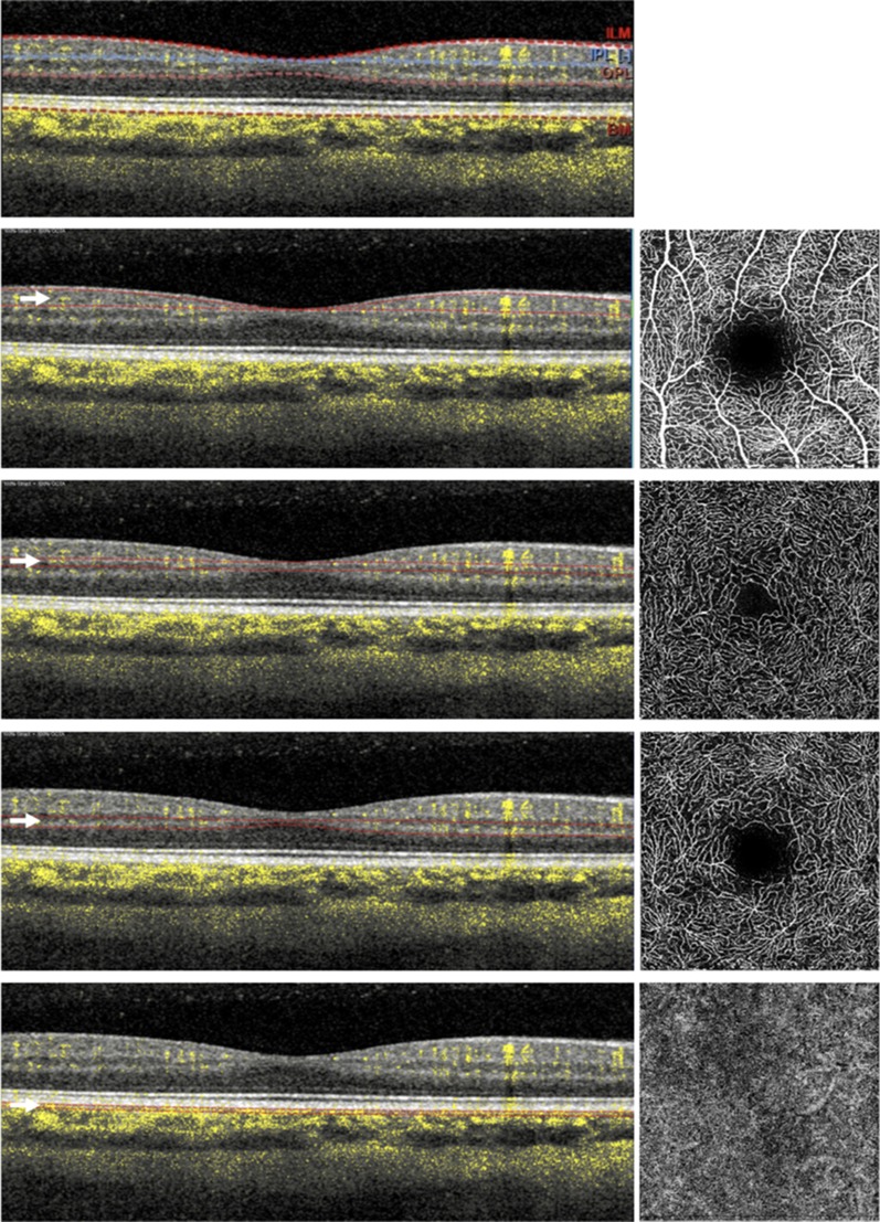
Optical coherence tomographic angiographic image of the macula of a healthy subject. The retinal vascularization at the macula includes four different plexuses: the superficial (SCP—second line from top), middle (MCP—third from top), and deep (DCP—second from bottom) retinal capillary plexuses, and choriocapillaris (CC—bottom). OCTA images are mainly displayed with en face visualization (images on the right) which is obtained by segmenting the volumetric OCTA scans at specific depths (indicated with white arrows). Using this strategy, the flow data within any slab, whose boundaries are red in the left images, are summed or projected into a two-dimensional en face image that can be viewed and studied. These boundaries follow predefined layers which can be differentiated on the basis of reflectivity, texture, or other attributes (top row). The layers are: (i) the inner limiting membrane (ILM), (ii) the inner border of the inner plexiform layer (IPL), (iii) the outer border of the outer plexiform layer (OPL), and (iv) Bruch’s membrane (BM)
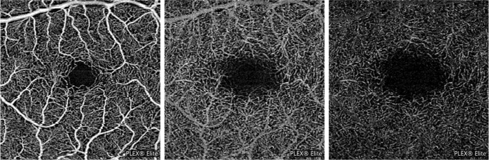
Example of projection artifacts. Superficial capillary plexus (SCP—left image) and deep capillary plexus (DCP—middle image) from a healthy subject and demonstrating the presence of projection artifacts from major SCP vessels on the DCP image. Projection artifacts from superficial retinal vessels are subtracted and replaced with dark versions of these vessels (right image)

Main OCTA metrics to quantify the retinal and choroidal vasculature. The OCTA images can be thresholded to obtain binarized and skeletonized images which are utilized to provide a quantitative analysis. Thresholding is used to create binary images from grayscale images, in which pixels over the applied threshold are displayed as black, and pixels falling under the threshold are shown as white. In this example, a MaxEntropy and Phansalkar thresholds were used to binarize retinal and CC vessels, respectively
References
-
- Borrelli E, Sarraf D, Freund KB, Sadda SR. OCT angiography and evaluation of the choroid and choroidal vascular disorders. Prog Retin Eye Res. 2018;67:30–55. - PubMed
-
- Tan PEZ, Yu PK, Balaratnasingam C, Cringle SJ, Morgan WH, McAllister IL, et al. Quantitative confocal imaging of the retinal microvasculature in the human retina. Investig Ophthalmol Vis Sci. 2012;53:5728–5736. - PubMed
-
- Chan G, Balaratnasingam C, Yu PK, Morgan WH, McAllister IL, Cringle SJ, et al. Quantitative morphometry of perifoveal capillary networks in the human retina. Investig Ophthalmol Vis Sci. 2012;53:5502–5514. - PubMed
-
- Ghasemi Falavarjani K, Al-Sheikh M, Akil H, Sadda SR. Image artefacts in swept-source optical coherence tomography angiography. Br J Ophthalmol [Internet]. 2016. http://bjo.bmj.com/lookup/doi/10.1136/bjophthalmol-2016-309104. Cited 19 Jan 2017. - DOI - PubMed
Publication types
LinkOut - more resources
Full Text Sources
