

Cerebellar, limbic, and midbrain volume alterations in sudden unexpected death in epilepsy
- PMID: 30868560
- PMCID: PMC6479118
- DOI: 10.1111/epi.14689
Cerebellar, limbic, and midbrain volume alterations in sudden unexpected death in epilepsy
Abstract
Objective: The processes underlying sudden unexpected death in epilepsy (SUDEP) remain elusive, but centrally mediated cardiovascular or respiratory collapse is suspected. Volume changes in brain areas mediating recovery from extreme cardiorespiratory challenges may indicate failure mechanisms and allow prospective identification of SUDEP risk.
Methods: We retrospectively imaged SUDEP cases (n = 25), patients comparable for age, sex, epilepsy syndrome, localization, and disease duration who were high-risk (n = 25) or low-risk (n = 23), and age- and sex-matched healthy controls (n = 25) with identical high-resolution T1-weighted scans. Regional gray matter volume, determined by voxel-based morphometry, and segmentation-derived structure sizes were compared across groups, controlling for total intracranial volume, age, and sex.
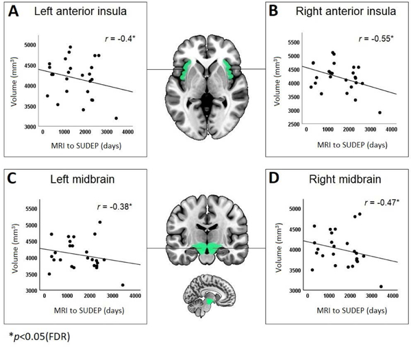
Results: Substantial bilateral gray matter loss appeared in SUDEP cases in the medial and lateral cerebellum. This was less prominent in high-risk subjects and absent in low-risk subjects. The periaqueductal gray, left posterior and medial thalamus, left hippocampus, and bilateral posterior cingulate also showed volume loss in SUDEP. High-risk subjects showed left thalamic volume reductions to a lesser extent. Bilateral amygdala, entorhinal, and parahippocampal volumes increased in SUDEP and high-risk patients, with the subcallosal cortex enlarged in SUDEP only. Disease duration correlated negatively with parahippocampal volume. Volumes of the bilateral anterior insula and midbrain in SUDEP cases were larger the closer to SUDEP from magnetic resonance imaging.
Significance: SUDEP victims show significant tissue loss in areas essential for cardiorespiratory recovery and enhanced volumes in areas that trigger hypotension or impede respiratory patterning. Those changes may shed light on SUDEP pathogenesis and prospectively detect patterns identifying those at risk.
Keywords: cerebellum; limbic; magnetic resonance imaging; midbrain; sudden unexpected death in epilepsy.
© 2019 The Authors. Epilepsia published by Wiley Periodicals, Inc. on behalf of International League Against Epilepsy.
Conflict of interest statement
Disclosure
No authors have any conflicts of interest to disclose.
Figures


References
-
- Ryvlin P, Nashef L, Lhatoo SD, et al. Incidence and mechanisms of cardiorespiratory arrests in epilepsy monitoring units (MORTEMUS): a retrospective study. Lancet Neurol 2013;12:966–977. - PubMed
-
- Surges R, Sander JW. Sudden unexpected death in epilepsy: mechanisms, prevalence, and prevention. Curr Opin Neurol 2012;25:201–207. - PubMed
Publication types
MeSH terms
Grants and funding
- G0802012/MRC_/Medical Research Council/United Kingdom
- MR/M00841X/1/MRC_/Medical Research Council/United Kingdom
- U01 NS090407/NS/NINDS NIH HHS/United States
- U01-NS090407/NS/NINDS NIH HHS/United States
- National Institute for Health Research University College London Hospitals Biomedical Research Centre/International
