. 2019 Jun;18(3):e12928.
doi: 10.1111/acel.12928. Epub 2019 Mar 13.

Increased intron retention is a post-transcriptional signature associated with progressive aging and Alzheimer's disease

Affiliations

Increased intron retention is a post-transcriptional signature associated with progressive aging and Alzheimer's disease

Swarnaseetha Adusumalli et al. Aging Cell. 2019 Jun.

Abstract

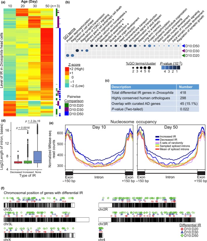
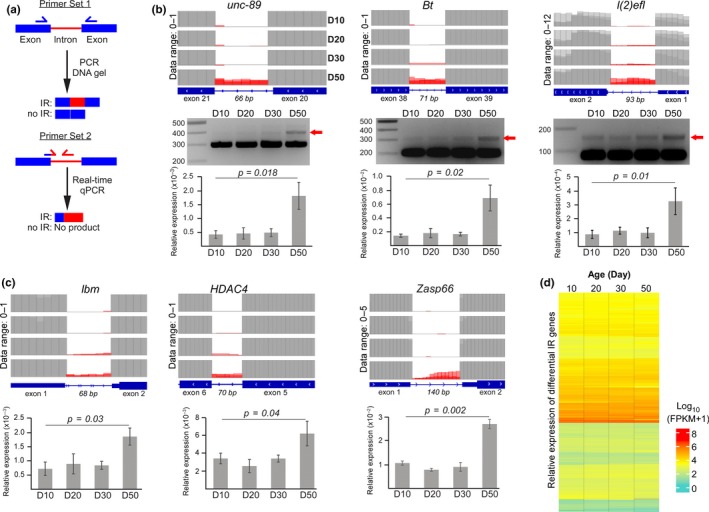
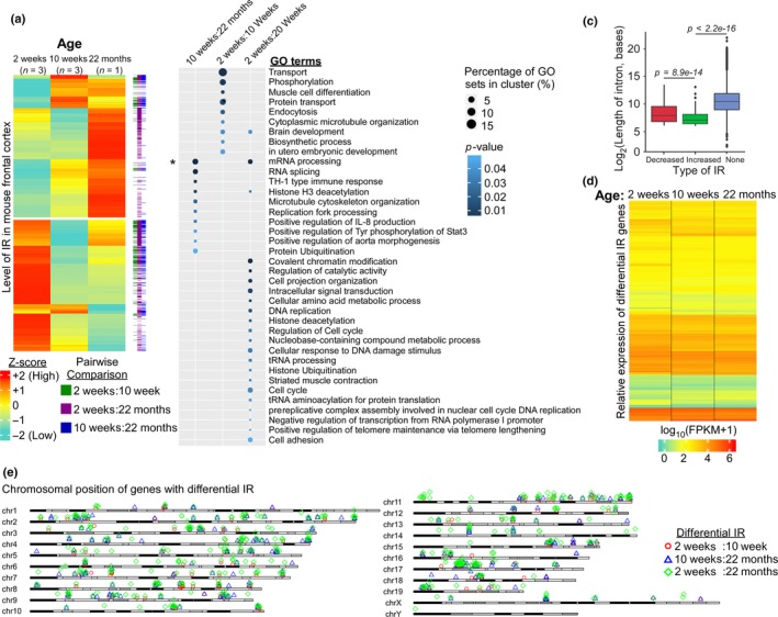
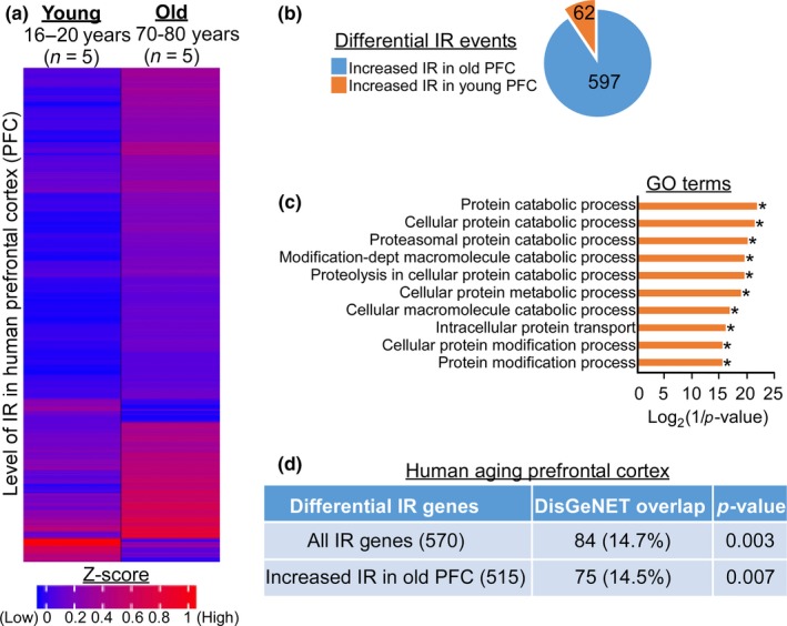
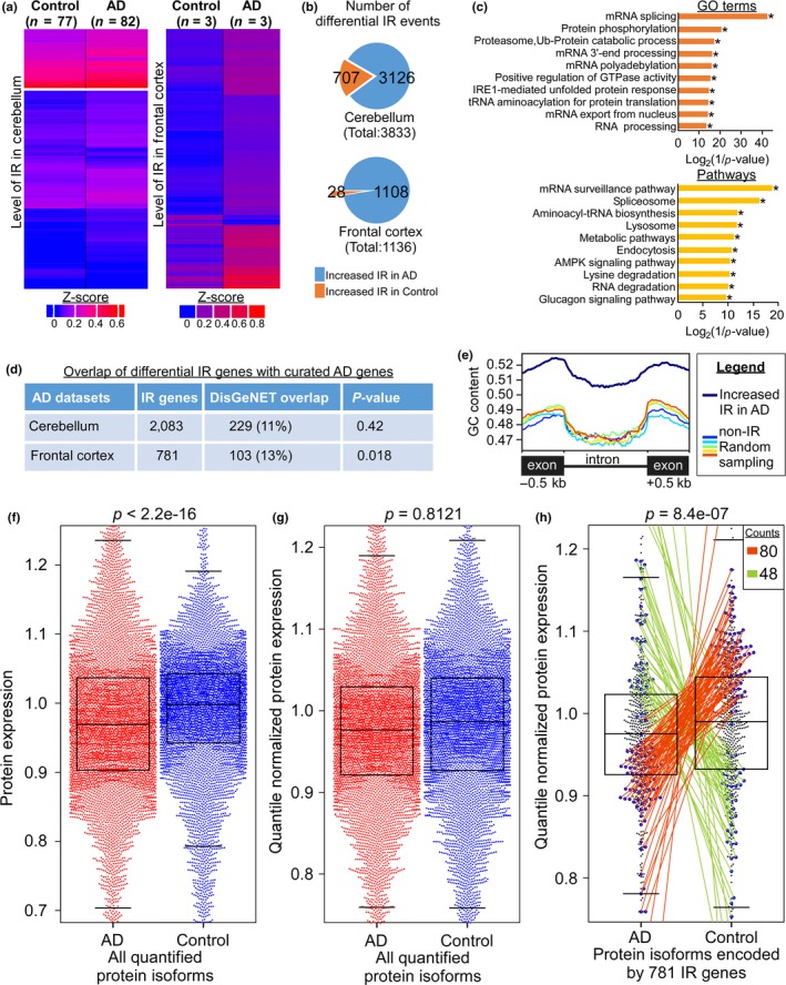
Intron retention (IR) by alternative splicing is a conserved regulatory mechanism that can affect gene expression and protein function during adult development and age-onset diseases. However, it remains unclear whether IR undergoes spatial or temporal changes during different stages of aging or neurodegeneration like Alzheimer's disease (AD). By profiling the transcriptome of Drosophila head cells at different ages, we observed a significant increase in IR events for many genes during aging. Differential IR affects distinct biological functions at different ages and occurs at several AD-associated genes in older adults. The increased nucleosome occupancy at the differentially retained introns in young animals suggests that it may regulate the level of IR during aging. Notably, an increase in the number of IR events was also observed in healthy older mouse and human brain tissues, as well as in the cerebellum and frontal cortex from independent AD cohorts. Genes with differential IR shared many common features, including shorter intron length, no perturbation in their mRNA level, and enrichment for biological functions that are associated with mRNA processing and proteostasis. The differentially retained introns identified in AD frontal cortex have higher GC content, with many of their mRNA transcripts showing an altered level of protein expression compared to control samples. Taken together, our results suggest that an increased IR is an conserved signature that is associated with aging. By affecting pathways involved in mRNA and protein homeostasis, changes of IR pattern during aging may regulate the transition from healthy to pathological state in late-onset sporadic AD.

Keywords: Alzheimer’s disease; Drosophila; Intron retention; aging; human; mouse.


Conflict of interest statement

None declared.

Figures

Figure 1
Figure 1
Differential intron retentions (IR) mark different stages of aging in Drosophila. (a) Expression heatmap of the differentially retained introns across various ages as represented by Z‐score. Pairwise comparison of the two age‐groups is highlighted by the color bar on the right. (b) Gene ontology analysis of the genes with differential IR in aging fly heads (p‐value <0.05, Fisher's exact test; *p‐value <0.05, Fisher's exact test with Benjamini–Hochberg correction). (c) The overlap between highly conserved human orthologues of fly differential IR genes and curated AD genes from DisGeNET. The p‐value was determined by two‐tailed chi‐square test with Yates’ continuity correction. (d) Boxplot for the length distribution of the various types of introns where D10 flies were used as the reference to determine the differential decreased or increased IR during aging. “None” refers to non‐retained, spliced introns (p < 0.05, Welch's t test). (e) Normalized MNase‐seq read counts across different types of introns in Day 10 and 50 fly heads showed that nucleosome occupancy over differentially retained introns was significantly different from the spliced introns (p < 0.05, Wilcoxon rank sum test). The increased or decreased IR referred to the differentially retained introns at Day 50 with respect to Day 10. Five sets of spliced introns with similar expression level were randomly picked as control. (f) Ideogram displaying the genome‐wide distribution of the differential IR genes in fly
Figure 2
Figure 2
Experimental validation of IR at specific Drosophila genes. (a) Two different primer sets were designed to validate differential IR. (b, c) Top panel: The expression level of retained intron (red) and flanking exons (gray) on Integrative Genome Browser where the respective data range is indicated. Middle panel: Visualization of retained introns of three specific genes with DNA gel electrophoresis. Bottom panel: Real‐time quantitative PCR of differential retained introns using three biological replicates of fly heads for each time‐point (p < 0.05, paired t test). (d) Relative expression of differential IR genes across various age‐groups as represented by Log10 (FPKM + 1) values
Figure 3
Figure 3
Intron retentions across three age‐groups in mouse frontal cortex. (a) Expression heatmap of differentially retained introns across three age‐groups (2, 10 weeks, and 22 months) in mouse frontal cortex. (b) Gene ontology analysis of the differential IR genes determined through pairwise comparison between two distinct time‐points (p‐value <0.05, Fisher's exact test; *p‐value <0.05, Fisher's exact test with Benjamini–Hochberg correction). (c) Boxplot for the length distribution of the differentially retained and spliced introns. The expression level of intron at 2 weeks was used as the reference to determine the decreased or increased IR during aging. “None” refers to spliced introns (p‐value <0.05, Welch's t test). (d) Relative expression of IR genes between different ages of mouse frontal cortex as represented by Log10 (FPKM + 1) values. (e) Ideogram displaying the distribution of the differential IR genes across the mouse genome
Figure 4
Figure 4
Genes with increased IR in old prefrontal cortex are linked to AD pathway. (a) Expression heatmap of differentially retained introns in young and old human prefrontal cortex (PFC). (b) Pie diagram showing the number of differentially increased IR events in young and old human PFC. (c) Gene ontology analysis of the genes with differential IR between young and old human PFC (*p‐value <0.05, Fisher's exact test with Benjamini–Hochberg correction). (d) The overlap of differential IR genes in aging PFC with curated AD genes from DisGeNET. The p‐value was determined by two‐tailed chi‐square test with Yates’ continuity correction
Figure 5
Figure 5
Increased number of IR events is observed in human AD brain tissues. (a) Expression heatmap of the differentially retained introns in the cerebellum (left, Mayo Clinic) and frontal cortex (right, University of Kentucky) from age‐matched control and AD subjects. (b) Pie diagrams indicating the number of increased IR events in cerebellum (top) and frontal cortex (bottom) of age‐matched control (orange) and AD patients (blue). (c) Gene ontology (*p‐value <0.05, Fisher's exact test with Benjamini–Hochberg correction) and pathway enrichment (*p‐value <0.05, hypergeometric test with FDR) analyses of the differential IR genes between control and AD cerebellum. (d) The overlap of differential IR genes identified in diseased brain tissues with curated AD genes from DisGeNET. The p‐value was determined by two‐tailed chi‐square test with Yates’ continuity correction. (e) GC content of the flanking exons and the differentially retained introns in AD frontal cortex. Five sets of non‐retained introns from genes with similar expression level were randomly picked as control. (f–g) Beeswarm boxplots illustrating the raw (f) and quantile normalized (g) expression of all 10,100 quantified protein isoforms in AD (red) and control (blue) frontal cortex. p‐value was calculated by Wilcoxon rank sum test with continuity correction. (h) The quantile normalized protein expression pattern of 781 differential IR genes is significantly different between AD and control frontal cortex (p‐value = 8.4e−07, Wilcoxon rank sum test with continuity correction). Blue dots represent genes whose protein level is significantly different between AD and control frontal cortex (p‐value <0.05, LIMMA t test). Among which, 73 (orange line) and 41 (green line) differential IR genes showed reduced and elevated protein level in AD frontal cortex, respectively

References

    1. Audic, S. , & Claverie, J. M. (1997). The significance of digital gene expression profiles. Genome Research, 7, 986–995. - PubMed
    1. Bai, B. , Hales, C. M. , Chen, P.‐C. , Gozal, Y. , Dammer, E. B. , Fritz, J. J. , … Peng, J. (2013). U1 small nuclear ribonucleoprotein complex and RNA splicing alterations in Alzheimer’s disease. Proceedings of the National Academy of Sciences, 110, 16562–16567. 10.1073/pnas.1310249110 - DOI - PMC - PubMed
    1. Baralle, F. E. , & Giudice, J. (2017). Alternative splicing as a regulator of development and tissue identity. Nature Reviews Molecular Cell Biology, 18, 437–451. 10.1038/nrm.2017.27 - DOI - PMC - PubMed
    1. Blanco, F. J. , & Bernabeu, C. (2011). Alternative splicing factor or splicing factor‐2 plays a key role in intron retention of the endoglin gene during endothelial senescence. Aging Cell, 10, 896–907. 10.1111/j.1474-9726.2011.00727.x - DOI - PubMed
    1. Brady, L. K. , Wang, H. , Radens, C. M. , Bi, Y. , Radovich, M. , Maity, A. , … Koumenis, C. (2017). Transcriptome analysis of hypoxic cancer cells uncovers intron retention in EIF2B5 as a mechanism to inhibit translation. PLOS Biology, 15, e2002623 10.1371/journal.pbio.2002623 - DOI - PMC - PubMed

Publication types