. 2019 Jun 20;47(11):e61.
doi: 10.1093/nar/gkz171.

A panel of colorimetric assays to measure enzymatic activity in the base excision DNA repair pathway

Affiliations

A panel of colorimetric assays to measure enzymatic activity in the base excision DNA repair pathway

Eleanor Healing et al. Nucleic Acids Res. .

Abstract

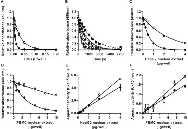
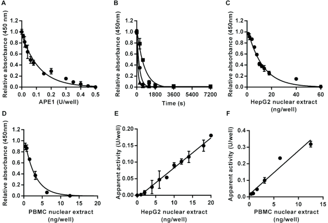
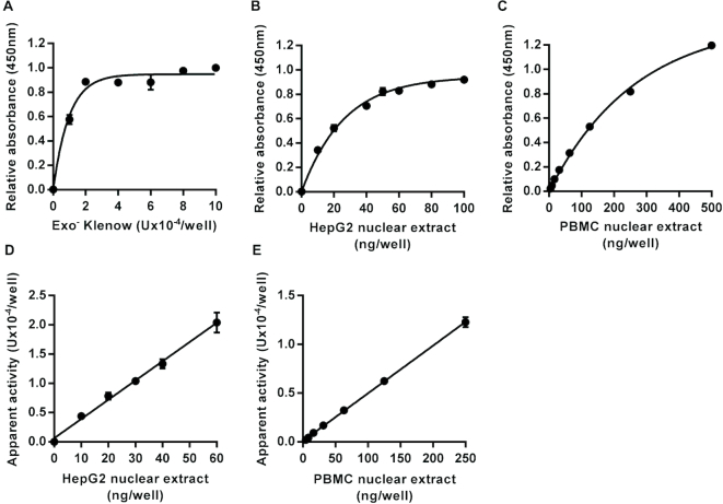
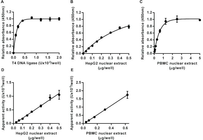
DNA repair is essential for the maintenance of genomic integrity, and evidence suggest that inter-individual variation in DNA repair efficiency may contribute to disease risk. However, robust assays suitable for quantitative determination of DNA repair capacity in large cohort and clinical trials are needed to evaluate these apparent associations fully. We describe here a set of microplate-based oligonucleotide assays for high-throughput, non-radioactive and quantitative determination of repair enzyme activity at individual steps and over multiple steps of the DNA base excision repair pathway. The assays are highly sensitive: using HepG2 nuclear extract, enzyme activities were quantifiable at concentrations of 0.0002 to 0.181 μg per reaction, depending on the enzyme being measured. Assay coefficients of variation are comparable with other microplate-based assays. The assay format requires no specialist equipment and has the potential to be extended for analysis of a wide range of DNA repair enzyme activities. As such, these assays hold considerable promise for gaining new mechanistic insights into how DNA repair is related to individual genetics, disease status or progression and other environmental factors and investigating whether DNA repair activities can be used a biomarker of disease risk.


Figures

Figure 1.
Figure 1.
Effect of uracil DNA glycosylase activity on oligonucleotide substrates containing a single U:A base pair or U:G mismatch. Incubation with recombinant Escherichia coli uracil DNA glycosylase led to enzyme concentration dependent (A) and time-dependent (B) decreases in the amount of intact U:A (open symbols) and U:G (closed symbols) containing substrate retained in the wells after alkaline denaturation. For the time course analysis squares, circles and triangles indicate data generated with 0.1, 0.2 or 0.4 units/well, respectively, for the U:A containing substrate or with 0.0125, 0.025 or 0.05 units/well, respectively, for the U:G containing substrate. Incubation with HepG2 nuclear extract (C) or peripheral blood mononuclear cell pooled extract (D) also caused concentration-dependent decreases in the amount of intact U:A and U:G containing substrate retained in the wells. For both substrates, the apparent uracil DNA glycosylase activity was directly proportional to the concentration of HepG2 (E) and peripheral blood mononuclear cell (F) nuclear extract over the entire range tested (r2 = 0.99 in both cases). Data shown represent the mean ± SD of triplicate technical replicates except for time course data for which duplicates were analyzed.
Figure 2.
Figure 2.
Effect of apurinic/apyrimidinic (AP) site incision activity on an oligonucleotide substrate containing a single tetrahydrofuran AP site analog. Incubation with recombinant human APE1 led to enzyme concentration-dependent (A) and time-dependent (B) decreases in the amount of intact substrate retained in the wells after neutral denaturation. For the time course analysis squares, circles and triangles indicate data generated with 0.1, 0.2 or 0.4 units/well, respectively. Incubation with HepG2 nuclear extract (C) or PBMC pooled nuclear extract (D) also caused concentration-dependent decreases in the amount of intact substrate retained in the wells. The apparent AP site incision activity was directly proportional to the concentration of nuclear extract over the range 0–0.02 μg/well for the HepG2 nuclear extract (r2 = 0.99) (E) and over the range 0–0.0125 μg/well for the peripheral blood mononuclear cell nuclear extract (r2 = 0.95) (F). Data shown represent the mean ± SD of triplicate technical replicates except for time course data for which duplicates were analyzed.
Figure 3.
Figure 3.
Assay for DNA polymerase repair activity. Incubation of an immobilized hairpin loop oligonucleotide substrate containing a single nucleotide gap on one strand within the double-stranded region with exonuclease minus Klenow fragment of Escherichia coli DNA polymerase I, in the presence of dNTPs, ATP and excess T4 DNA ligase, led to an increase in intact substrate retained in the microplate wells following neutral denaturation that was dependent on the concentration of the polymerase (A). Incubation with HepG2 nuclear extract (B) or peripheral blood mononuclear cell pooled nuclear extract (C) also caused concentration-dependent increases in the amount of intact substrate retained in the wells. The apparent DNA polymerase activity was directly proportional to the concentration of nuclear extract over the range 0–0.06 μg/well for the HepG2 nuclear extract (r2 = 0.98) (D) and over the range 0–0.25 μg/well for the peripheral blood mononuclear cell nuclear extract (r2 = 0.99) (E). Data shown represent the mean ± SD of triplicate technical replicates.
Figure 4.
Figure 4.
Assay for DNA ligase activity. Incubation of an immobilized hairpin loop oligonucleotide substrate containing a single ligatable nick in one strand within the double-stranded region with T4 DNA ligase, in the presence of ATP, led to an increase in intact substrate retained in the microplate wells following neutral denaturation that was dependent on the concentration of the polymerase (A). Incubation with HepG2 nuclear extract (B) and peripheral blood mononuclear cell pooled nuclear extract (C) also caused concentration-dependent increases in the amount of intact substrate retained in the wells. The apparent DNA ligase activity was directly proportional to the concentration of HepG2 (D) and PBMC (E) nuclear extract over the range 0–0.5 μg/well for the HepG2 nuclear extract (r2 = 0.99) and over the range 0–0.63 μg/well for the peripheral blood mononuclear cell nuclear extract (r2 = 0.99). Data shown represent the mean ± SD of triplicate technical replicates.
Figure 5.
Figure 5.
Within and between cell line variability in DNA repair enzyme activity of HepG2 and Caco-2 cells. (A) Uracil DNA glycosylase activity was higher in Caco-2 nuclear extract than HepG2 nuclear extract. (B) No significant differences were found in apurinic/apyrimidinic site incision activity between the two cell lines. (C) DNA polymerase activity was lower in Caco-2 nuclear extract than HepG2 nuclear extract. (D) DNA ligase activity was lower in Caco-2 nuclear extract than HepG2 nuclear extract. Bars indicate mean ± SD of six independent replicates. The symbols * and *** indicate significant differences between the enzyme activities of the cell lines at P < 0.05 and P < 0.001, respectively (based on two-tailed unpaired t-tests).
Figure 6.
Figure 6.
Base excision repair enzyme activity is correlated between some, but not all, enzymes of the pathway in nuclear extracts of peripheral blood mononuclear cells from different individuals. (A) Uracil DNA glycosylase (UDG) activity was positively correlated with apurinic/apyrimidinic (AP) site incision activity (r = 0.61, P < 0.05). (B) UDG activity was not significantly correlated with DNA polymerase activity (r = 0.58, P = 0.053). (C) UDG activity was positively correlated with DNA ligase activity (r = 0.81, P < 0.001). (D) AP site incision activity was not significantly correlated with DNA polymerase activity (r = 0.40, P = 0.177). (E) AP site incision activity was positively correlated with DNA ligase activity (r = 0.73, P < 0.01). (F) DNA polymerase activity was positively correlated with DNA ligase activity (r = 0.67, P < 0.05).
Figure 7.
Figure 7.
Complete repair of an apurinic/apyrimidinic (AP) site or a tetrahydrofuran AP site analog by HepG2 nuclear extract. The data presented in (A) demonstrate that color development observed in the assay plate wells is directly proportional to the amount of fluorescein modified oligonucleotide retained in the wells (r2 = 0.99). These data were generated by (i) coating the wells of a Nunc® Immobilizer™ plate with 0.5 pmol/well of mixtures containing varying proportions of oligonucleotides URA03 (containing a single uracil lesion) and Contr01 (lesion free control), (ii) hybridizing and ligating a complementary oligonucleotide (Loop01Aflc) to the immobilized URA03/Contr01 mixtures to form intact hairpin loop structures covalently attached to the plate at the 3′ end with a fluorescein moiety at the 5′ end, (iii) incubating the immobilized substrates with excess Escherichia coli uracil DNA glycosylase to excise the uracil from those oligonucleotide carrying them to create AP sites, (iv) subjecting the substrate to hot alkaline denaturation to convert all AP sites into single strand breaks and remove any oligonucleotides released as a result and (v) detecting fluorescein retained in the wells using anti-fluorescein horseradish peroxidase conjugate and TMB substrate. Based on this observation, percentage repair was determined by linear interpolation from the absorbance values for 0 and 100% repair control wells. (B) Effects of varying concentrations of HepG2 nuclear extract on the extent of complete AP site repair in the absence (open circles) and presence (closed circles) of excess T4 DNA ligase. (C) Comparison of extent of complete repair by HepG2 nuclear extract of an oligonucleotide complex containing an AP site that had either been pre-treated or not with excess recombinant human AP endonuclease 1. (D) Comparison of the extent of complete repair of an oligonucleotide containing either a normal AP site (black bars) or the tetrahydrofuran AP site analog (white bars) by 800 or 400 ng/well of HepG2 nuclear extract in the presence of either 30 μM of all four deoxynucleotides (dATP, dCTP, dGTP and dTTP) or only 30 μM dTTP. Error bars indicate mean ± SD of triplicate technical replicates.
Figure 8.
Figure 8.
Assay for alkyladenine DNA glycosylase (AAG) activity. Incubation with recombinant human AAG led to enzyme concentration- (A) and time-dependent (B) decreases in the amount of intact oligonucleotide substrate containing an I:T base pair following alkaline denaturation. For the time course analysis squares, circles and triangles indicate data generated with 0.1, 0.2 or 0.4 units/well, respectively. Incubation with HepG2 nuclear extract (C) or peripheral blood mononuclear cell pooled extract (D) also caused concentration-dependent decreases in the amount of intact substrate retained in the wells. The apparent AAG activity was directly proportional to the sample concentration over the range 0–10 μg/well for HepG2 (E) and peripheral blood mononuclear cell (F) nuclear extracts (r2 = 0.94 in both cases). Data shown represent the mean ± SD of triplicate technical replicates except for time course data for which duplicates were analyzed.

Similar articles

Cited by

References

    1. Iyama T., Wilson D.M. 3rd. DNA repair mechanisms in dividing and non-dividing cells. DNA Repair. 2013; 12:620–636. - PMC - PubMed
    1. Jeggo P.A., Pearl L.H., Carr A.M.. DNA repair, genome stability and cancer: a historical perspective. Nat. Rev. Cancer. 2016; 16:35–42. - PubMed
    1. Krokan H.E., Bjoras M.. Base excision repair. Cold Spring Harb. Perspect. Biol. 2013; 5:a012583. - PMC - PubMed
    1. Hegde M.L., Hazra T.K., Mitra S.. Early steps in the DNA base excision/single-strand interruption repair pathway in mammalian cells. Cell Res. 2008; 18:27–47. - PMC - PubMed
    1. D’Errico M., Parlanti E., Pascucci B., Fortini P., Baccarini S., Simonelli V., Dogliotti E.. Single nucleotide polymorphisms in DNA glycosylases: from function to disease. Free Radic. Biol. Med. 2016; 107:278–291. - PubMed

Publication types