Review
. 2020 Apr 16;59(16):6342-6366.
doi: 10.1002/anie.201900585. Epub 2020 Jan 30.

Targeting the Small GTPase Superfamily through Their Regulatory Proteins

Affiliations
Review

Targeting the Small GTPase Superfamily through Their Regulatory Proteins

Janine L Gray et al. Angew Chem Int Ed Engl. .

Abstract

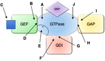
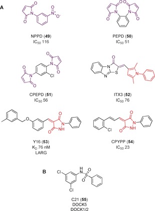
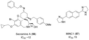
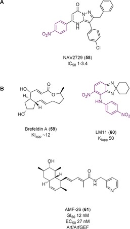
The Ras superfamily of small GTPases are guanine-nucleotide-dependent switches essential for numerous cellular processes. Mutations or dysregulation of these proteins are associated with many diseases, but unsuccessful attempts to target the small GTPases directly have resulted in them being classed as "undruggable". The GTP-dependent signaling of these proteins is controlled by their regulators; guanine nucleotide exchange factors (GEFs), GTPase activating proteins (GAPs), and in the Rho and Rab subfamilies, guanine nucleotide dissociation inhibitors (GDIs). This review covers the recent small molecule and biologics strategies to target the small GTPases through their regulators. It seeks to critically re-evaluate recent chemical biology practice, such as the presence of PAINs motifs and the cell-based readout using compounds that are weakly potent or of unknown specificity. It highlights the vast scope of potential approaches for targeting the small GTPases in the future through their regulatory proteins.

Keywords: drug discovery; peptides; protein-protein interactions; small GTPases; small molecules.


Conflict of interest statement

The authors declare no conflict of interest.

Figures

Figure 1
Figure 1
A. Phylogenetic tree of the Ras superfamily. Nine unclassified members are not included. The Rab subfamily is shown in blue, Ran in orange, Rho in green, Ras in red and Arf in purple. Grey dots indicate the X‐ray crystal structure has been solved. A yellow dot indicates an NMR structure. B. Comparison of different subfamily sequences containing the switch I and switch II regions. Characteristic “G‐boxes” G1, G2 and G3, which are involved with nucleotide and magnesium ion binding, are mainly conserved across the subfamilies. Whilst some key residues are maintained across the superfamily, the divergence in sequence between the subfamilies could be exploited for selectivity by therapeutics.1
Figure 2
Figure 2
Small GTPase activation cycle with GEFs, GAPs, and GDIs.
Figure 3
Figure 3
Structures showing conformational changes during nucleotide exchange. Switch I shown in orange, switch II in red, P‐loop in light green, GDP/GTP as purple sticks. A) PDB 4Q21. Ras bound to GDP. The switch II region is relatively disordered. B) PDB 1NVU. Nucleotide‐free Ras bound to Sos1 (interacting residues shown in dark green). The switch I loop has moved significantly and the switch II loop is more ordered. C) PDB 3L8Z. Ras bound to GTP analogue GPPNHP. The switch I loop has moved back but the switch II remains more helical.5
Figure 4
Figure 4
Ras, Rho, and Arf subfamilies’ GAP mechanism. A) PDB 1FTN. In RhoA‐GTP, the key catalytic glutamine (Q63, RhoA nomenclature) is oriented away from the nucleotide. B) PDB ITX4. Upon RhoGAP binding, is Q63 oriented towards a water molecule ready to catalyze hydrolysis of GTP. The arginine finger (R85, RhoGAP nomenclature) helps to stabilize the negative charge of the transition state (modelled as GDP and ALF4).
Figure 5
Figure 5
Methods of targeting GEFs, GAPs and GDIs. A) Binding to GTPase and blocking GEF binding. B) Binding to GEF and blocking GTPase binding. C) Preventing GEF activation. D) Binding to the GEF/GTPase complex. E) Targeting the effector binding site on the GTPase that overlaps with the GEF binding site. F) Binding to the GDI and blocking GTPase binding. G) Binding to the GDI/GTPase complex. H) Binding to the GAP/GTPase complex. I) Binding to the GAP. J) Binding to the GTPase to mimic GAP. Relevant references for each example are shown in Table 2.
Figure 6
Figure 6
Compounds that block RasGEF–Sos interaction with Ras GTPase. Toxicophores/structural alerts are shown in purple.
Figure 7
Figure 7
A) Covalent inhibitors of KRas G12C. B) Binding mode of 9 to KRas G12C (blue, PDB 5F2E) compared to the apo KRas (grey, PDB 4OBE). The binding of 9 caused the α2 helix and Met72 of switch II to move. The carbonyl of the acrylamide is situated where the γ‐phosphate of GTP would be situated. These changes result in the GDP‐state of KRas being favored and prevent nucleotide exchange.
Figure 8
Figure 8
Binding interactions of the Ras‐GEF PPI inhibitors. The purple surface corresponds to the binding site of 4, 5, 6, and 7. Pink corresponds to the residues interacting with 8. Yellow corresponds to the residues interacting with 1, 2, 3, 9, and 10. The GDP binding site is shown with the GDP as purple sticks, PDB 4EPY.35
Figure 9
Figure 9
Inhibitors of Ral‐GDP.
Figure 10
Figure 10
RasGEF inhibitors. PAINs motifs are shown in red and toxicophores/structural alerts in purple.
Figure 11
Figure 11
A) Compounds discovered to bind to Sos1. B) 19 bound to a pocket in Sos1 (green) adjacent to KRas (blue) (PDB 6EPM). It formed hydrogen bonds with Tyr884 and Asp887 and forced a Phe890‐out conformation in comparison to the Phe890‐in conformation seen in the apo structure (pink, PDB 6EPL). C) 21 bound to Sos1 (green, PDB 5OVI), maintaining the Phe‐out conformation of 19 but expanding into the neighbouring pocket where 20 is found to bind. Key interacting residues are labelled.
Figure 12
Figure 12
Inhibitors of Epac activation by blocking the interaction with regulatory cAMP. PAINs motifs highlighted in red.
Figure 13
Figure 13
Compounds that activate Sos‐catalyzed nucleotide exchange and induce biphasic responses in cells.
Figure 14
Figure 14
Inhibitors of Ras‐effectors interactions, in which regulator binding is also affected. PAINs motifs highlighted in red and toxicophores/structural alerts in purple.
Figure 15
Figure 15
A) Elaboration of initial hit NSC23766 using structure guided design to yield potent derivatives. Structures similar to known aggregators are shown in blue. Toxicophores/structural alerts are shown in purple. B) Crystal structure of NSC23766 bound to Rac1. Important residues forming the pocket, including critical residue W56, are shown.75
Figure 16
Figure 16
Small molecules developed for targeting Cdc42 and RhoA.
Figure 17
Figure 17
A) RhoGEF inhibitors that are highly reactive or contain a PAIN motif (red). Toxicophores of reactive motifs are shown in purple. B) C21, inhibitor of DOCK5, a DHR RhoGEF.
Figure 18
Figure 18
RhoGDI and RhoGAP inhibitors.
Figure 19
Figure 19
A) NAV2729, a small molecule inhibitor developed for the Arf GTPases. B) Interfacial inhibitors targeting the Arf/ArfGEF subfamily. Toxicophores/structural alerts are shown in purple.
Figure 20
Figure 20
A) PDB 1RE0. Arf1 (blue) bound to BFA‐sensitive ARNO4M (green). The residues critical for BFA binding are labelled. B) Arf1 (blue) bound to BFA‐bound ARNO4M (green) (PDB 1RE0) and Arf1 (grey) bound to IQSEC2 (green) (PDB 6FAE). Binding of BFA prevents reorganization allowing the glutamic finger (E156 in ARNO, E849 in IQSEC2) to be oriented towards GDP, preventing nucleotide exchange.
Figure 21
Figure 21
A) ArfGEF inhibitors. B) ArfGAP inhibitor. Structures similar to aggregators shown in blue. PAINs motifs are shown in red. Toxicophores/structural alerts are shown in purple.

References

    1. Wennerberg K., Rossman K. L., Der C. J., J. Cell Sci. 2005, 118, 843–846. - PubMed
    1. Rojas A. M., Fuentes G., Rausell A., Valencia A., J. Cell Biol. 2012, 196, 189–201. - PMC - PubMed
    1. None
    1. Leicht D. T., Balan V., Kaplun A., Singh-Gupta V., Kaplun L., Dobson M., Tzivion G., Biochim. Biophys. Acta Mol. Cell Res. 2007, 1773, 1196–1212; - PMC - PubMed
    1. Bishop A. L., Hall A., Biochem. J. 2000, 348, 241–255; - PMC - PubMed

Publication types

MeSH terms

LinkOut - more resources