. 2019 Mar 14;129(6):2222-2236.
doi: 10.1172/JCI124694.

Anti-Neurofascin-155 IgG4 antibodies prevent paranodal complex formation in vivo

Affiliations

Anti-Neurofascin-155 IgG4 antibodies prevent paranodal complex formation in vivo

Constance Manso et al. J Clin Invest. .

Abstract

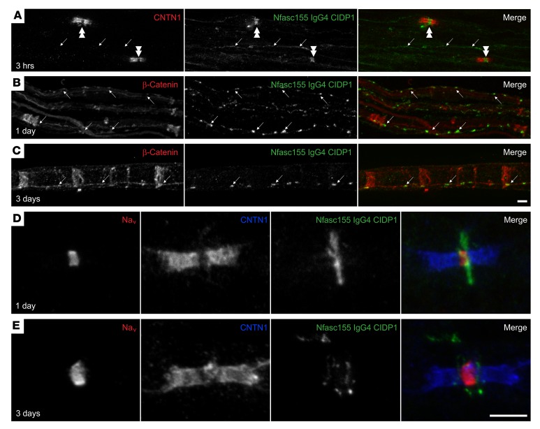
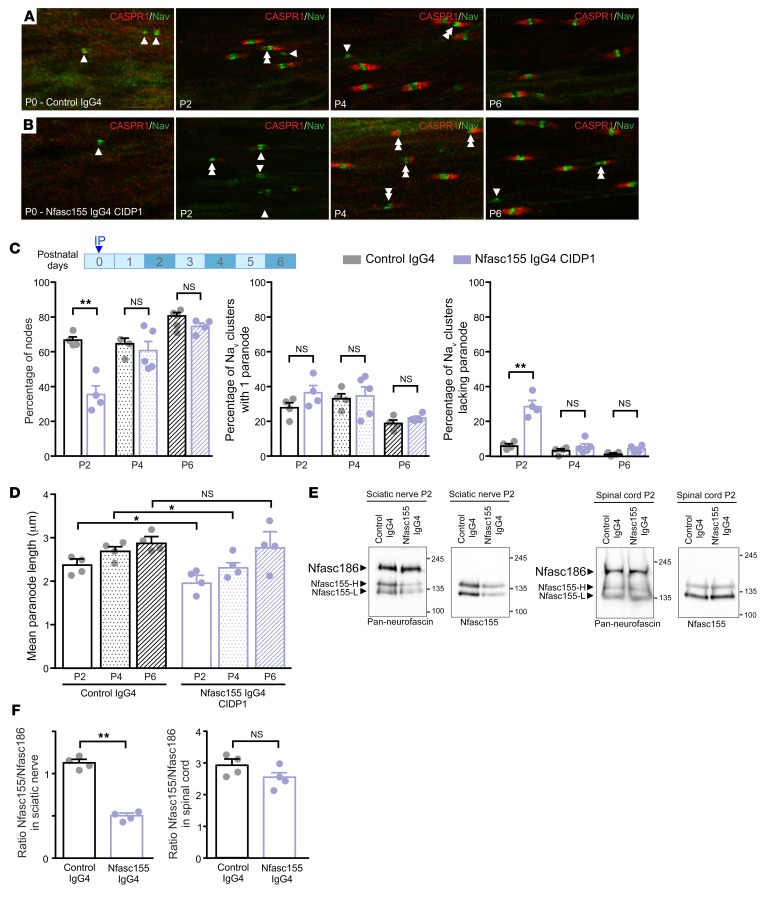
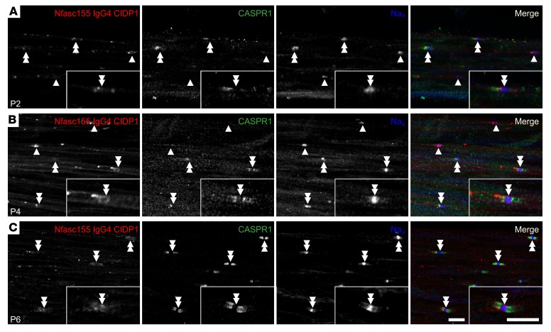
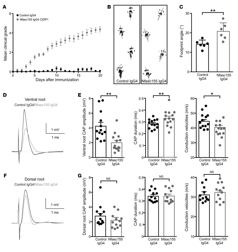
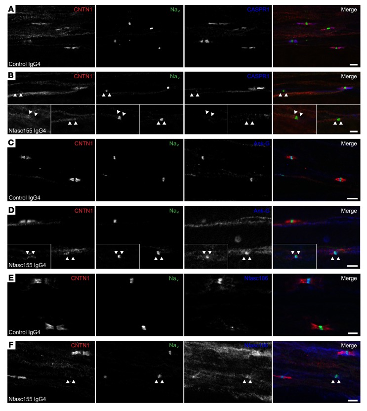
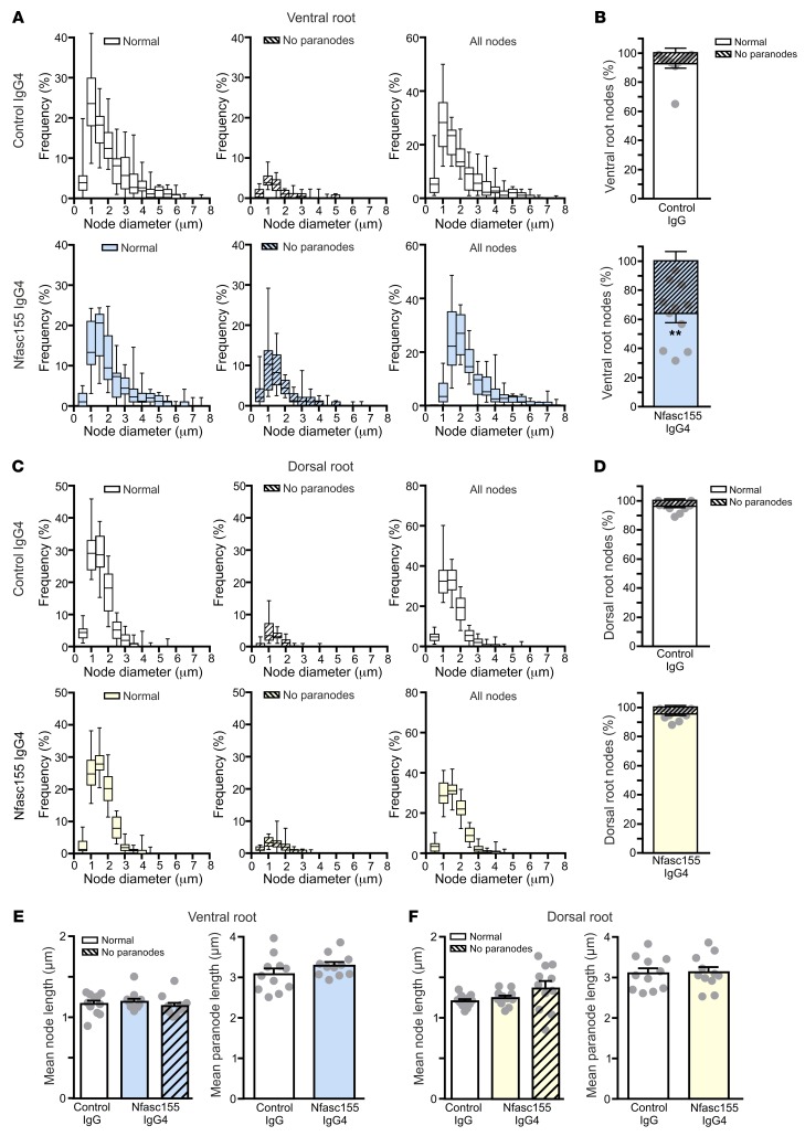
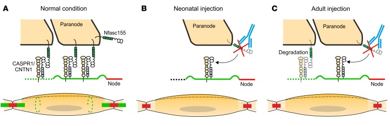
Neurofascin-155 (Nfasc155) is an essential glial cell adhesion molecule expressed in paranodal septate-like junctions of peripheral and central myelinated axons. The genetic deletion of Nfasc155 results in the loss of septate-like junctions and in conduction slowing. In humans, IgG4 antibodies against Nfasc155 are implicated in the pathogenesis of chronic inflammatory demyelinating polyneuropathy (CIDP). These antibodies are associated with an aggressive onset, a refractoriness to intravenous immunoglobulin, and tremor of possible cerebellar origin. Here, we examined the pathogenic effects of patient-derived anti-Nfasc155 IgG4. These antibodies did not inhibit the ability of Nfasc155 to complex with its axonal partners contactin-1/CASPR1 or induce target internalization. Passive transfer experiments revealed that IgG4 antibodies target Nfasc155 on Schwann cell surface, and diminished Nfasc155 protein levels and prevented paranodal complex formation in neonatal animals. In adult animals, chronic intrathecal infusions of antibodies also induced the loss of Nfasc155 and of paranodal specialization and resulted in conduction alterations in motor nerves. These results indicate that anti-Nfasc155 IgG4 perturb conduction in absence of demyelination, validating the existence of paranodopathy. These results also shed light on the mechanisms regulating protein insertion at paranodes.

Keywords: Autoimmune diseases; Autoimmunity; Neurological disorders; Neuromuscular disease; Neuroscience.


Conflict of interest statement

Conflict of interest: JJD received a research grant from CSL Behring. II holds a patent for dysferlin detection in monocytes (US20030165937A1), and has consulted for Grifols, Genzyme, Alexion, and UCB.

Figures

Figure 1
Figure 1. Antibodies to Nfasc155 do not alter the interaction between Nfasc155 and its axonal partners CNTN1 and CASPR1.
(AC) For aggregation assays, HEK293 cells were cotransfected with mCherry-tagged Nfasc155 or with GFP-tagged CASPR1 and CNTN1. Cells were then incubated under gentle agitation for 2 hours in the presence of 10 μg of control IgG4, anti-CNTN1 IgG4, or anti-Nfasc155 IgG4 from 3 patients (CIDP1–3). As negative controls, Nfasc155-expressing HEK293 cells were incubated with cells expressing GFP alone (top left panel). Cells were examined with a fluorescence microscope at ×10 objective. Representative fields are shown in A (n = 3–4 experiments for each condition). Dashed circles highlight cell aggregates with contacts between red and green cells. The percentage of cell clusters with contacts between green and red cells was quantified (B), as well as the relative frequency of green cells per aggregate (C) (n = 3–4 experiments for each condition). CASPR1/CNTN1– and Nfasc155-expressing cells form clusters. Anti-CNTN1 IgG4 significantly prevented the formation of cell aggregates (**P < 0.005 by unpaired 2-tailed Student’s t tests for 2 samples of equal variance and by 1-way ANOVA followed by Bonferroni’s post hoc tests). By contrast, anti-Nfasc155 IgG4 did not affect the interaction between Nfasc155 and CASPR1/CNTN1. Bars represent mean and SEM. Scale bar: 50 μm.
Figure 2
Figure 2. Anti-Nfasc155 autoantibodies target surface Schwann cell antigens.
(A) Sciatic nerve fibers were incubated in vitro with purified anti-Nfasc155 IgG4 from patient CIDP1 for 3 hours, and immunolabeled for IgG4 (green) and CNTN1 (red). (BE) Sciatic nerves were fixed 1 day (B and D) or 3 days (C and E) after intraneural injections of anti-Nfasc155 IgG4, and immunolabeled for IgG4 (green) and β-catenin (red; B and C) or CNTN1 (red; D and E) and Nav channels (blue; D and E). Note that anti-Nfasc155 IgG4 bound to the surface of the Schwann cells and deposited at the vicinity of the node of Ranvier (double arrowheads) and at adherens junctions along the internode stained here with β-catenin (arrows). However, no penetration across the paranodal region was observed (images are representative of n = 3 independent experiments). Scale bars: 10 μm.
Figure 3
Figure 3. Passive transfer of anti-Nfasc155 IgG4 affects the formation of paranodal axoglial unit during development.
(AD) Newborn rat pups received an i.p. injection of 250 μg of control IgG4 (A) or anti-Nfasc155 IgG4 from patient CIDP1 (B) on the day of birth (n = 4 animals for each condition and age). Sciatic nerve fibers were fixed and immunolabeled for voltage-gated sodium channels (Nav; green) and CASPR1 (red) at postnatal days 0 (P0), 2, 4, and 6. The percentages of Nav clusters with 1 or 2 flanking CASPR1-positive paranodes (double arrowheads) or without CASPR1-positive paranodes (arrowheads) were quantified at each age (C), as well as the paranodal length (D) (n = 200–300 nodes or paranodes for each condition and age). Injection of anti-Nfasc155 IgG4 importantly delayed the formation of CASPR1-positive paranodes, and a significantly higher percentage of heminodes without flanking paranodes was observed at P2 (**P < 0.005 by 1-way ANOVA followed by Bonferroni’s post hoc tests). Paranodal length was also significantly shorter 2 and 4 days after injection of anti-Nfasc155 IgG4 (*P < 0.05 by 1-way ANOVA followed by Bonferroni’s post hoc tests). Scale bar: 10 μm. (E and F) Sciatic nerve and spinal cord proteins (100 μg) from P2 animals injected with control IgG4 (n = 4) or anti-Nfasc155 IgG4 (n = 4) at P0 were immunoblotted with antibodies recognizing all neurofascin isoforms (Pan-neurofascin) or specifically Nfasc155. The level of Nfasc155 was quantified relatively to that of Nfasc186 in both sciatic nerve and spinal cord (F). Nfasc155 level was significantly decreased in sciatic nerves of animals treated with anti-Nfasc155 IgG4, but not in spinal cord (**P < 0.005 by unpaired 2-tailed Student’s t tests for 2 samples of equal variance). Molecular weight markers are shown on the right (in kilodaltons). Bars represent mean and SEM.
Figure 4
Figure 4. Anti-Nfasc155 IgG4 deposits at proximity of nodes in neonatal animals.
(AC) Newborn rat pups received daily i.p. injection of 250 μg of control IgG4 (A) or anti-Nfasc155 IgG4 from patient CIDP1 (B) from the day of birth to P6 (images are representative of n = 3 independent experiments for each age). Sciatic nerve fibers were immunolabeled for voltage-gated sodium channels (Nav; blue), CASPR1 (green), and IgG (red) at P2 (A), P4 (B), and P6 (C). At P2 and P4, IgG deposits were mostly detected at the borders of Nav clusters, even in Nav clusters lacking CASPR1-positive paranodes (arrowheads). No colocalization with CASPR1 was found even in Nav clusters flanked by 1 or 2 paranodes (double arrowheads). At P6, IgG deposits appeared fainter and were mostly detected at the node vicinity. A weak IgG labeling can be observed at paranodes in some fibers. Isolated node labeling is shown at a higher magnification in the insets. Scale bars: 10 μm.
Figure 5
Figure 5. The chronic intrathecal infusion of autoantibodies induces gait abnormalities and motor nerve conduction slowing.
(AC) Adult Lewis rats received daily intrathecal infusions (100 μg/d) of control IgG4 (black circles; n = 15 animals) or anti-Nfasc155 IgG4 from patient CIDP1 (gray circles; n = 15 animals) during 3 weeks. The clinical score was monitored daily and averaged. The passive infusion of anti-Nfasc155 IgG4 induced progressive clinical symptoms. (B and C) Footprint analysis revealed abnormal spreading of hind limbs in animals treated with anti-Nfasc155 IgG4 compared with control animals. The footprint angle (gray lines) was significantly increased in animals treated with anti-Nfasc155 IgG4 compared with controls. (DG) L6 ventral and dorsal roots from animals injected with control IgG4 or anti-Nfasc155 IgG4 were recorded on day 21 after the beginning of the injections (n = 12–14 nerves from 12–14 animals). Representative CAPs from control IgG4–treated (black traces) and anti-Nfasc155 IgG4–treated (gray traces) animals are shown in D and F. The peak amplitude, CAP duration, and conduction velocities at peak amplitude are represented in E and G for ventral and dorsal roots, respectively. The ventral spinal nerves of animals treated with anti-Nfasc155 IgG4 showed a significant decrease in CAP amplitude and conduction velocity compared with controls. This was associated with a significant increase in CAP duration. By contrast, nerve activity was not significantly affected in dorsal root. *P < 0.05, **P < 0.005 by unpaired 2-tailed Student’s t tests for 2 samples of equal variance. Bars represent mean and SD.
Figure 6
Figure 6. Anti-Nfasc155 IgG4 disrupts paranodal specialization in ventral spinal nerve.
(AF) Teased fibers from L6 ventral roots of animals treated with control IgG4 (n = 15; A, C, and E) or anti-Nfasc155 IgG4 from patient CIDP1 (n = 15; B, D, and F) were stained for Nav channels (green), CNTN1 (red), and CASPR1 (blue, A and B), ankyrin-G (Ank-G; blue, C and D), or Nfasc186 (blue, E and F). Most nodes of Ranvier were positive for Nav channel, ankyrin-G, and Nfasc186 clusters and were bordered by paranodes stained for CASPR1 and CNTN1 in control animals. By contrast, many nodes lacked CASPR1 or CNTN1 staining at paranodes (arrowheads) in animals chronically injected with anti-Nfasc155 IgG4. Insets in B and D show representative affected nodes. Scale bars: 10 μm.
Figure 7
Figure 7. Autoantibodies preferentially affect motor axons.
(AD) The distribution of the axonal population according to the nodal diameters of axons was examined in L6 ventral roots (A and B) and dorsal roots (C and D) of animals treated with control IgG4 (n = 1297 nodes for ventral roots and 1061 nodes for dorsal roots from 11 animals) or anti-Nfasc155 IgG4 from patient CIDP1 (n = 1376 nodes for ventral roots and 875 nodes for dorsal roots from 11 animals). The respective proportion of normal nodes (open boxes) or nodes lacking paranodes (hatched boxes) is represented on the left, and the distribution of the total axonal population is represented on the right. The percentage of nodes showing paranodal alterations was calculated in ventral (B) and dorsal roots (D). Scatter plots represent the percentage of normal nodes in each animal. Paranodes were more significantly affected in ventral root axons than in dorsal roots (**P < 0.005 by unpaired 2-tailed Student’s t tests for 2 samples of equal variance). (E and F) The mean length of nodes lacking paranodes (hatched boxes) or appearing normal (open boxes) was measured in 11 animals for each group, as well as the mean paranodal length. No significant alterations in node or paranode length were observed between control and anti-Nfasc155 IgG4–treated animals (unpaired 2-tailed Student’s t tests and 1-way ANOVA followed by Bonferroni’s post hoc tests). In B and DF, bars represents mean and SEM. In A and C, box bounds represent the first and third quartiles, lines within the box represent the median, and whiskers show the minimal and maximal values.
Figure 8
Figure 8. Autoantibodies induce the selective depletion of Nfasc155 in ventral spinal nerve.
(A) Ventral and dorsal spinal nerve proteins (250 μg) from adult animals injected with control IgG4 (n = 4–6) or anti-Nfasc155 IgG4 from patient CIDP1 (n = 4–6) for 3 weeks were separated on SDS-PAGE gels and immunoblotted with antibodies against Nfasc155, CASPR1, neurofilament-200, and E-cadherin. Arrowheads indicate Nfasc155-H and Nfasc155-L isoforms. (B) The levels of Nfasc155, CASPR1, neurofilament-200, and E-cadherin were evaluated in animals treated with anti-Nfasc155 IgG4 relatively to those in control animals in both ventral and dorsal spinal roots. Nfasc155 levels were significantly decreased in ventral spinal nerves, but not in dorsal spinal nerves (*P < 0.05 by unpaired 2-tailed Student’s t tests for 2 samples of equal variance). Molecular weight markers are shown on the left (in kilodaltons). Bars represent mean and SEM.
Figure 9
Figure 9. Schematic representation of the pathogenic mechanisms of anti–paranodal protein autoantibodies.
(A) Representation of a mature node of Ranvier and myelinated axon. Myelin (yellow) covers the axon except at the node of Ranvier (red). At paranodal junctions (green), Nfasc155 interacts with its axonal partners CASPR1/CNTN1. Nfasc155 is also found at Schmidt-Lanterman incisures (dashed green lines). The Schwann cell nucleus is shown in gray. (B) In neonatal animals, the progressive enwrapping of axons by Schwann cells induces the formation of paranodal regions at node borders. The injection of anti-Nfasc155 IgG4 (blue) during the neonatal period does not affect myelination or node/paranode formation, but induces the depletion of Nfasc155, and thereby alters the formation of paranodal septate-like junctions. (C) At adult age, evidence suggests that the Nfasc155/CASPR1/CNTN1 complex is constantly renewed at paranodes possibly through degradation and replenishment mechanisms. The chronic infusion of anti-Nfasc155 IgG4 (blue) may preclude the regeneration of the paranodal axoglial junction by inducing Nfasc155 depletion, and thereby alter paranode structure and conduction.

Similar articles

Cited by

References

    1. Dalakas MC, Medscape Advances in the diagnosis, pathogenesis and treatment of CIDP. Nat Rev Neurol. 2011;7(9):507–517. - PubMed
    1. Querol L, et al. Antibodies against peripheral nerve antigens in chronic inflammatory demyelinating polyradiculoneuropathy. Sci Rep. 2017;7(1):14411. doi: 10.1038/s41598-017-14853-4. - DOI - PMC - PubMed
    1. Delmont E, et al. Autoantibodies to nodal isoforms of neurofascin in chronic inflammatory demyelinating polyneuropathy. Brain. 2017;140(7):1851–1858. doi: 10.1093/brain/awx124. - DOI - PubMed
    1. Devaux JJ, et al. Neurofascin-155 IgG4 in chronic inflammatory demyelinating polyneuropathy. Neurology. 2016;86(9):800–807. doi: 10.1212/WNL.0000000000002418. - DOI - PMC - PubMed
    1. Doppler K, et al. Auto-antibodies to contactin-associated protein 1 (Caspr) in two patients with painful inflammatory neuropathy. Brain. 2016;139(pt 10):2617–2630. - PubMed

Publication types

MeSH terms