. 2019 Mar 14;14(3):e0212642.
doi: 10.1371/journal.pone.0212642. eCollection 2019.

Peripheral blood-derived mesenchymal stem cells demonstrate immunomodulatory potential for therapeutic use in horses

Affiliations

Peripheral blood-derived mesenchymal stem cells demonstrate immunomodulatory potential for therapeutic use in horses

Ana Leda F Longhini et al. PLoS One. .

Abstract

Previously, we showed that mesenchymal stem cells (MSC) can be mobilized into peripheral blood using electroacupuncture (EA) at acupoints, LI-4, LI-11, GV-14, and GV-20. The purpose of this study was to determine whether EA-mobilized MSC could be harvested and expanded in vitro to be used as an autologous cell therapy in horses. Peripheral blood mononuclear cells (PBMC) isolated from young and aged lame horses (n = 29) showed a marked enrichment for MSCs. MSC were expanded in vitro (n = 25) and administered intravenously at a dose of 50 x 106 (n = 24). Treatment resulted in significant improvement in lameness as assessed by the American Association of Equine Practitioners (AAEP) lameness scale (n = 23). MSCs exhibited immunomodulatory function by inhibition of lymphocyte proliferation and induction of IL-10. Intradermal testing showed no immediate or delayed immune reactions to MSC (1 x 10⁶ to 1 x 10⁴). In this study, we demonstrated an efficient, safe and reproducible method to mobilize and expand, in vitro, MSCs in sufficiently high concentrations for therapeutic administration. We confirm the immunomodulatory function of these cells in vitro. This non-pharmacological and non-surgical strategy for stem cell harvest has a broad range of biomedical applications and represents an improved clinically translatable and economical cell source for humans.


Conflict of interest statement

The authors have declared that no competing interests exist.

Figures

Fig 1
Fig 1. MSCs derived from horse peripheral blood expressed typical MSC markers and reduced lameness in horses with osteoarthritis.
(A) Representative phenotype of MSC, gray line represents unstained cells; the red filled line represents cells stained with individual specific antibodies (CD105, CD146, CD90, CD73). (B) The AAEP lameness scale was used for grading lameness before and after six weeks. Autologous MSCs were injected intravenously in 24 horses and lameness was graded before and at 6 weeks following MSC infusion (p<0.0001, n = 24). No grading difference was observed between the initial evaluation and the 6-week assessment in horses not injected with MSCs (n = 5). Abbreviation: AAEP, American Association of Equine Practitioners. MSC, Mesenchymal stem cell.
Fig 2
Fig 2. EA-MSC inhibiting lymphocyte proliferation.
Cell tracer stained PBMC were stimulated in vitro with PHA (2μg/mL) in the presence or absence of autologous MSC in a ratio of 1 MSC: 10 PBMC for 72 hours (A) and 96 hours (B). Results are shown as Mean + SEM (n = 4). * indicates significant difference (p < 0.05). Abbreviations: PBMC, peripheral blood mononuclear cell; MSC, mesenchymal stem cell.
Fig 3
Fig 3. EA-MSC stimulate PBMCs to produce IL-10.
PBMC were stimulated with PHA in the presence or absence of autologous MSC (1 MSC: 10 PBMC) for 72 hours (A) and 96 hours (B) and the IL-10 concentration was measured in the supernatant. Results are shown as Mean + SEM (n = 4). *indicates significant difference (p < 0.05). Abbreviations: PBMC, peripheral blood mononuclear cell; MSC, mesenchymal stem cell.
Fig 4
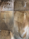
Fig 4. Intradermal testing.
A) 20 minutes post-injection. Negative control (i), PHA (ii), histamine(iii), FBS (iv), and a linear decrease in reaction of the MSC (v to ix). B) Four hours post-injection shows the reaction elicited by the FBS. All the other injection sites were negative. C) Twenty-four hours post-injection shows an increase in the size of the FBS reaction with subcutaneous edema involving the lateral neck (black arrow). D) Forty-eight hours post-injection shows the increase in the size of the FBS reaction with subcutaneous edema starting from the injection site extending to the ventral lateral neck (dripping effect) (black arrow), and persistent localized swelling consistent with delayed hypersensitivity reaction (white arrow). E) Seventy-two hours post-injection showing the dramatic reduction in the subcutaneous edema and the persistent reaction due to the FBS (white arrow).
Fig 5
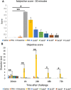
Fig 5. FBS induces an immediate and a delayed-type hypersensitivity reaction in previously exposed horses on intradermal testing.
Eight horses were challenged with 5 different concentrations of autologous MSC, histamine, PHA, saline, and FBS. The acute reaction was measured at five different times after the initial challenge and is shown in cm. FBS caused a significant reaction at 4, 24, 48 and 72 hours. (A) Subjective score and (B) Objective score. Results are shown as means ± SEM (n = 8). * indicates significant difference (p < 0.05); ** indicates significant difference (p < 0.005). Abbreviations: MSC, mesenchymal stem cell; PHA: phytohemmagglutinin; FBS: fetal bovine serum.
Fig 6
Fig 6. Basal medium with autologous plasma (AP) or autologous platelet lysate (APL) results in loss of cell surface marker phenotype and impaired growth respectively.
Flow cytometry analysis shows lack of CD90 expression by MSC cultured for 4 days in basal medium + AP (B) compared to MSCs cultured in basal medium + FBS (A) and compared to MSCs cultured in basal medium + APL (C). (D): MSC cultured for 4 days in basal medium + FBS, (E): MSC cultured for 4 days in basal medium + APL. (F): concentration of harvested cells was determined 4 days after the cells were plated and shows a decrease in the growth rate when the cells were cultured in basal medium + APL compared to cells cultured in basal medium + FBS (n = 2). Abbreviations: MSC, mesenchymal stem cell; FBS: fetal bovine serum.
Fig 7
Fig 7. Horse recombinant media (HRM) supplemented with autologous serum (AS) exhibits similar growth promoting characteristics as basal medium supplemented with FBS.
MSC cultured in basal medium + FBS (A) and HRM + AS (B), after 2 days of culture exhibit no difference in confluence or in the expression of mesenchymal markers.

References

    1. Katsuda T, Kosaka N, Takeshita F, Ochiya T. The therapeutic potential of mesenchymal stem cell-derived extracellular vesicles. Proteomics. 2013;13(10–11):1637–53. 10.1002/pmic.201200373 . - DOI - PubMed
    1. Le Blanc K, Ringden O. Immunomodulation by mesenchymal stem cells and clinical experience. J Intern Med. 2007;262(5):509–25. 10.1111/j.1365-2796.2007.01844.x . - DOI - PubMed
    1. Sangiorgi B, Panepucci RA. Modulation of Immunoregulatory Properties of Mesenchymal Stromal Cells by Toll-Like Receptors: Potential Applications on GVHD. Stem Cells Int. 2016;2016:9434250 10.1155/2016/9434250 - DOI - PMC - PubMed
    1. Crovace A, Lacitignola L, Rossi G, Francioso E. Histological and immunohistochemical evaluation of autologous cultured bone marrow mesenchymal stem cells and bone marrow mononucleated cells in collagenase-induced tendinitis of equine superficial digital flexor tendon. Vet Med Int. 2010;2010:250978 10.4061/2010/250978 - DOI - PMC - PubMed
    1. Godwin EE, Young NJ, Dudhia J, Beamish IC, Smith RK. Implantation of bone marrow-derived mesenchymal stem cells demonstrates improved outcome in horses with overstrain injury of the superficial digital flexor tendon. Equine Vet J. 2012;44(1):25–32. 10.1111/j.2042-3306.2011.00363.x . - DOI - PubMed

Publication types