. 2019 Mar 14;15(3):e1007618.
doi: 10.1371/journal.ppat.1007618. eCollection 2019 Mar.

The small non-coding RNA RsaE influences extracellular matrix composition in Staphylococcus epidermidis biofilm communities

Affiliations

The small non-coding RNA RsaE influences extracellular matrix composition in Staphylococcus epidermidis biofilm communities

Sonja M K Schoenfelder et al. PLoS Pathog. .

Abstract

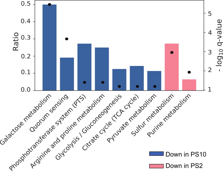
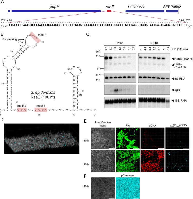
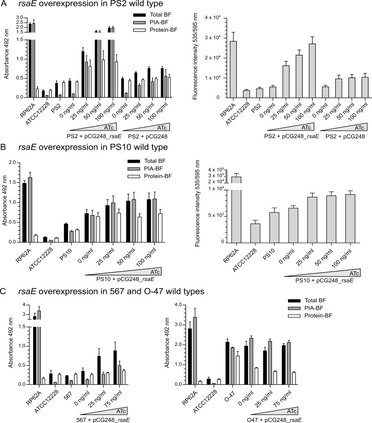
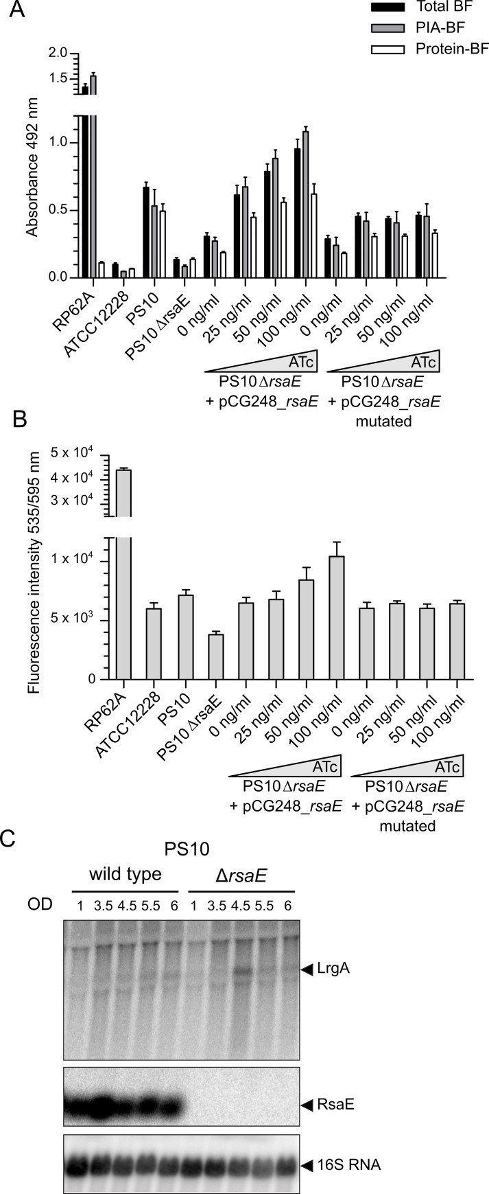
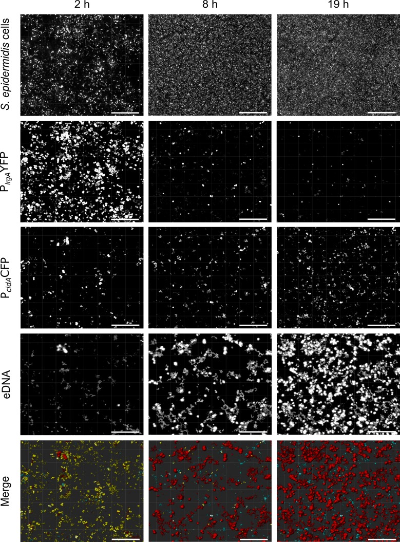
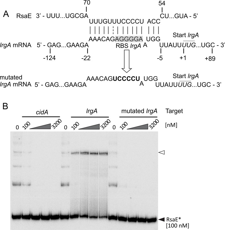
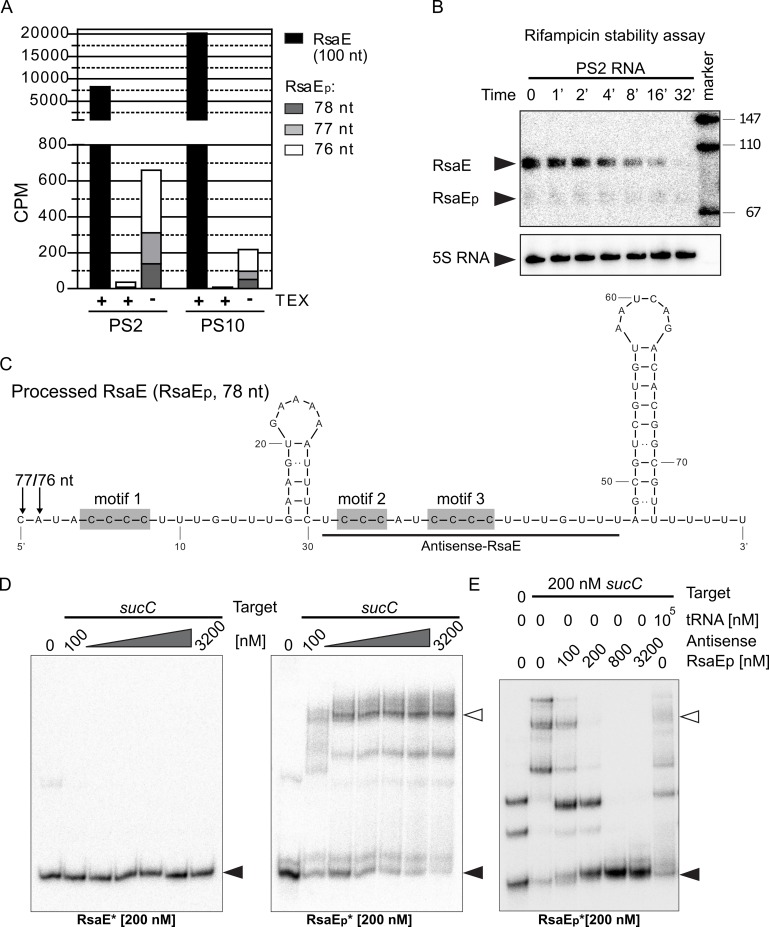
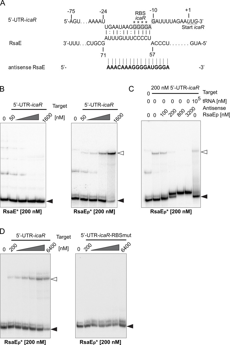
RsaE is a conserved small regulatory RNA (sRNA) which was previously reported to represent a riboregulator of central carbon flow and other metabolic pathways in Staphylococcus aureus and Bacillus subtilis. Here we show that RsaE contributes to extracellular (e)DNA release and biofilm-matrix switching towards polysaccharide intercellular adhesin (PIA) production in a hypervariable Staphylococcus epidermidis isolate. Transcriptome analysis through differential RNA sequencing (dRNA-seq) in combination with confocal laser scanning microscopy (CLSM) and reporter gene fusions demonstrate that S. epidermidis protein- and PIA-biofilm matrix producers differ with respect to RsaE and metabolic gene expression. RsaE is spatiotemporally expressed within S. epidermidis PIA-mediated biofilms, and its overexpression triggers a PIA biofilm phenotype as well as eDNA release in an S. epidermidis protein biofilm matrix-producing strain background. dRNA-seq and Northern blot analyses revealed RsaE to exist as a major full-length 100-nt transcript and a minor processed species lacking approximately 20 nucleotides at the 5'-end. RsaE processing results in expansion of the mRNA target spectrum. Thus, full-length RsaE interacts with S. epidermidis antiholin-encoding lrgA mRNA, facilitating bacterial lysis and eDNA release. Processed RsaE, however, interacts with the 5'-UTR of icaR and sucCD mRNAs, encoding the icaADBC biofilm operon repressor IcaR and succinyl-CoA synthetase of the tricarboxylic acid (TCA) cycle, respectively. RsaE augments PIA-mediated biofilm matrix production, most likely through activation of icaADBC operon expression via repression of icaR as well as by TCA cycle inhibition and re-programming of staphylococcal central carbon metabolism towards PIA precursor synthesis. Additionally, RsaE supports biofilm formation by mediating the release of eDNA as stabilizing biofilm matrix component. As RsaE itself is heterogeneously expressed within biofilms, we consider this sRNA to function as a factor favoring phenotypic heterogeneity and supporting division of labor in S. epidermidis biofilm communities.


Conflict of interest statement

The authors have declared that no competing interests exist.

Figures

Fig 1
Fig 1. Enrichment of gene ontology (GO) terms among differentially expressed genes between S. epidermidis PS2 and PS10.
The analysis considers differentially expressed genes in the TEX-untreated library of the dRNA-seq experiment with an absolute log2(fold-change) ≥ 1 (p-value < 0.05). Bars display the ratio of the number of differentially expressed genes in relation to the total number of genes associated to the respective GO term, while dots represent -log10 q-values.
Fig 2
Fig 2. Genetic localization, structure and expression of RsaE in S. epidermidis.
(A) Genetic localization (position 576.475 to 576.574) and nucleotide sequence of the rsaE gene in the S. epidermidis RP62A reference genome (NC_002976). (B) Secondary structure of RsaE. Arrows mark putative processing sites of RsaE (see text for details) and red boxes highlight unpaired C-rich motifs prone to mediate mRNA binding [19]. Circled nucleotides next to the sequence indicate positions of two varying nucleotides present in S. aureus RsaE. (C) Northern blot detection of rsaE and lrgA expression during growth of S. epidermidis PS2 and PS10 in batch cultures at 37°C in TSB. Optical densities (OD600) at which samples were taken are indicated, with the last samples taken after 24 hours. Arrowheads mark full-length and processed (RsaEp) RsaE species, lrgA transcript as well as 5S and 16S rRNA used as loading controls. (D) 3D-CLSM image of a S. epidermidis PS10 biofilm population (grey cells) grown in chamber slides for 20 hours. The strain carries plasmid p_(PrsaECFP) in which the rsaE promoter is fused to the blue-fluorescent Cerulean protein gene cfp as reporter. Light blue cells represent bacteria with an active rsaE promoter. (E) CLSM images of an S. epidermidis PS10 p_(PrsaECFP) biofilm during growth (10 and 20 hours) in chamber slides. The total bacterial cell mass is visualized by transmission microscopy (left panels), PIA matrix is stained in green by Alexa Fluor 488 wheat-germ agglutinin (middle left), and eDNA (in red) by Ethidium Homodimer III (middle right). Cerulean expression (light blue) highlights rsaE-expressing bacterial cells (right panels). (F) CLSM images of S. epidermidis PS10 carrying control plasmid pCerulean in which cfp is under control of an ATc-inducible promoter. Images were taken after 20 hours of growth in chamber slides in TSB supplemented with 100 ng/ ml ATc. The total bacterial cell mass is visualized by transmission microscopy (left panels), Cerulean expression (light blue) highlights CFP expressing bacterial cells (right panels). Bars represent 20 μm.
Fig 3
Fig 3. RsaE effects on biofilm production and eDNA release.
(A) Left panel. Analysis of biofilm production of S. epidermidis PS2 (pCG248_rsaE) by static 96-well microtiter plate biofilm assays. Plasmid pCG248_rsaE harbours the rsaE gene under the control of an anhydrotetracycline (ATc)-inducible promoter. As control, a strain carrying an empty plasmid (pCG248) was included in the analyses. Expression of rsaE was induced by increasing ATc concentrations (25 to 100 ng/ml). Total biofilm (BF) mass as well as PIA- and protein-mediated biofilm proportions were determined by sodium-periodate and proteinase K treatments, respectively, as described in Methods. Sterile TSB medium served as background control. The strong PIA-producer S. epidermidis RP62A (ica locus positive) as well as S. epidermidis ATCC12228 (ica locus negative) were used as positive and negative controls, respectively. Biofilm production was also determined in the S. epidermidis PS2 wildtype. (A) Right panel. Detection of eDNA content in S. epidermidis biofilms by Ethidium Homodimer III staining and fluorescence intensity measurements at 535/595 nm. Biofilms were grown in 96-well microtiter plates using the same strains and conditions as in the left panel. (B) Analysis of biofilm production and eDNA release of S. epidermidis PS10 (pCG248_rsaE) by static 96-well microtiter plate biofilm assays. Biofilms were grown in 96-well microtiter plates using the same conditions as in panel A. (C) Analysis of biofilm production of S. epidermidis 567 and O47 carrying plasmid pCG248_rsaE by static 96-well microtiter plate biofilm assays. Biofilms were grown in 96-well microtiter plates using the same conditions as in panel A.
Fig 4
Fig 4. Effect of RsaE deletion on biofilm production and eDNA release.
(A) Analysis of biofilm production of S. epidermidis PS10 wildtype, rsaE deletion mutant and complemented strains carrying either a wildtype copy of rsaE or a mutated version of RsaE (pCG248_rsaE_mutated) under ATc-inducible tet promoter control, respectively. Expression of rsaE was induced by increasing ATc concentrations (25 to 100 ng/ml). Total biofilm (BF) mass as well as PIA- and protein-mediated biofilm proportions were determined by sodium-periodate and proteinase K treatments, respectively, as described in Methods. Sterile TSB medium served as background control. The strong PIA-producer S. epidermidis RP62A (ica locus positive) as well as S. epidermidis ATCC12228 (ica locus negative) were used as positive and negative controls, respectively. (B) Detection of eDNA content in S. epidermidis biofilms by Ethidium Homodimer III staining and fluorescence intensity measurements at 535/595 nm. Biofilms were grown in 96-well microtiter plates using the same strains and conditions as in the panel A. (C) Northern blot detection of rsaE and lrgA expression during growth of S. epidermidis PS10 wildtype and rsaE deletion mutant in batch culture at 37°C in TSB. Optical densities (OD600) at which samples were taken are indicated.
Fig 5
Fig 5. CLSM live cell imaging of lrgA/cidA expression and eDNA release in a S. epidermidis biofilm during growth in chamber slides.
The strain harbours plasmid p_(PcidACFP/PlrgAYFP) carrying the lrgA promoter fused to yfp (encoding yellow-fluorescent protein) and the cidA promoter fused to cfp (encoding blue-fluorescent Cerulean protein). eDNA was stained in red by Ethidium Homodimer III. Bars represent 20 μm.
Fig 6
Fig 6. Interaction of RsaE with lrgA mRNA.
(A) IntaRNA-based interaction prediction [29, 30] of lrgA mRNA with RsaE (top) as well as nature and positions of the nucleotide exchanges in the mutated lrgA mRNA (bottom). Numbering refers to the upstream (-124) and downstream (+89) region around the lrgA mRNA start codon (+1). Ribosomal binding site (RBS) of lrgA is highlighted in grey. (B) RNA/RNA electromobility gel shift assays (EMSAs) employing 100 nM of radioactively labeled (*) RsaE and increasing amounts of cidA (left), lrgA (middle) and mutated lrgA (right) target RNAs as binding partners. Open and filled triangles mark RsaE/target RNA complexes and labeled unbound RsaE, respectively.
Fig 7
Fig 7. RsaE processing, stability and interaction with sucC mRNA.
(A) Quantification of full-length and processed RsaE (RsaEp) transcripts in PS2 and PS10 by dRNA-seq analysis, with (+) and without (-) TEX treatment. CPM: counts per million reads, calculated as NOAR (number of aligned reads)*106/TNOAR (total number of aligned reads). Black columns indicate the number of transcription starts of full-length RsaE (100 nt); the number of processed RsaE species (RsaEp, 76–78 nt) are shown as white, light grey and dark grey columns, respectively. (B) Stability determination of full-length (RsaE) and processed (RsaEp) RsaE species. S. epidermidis PS2 was grown in TSB to early-exponential growth stage and RNA was isolated before (0) and at the time points indicated after transcription blocking by rifampicin. RNA samples were subjected to Northern blot analyses using radioactively labeled ssDNA-oligonucleotide probes targeting either RsaE or 5S rRNA as loading control. (C) Proposed secondary structure of the processed RsaE species (RsaEp, 78 nt) according to mFold-4.7-based prediction [65]. Grey boxes highlight positions of the C-rich motifs within the processed RsaE molecule. Nucleotides complementary to the antisense-RsaE RNA probe used in (D) are underlined. Arrows indicate alternative RsaE processing sites identified by dRNA-seq. (D) EMSAs using increasing amounts of sucC target RNA and radioactively labeled (*) full-length (RsaE, left panel) or processed RsaE (RsaEp, right panel) as binding partners. (E) sucC/RsaEp EMSA upon competition with an antisense-RsaE RNA oligonucleotide or unspecific tRNAs during complex formation. 200 nM of sucC target RNA was mixed with increasing amounts of antisense-RsaE RNA oligonucleotide (lanes 3–6) or a 500-fold excess of yeast tRNAs (lane 7) prior to addition of 200 nM radioactively (*) labeled RsaEp to the samples. Filled triangles in (D) and (E) indicate labeled unbound RsaE and open triangles mark RsaE/target complexes.
Fig 8
Fig 8. Interaction of processed RsaE (RsaEp) with 5'-UTR of icaR.
(A) IntaRNA-based prediction of interaction between RsaE and the 5'-UTR-icaR [29, 30]. The icaR ribosomal binding site (RBS) is highlighted in grey and the icaR start codon is underlined. Asterisks mark G to U mutations used to create 5'-UTR-icaR-RBS-mut in (D). The nucleotide sequence of the antisense RsaE RNA oligonucleotide is shown at the bottom. (B) EMSAs using increasing amounts of 5'-UTR-icaR target RNA and radioactively labeled (*) full-length (RsaE, left panel) or processed RsaE (RsaEp, right panel) as binding partners. (C) EMSA with 5'-UTR-icaR and RsaEp upon competition with an antisense-RsaE RNA oligonucleotide or unspecific tRNAs during complex formation. 200 nM of the 5'-UTR-icaR target RNA was mixed with increasing amounts of antisense-RsaE RNA oligonucleotide (lanes 3–6) or a 500-fold excess of yeast tRNAs (lane 7) prior to addition of 200 nM radioactively (*) labeled RsaEp to the samples. (D) EMSA employing 200 nM radioactively (*) labeled RsaEp with increasing amounts of either wildtype 5'-UTR-icaR (left) or 5'-UTR-icaR-RBS-mut (right) carrying mutations in the ribosomal binding site of icaR mRNA (GGGG to UUUU, marked by asterisks in (A)). Filled triangles in B-D indicate labeled unbound RsaE and open triangles mark RsaE/target complexes.

Similar articles

Cited by

References

    1. de la Fuente-Nunez C, Reffuveille F, Fernandez L, Hancock RE. Bacterial biofilm development as a multicellular adaptation: antibiotic resistance and new therapeutic strategies. Curr Opin Microbiol. 2013;16(5):580–9. 10.1016/j.mib.2013.06.013 . - DOI - PubMed
    1. Claessen D, Rozen DE, Kuipers OP, Sogaard-Andersen L, van Wezel GP. Bacterial solutions to multicellularity: a tale of biofilms, filaments and fruiting bodies. Nature Rev. 2014;12(2):115–24. 10.1038/nrmicro3178 . - DOI - PubMed
    1. Smits WK, Kuipers OP, Veening JW. Phenotypic variation in bacteria: the role of feedback regulation. Nature Rev. 2006;4(4):259–71. Epub 2006/03/17. nrmicro1381 [pii] 10.1038/nrmicro1381 . - DOI - PubMed
    1. Otto M. Staphylococcus epidermidis—the 'accidental' pathogen. Nature Rev. 2009;7(8):555–67. 10.1038/nrmicro2182 - DOI - PMC - PubMed
    1. Buttner H, Mack D, Rohde H. Structural basis of Staphylococcus epidermidis biofilm formation: mechanisms and molecular interactions. Front Cell Infect Microbiol. 2015;5:14 10.3389/fcimb.2015.00014 - DOI - PMC - PubMed

Publication types

MeSH terms

Substances