. 2019 Mar 13;25(3):404-417.e6.
doi: 10.1016/j.chom.2019.02.004.

Commensal Candida albicans Positively Calibrates Systemic Th17 Immunological Responses

Affiliations

Commensal Candida albicans Positively Calibrates Systemic Th17 Immunological Responses

Tzu-Yu Shao et al. Cell Host Microbe. .

Abstract

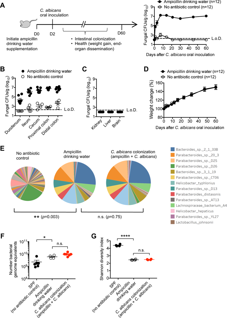
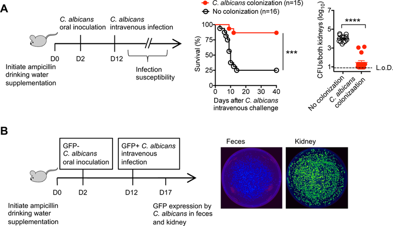
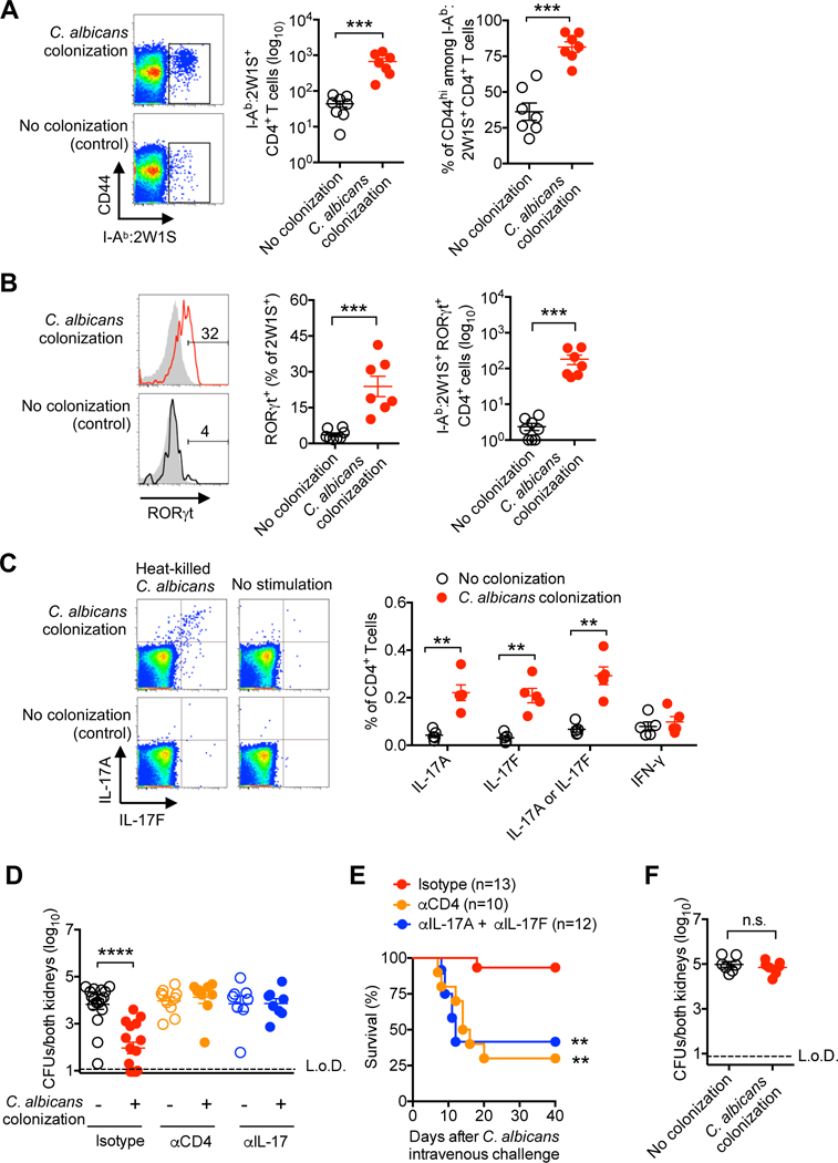
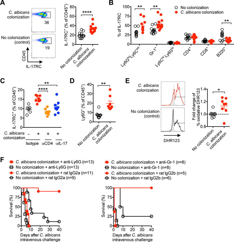
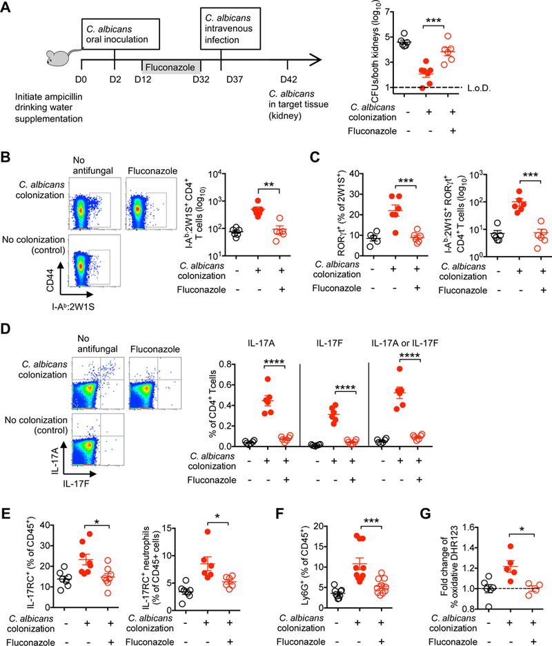
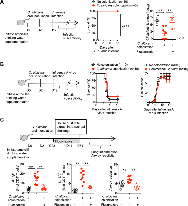
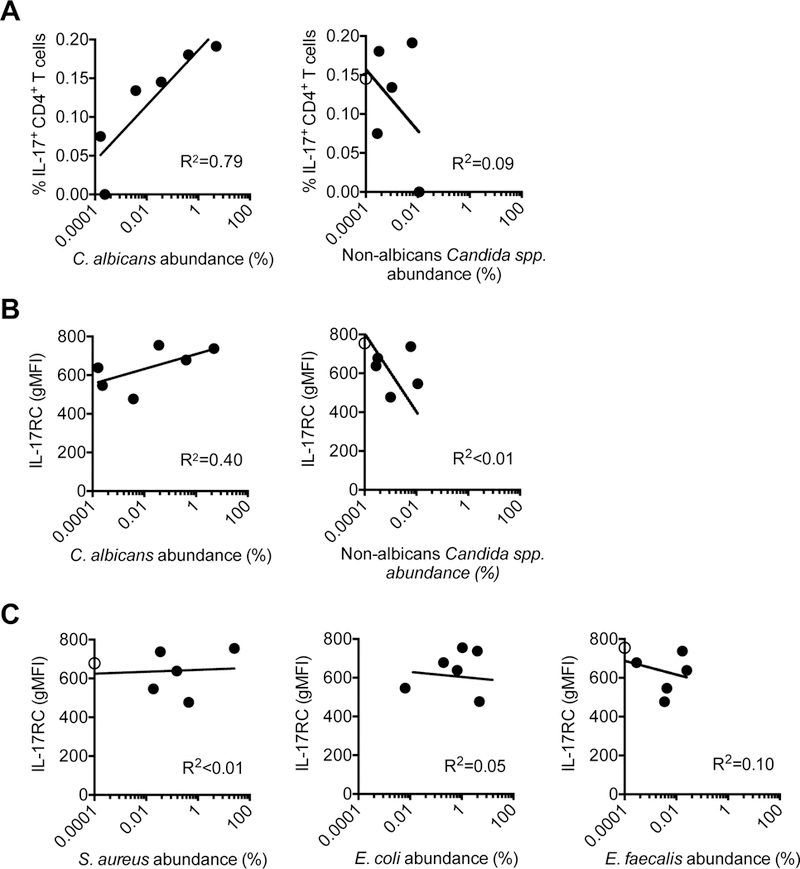
Mucosal barriers are densely colonized by pathobiont microbes such as Candida albicans, capable of invasive disseminated infection. However, systemic infections occur infrequently in healthy individuals, suggesting that pathobiont commensalism may elicit host benefits. We show that intestinal colonization with C. albicans drives systemic expansion of fungal-specific Th17 CD4+ T cells and IL-17 responsiveness by circulating neutrophils, which synergistically protect against C. albicans invasive infection. Protection conferred by commensal C. albicans requires persistent fungal colonization and extends to other extracellular invasive pathogens such as Staphylococcus aureus. However, commensal C. albicans does not protect against intracellular influenza virus infection and exacerbates allergic airway inflammation susceptibility, indicating that positively calibrating systemic Th17 responses is not uniformly beneficial. Thus, systemic Th17 inflammation driven by CD4+ T cells responsive to tonic stimulation by commensal C. albicans improves host defense against extracellular pathogens, but with potentially harmful immunological consequences.

Keywords: CD4 T cells; IL-17; asthma; commensal; fungi; immunity; infection; inflammation; neutrophil; pathobiont.


Conflict of interest statement

DECLARATION OF INTERESTS

The authors declare no competing interests.

Figures

Figure 1.
Figure 1.. Persistent C. albicans intestinal colonization facilitated by drinking water ampicillin supplementation
(A) Recoverable C. albicans in the feces of mice with ampicillin supplementation in the drinking water compared with no antibiotic controls after oral C. albicans inoculation. (B) Recoverable C. albicans in each intestinal segment 7 days after oral C. albicans inoculation for the mice described in (A). (C) Recoverable C. albicans in each tissue seven days after oral C. albicans inoculation for mice with ampicillin drinking water supplementation. (D) Weight change after oral C. albicans inoculation for the mice described in (A). (E) Diversity and abundance of fecal bacterial species for each group of mice after drinking water ampicillin supplementation (12 days) with or without oral C. albicans inoculation (2 days after initiating ampicillin drinking water supplementation) determined by shotgun sequencing. Pie-chart area is directly proportional to the absolute abundance of fecal bacterial genomic DNA for each group of mice. (F) Bacterial genomic equivalents for each group of mice described in (E). (G) Diversity of fecal bacteria for each group of mice described in (E). *p<0.05, ****p<0.0001, Bar, mean ± SEM. L.o.D., limit of detection. See also Figure S1
Figure 2.
Figure 2.. C. albicans intestinal colonization protects against systemic C. albicans invasive infection
(A) Percent survival and recoverable C. albicans 5 days after recombinant C. albicans intravenous infection (5 × 10⁴ CFUs) for mice with recombinant C. albicans intestinal colonization or control mice maintained on ampicillin supplemented drinking water. (B) Fluorescence intensity of C. albicans recovered from the feces compared with kidneys 5 days after intravenous infection with GFP+ C. albicans for mice with prior GFP- C. albicans oral inoculation. ***p<0.001, ****p<0.0001, Bar, mean ± SEM. L.o.D., limit of detection. See also Figure S2
Figure 3.
Figure 3.. Systemic expansion of protective Th17 CD4+ T cells with commensal C. albicans specificity
(A) Number and percent CD44hi among I-Ab:2W1S tetramer positive CD4+ T cells from spleen and peripheral lymph nodes of mice with recombinant C. albicans intestinal colonization compared with no colonization controls. (B) Percent and number RORγt+ among I-Ab:2W1S positive (solid line) or negative (gray shaded) CD4+ T cells for mice described in (A). (C) Percent IL-17A, IL-17F or IFN-γ production after heat-killed WT C. albicans stimulation by CD4+ splenocyte and lymph nodes cells for mice described in (A). (D) Recoverable C. albicans five days after recombinant C. albicans intravenous infection (5 × 10⁴ CFUs) amongst recombinant C. albicans colonized compared with control mice administered rat IgG (isotype), anti-CD4, or anti-IL-17A plus anti-IL-17F antibodies beginning one day prior to infection. (E) Percent survival for mice described in (D). (F) Recoverable C. albicans 5 days after recombinant C. albicans intravenous infection (5 × 10⁴ CFUs) for Rag1-deficient mice colonized with recombinant C. albicans or no colonization controls. ** p<0.01, *** p<0.001, ****p<0.0001, Bar, mean ± SEM. L.o.D., limit of detection. See also Figures S3-S5
Figure 4.
Figure 4.. C. albicans intestinal colonization stimulates accumulation, activation and IL-17 responsiveness by circulating neutrophils
(A) Percent IL-17RC+ amongst CD45+ leukocytes in the peripheral blood of mice with recombinant C. albicans intestinal colonization compared with no colonization controls. (B) Percent IL-17RC+ amongst each leukocyte subsets for mice described in (A). (C) Percent IL-17RC+ amongst CD45+ leukocytes in the peripheral blood of recombinant C. albicans colonized mice 7 days after the administration of rat IgG (isotype), anti-CD4, or anti-IL-17A plus anti-IL-17F antibodies. (D) Percent of Ly6G+Ly6Cint neutrophils amongst CD45+ leukocytes in the peripheral blood for each group of mice described in panel (A). (E) Representative plots (solid line histogram, C. albicans extract stimulation; shaded histogram, no stimulation controls) and composite data showing the relative proportion of dihydrorhodamine (DHR)123 fluorescence amongst Ly6G+Ly6Cint neutrophils in the peripheral blood for each group of mice described in panel (A). (F) Percent survival after recombinant C. albicans intravenous infection (5 × 10⁴ CFUs) amongst recombinant C. albicans colonized compared with or no colonization control mice administered anti-Ly6G or anti-Gr-1 antibodies, along with each respective rat IgG isotype control antibody, beginning one day prior to infection. *p<0.05, ** p<0.01, ****p<0.0001, Bar, mean ± SEM. L.o.D., limit of detection.
Figure 5.
Figure 5.. Protection against systemic C. albicans invasive infection requires persistent C. albicans intestinal colonization
(A) Recoverable C. albicans 5 days after recombinant C. albicans intravenous infection (5 × 10⁴ CFUs) for recombinant C. albicans colonized mice treated with fluconazole, no antifungal treatment, or no C. albicans colonization control mice. (B) Number of I-Ab:2W1S tetramer positive CD4+ T cells from spleen and peripheral lymph nodes for recombinant C. albicans colonized mice treated with fluconazole for 20 days, no antifungal treatment, or no colonization control mice. (C) Percent and number RORγt+ among I-Ab:2W1S positive CD4+ T cells for mice described in (B). (D) Percent IL-17A or IL-17F production by CD4+ splenocyte and lymph nodes cells after heat-killed WT C. albicans stimulation for mice described in (B). (E) Percent IL-17RC+ amongst CD45+ leukocytes or Ly6G+Ly6Cint neutrophils in the peripheral blood for mice described in (B). (F) Percent of Ly6G+Ly6Cint neutrophils amongst CD45+ leukocytes in the peripheral blood for mice described in (B). (G) Relative intensity of dihydrorhodamine (DHR)123 fluorescence amongst Ly6G+Ly6Cint neutrophils in the peripheral blood after C. albicans extract stimulation for mice described in (B). *p<0.05, ** p<0.01, *** p<0.001, ****p<0.0001, Bar, mean ± SEM. L.o.D., limit of detection. See also Figure S6
Figure 6.
Figure 6.. Commensal C. albicans protects against S. aureus infection and promotes susceptibility to Th17 airway inflammation
(A) Percent survival after S. aureus intravenous infection (strain USA300; 108 [left panel]), and recoverable S. aureus 5 days after infection with a reduced dosage (strain USA300; 3 × 10⁷ CFUs [right panel]) for mice with recombinant C. albicans intestinal colonization, C. albicans colonized mice treated with fluconazole, or no colonization control mice. (B) Percent survival and clinical score progression after influenza A virus intranasal infection (strain PR8, 2 × 10⁵ PFU) for mice with recombinant C. albicans intestinal colonization or no colonization control mice. (C) Airway resistance, percent RORγt+ or IL-17A production by CD4+ T cells recovered from the lungs of recombinant C. albicans colonized mice treated with fluconazole, no antifungal treatment, or no C. albicans colonization control mice. Mice were intratracheally sensitized and challenged with house dust mite extract, and airway resistance represent change over baseline after inhaled methacholine challenge. ** p<0.01, *** p<0.001, ****p<0.0001, Bar, mean ± 1 SEM. L.o.D., limit of detection.
Figure 7.
Figure 7.. C. albicans fecal colonization density positively correlates with systemic levels of fungal-specific Th17 inflammation
(A) Regression analysis comparing IL-17A or IL-17F production by CD4+ peripheral blood cells of after heat-killed C. albicans stimulation compared with the fecal relative abundance of C. albicans or non-albicans Candida spp. determined by shotgun sequencing for each individual. (B) Regression analysis comparing intensity of IL-17RC staining by CD15+CD16+ neutrophils in peripheral blood cells compared with the fecal relative abundance of C. albicans or non-albicans Candida spp. for each individual. (C) Regression analysis comparing intensity of IL-17RC staining by CD15+CD16+ neutrophils in peripheral blood cells compared with the fecal relative abundance of S. aureus, E. coli, or E. faecalis for each individual. Levels below the limits of detection are shown as open circles on the y-axis. See also Figure S7

References

    1. Acosta-Rodriguez EV, Rivino L, Geginat J, Jarrossay D, Gattorno M, Lanzavecchia A, Sallusto F, and Napolitani G (2007). Surface phenotype and antigenic specificity of human interleukin 17–producing T helper memory cells. Nature immunology 8, 639–646. - PubMed
    1. Acton D, Plat-Sinnige MT, van Wamel W, de Groot N, and van Belkum A (2009). Intestinal carriage of Staphylococcus aureus: how does its frequency compare with that of nasal carriage and what is its clinical impact? European journal of clinical microbiology & infectious diseases 28, 115. - PubMed
    1. Alcorn JF, Crowe CR, and Kolls JK (2010). TH17 cells in asthma and COPD. Annual review of physiology 72, 495–516. - PubMed
    1. Baaten BJG, Tinoco R, Chen AT, and Bradley LM (2012). Regulation of antigen-experienced T cells: lessons from the quintessential memory marker CD44. Frontiers in immunology 3, 23. - PMC - PubMed
    1. Bacher P, Hohnstein T, Beerbaum E, Rocker M, Blango MG, Kaufmann S, Rohmel J, Eschenhagen P, Seidel K, Rickerts V, et al. (2019). Instruction of human anti-fungal Th17 immunity and immune pathology by cross-reactivity against a single member of the microbiota. Cell (in press). - PubMed

MeSH terms