

Large single cutaneous metastasis of colon adenocarcinoma mimicking a squamous cell carcinoma of the skin: A case report
- PMID: 30870738
- PMCID: PMC6424055
- DOI: 10.1016/j.ijscr.2019.02.043
Large single cutaneous metastasis of colon adenocarcinoma mimicking a squamous cell carcinoma of the skin: A case report
Abstract
Introduction: Metastases represent one of the most outstanding characteristics of malignant neoplasms and are relatively rare in the skin, in spite of the great extension of the cutaneous organs. Development of cutaneous metastases from colon cancer is a rare event, usually occurring in widely disseminated disease and commonly leading to a poor prognosis. As to location, cutaneous metastases often favor areas close to the primary malignancy, such as lung cancer and skin metastases on the trunk. However, remote sites as the scalp may be also involved.
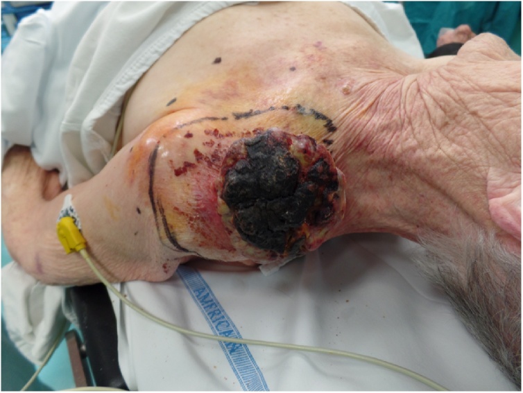
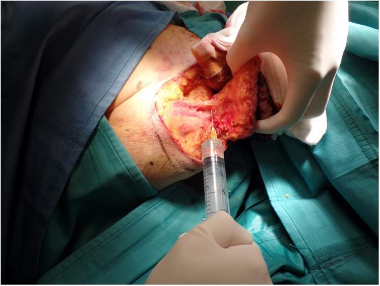
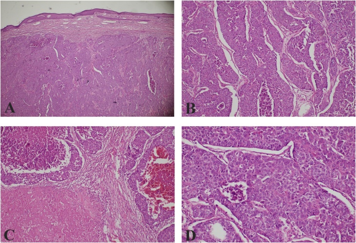
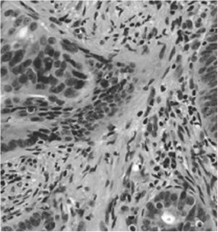
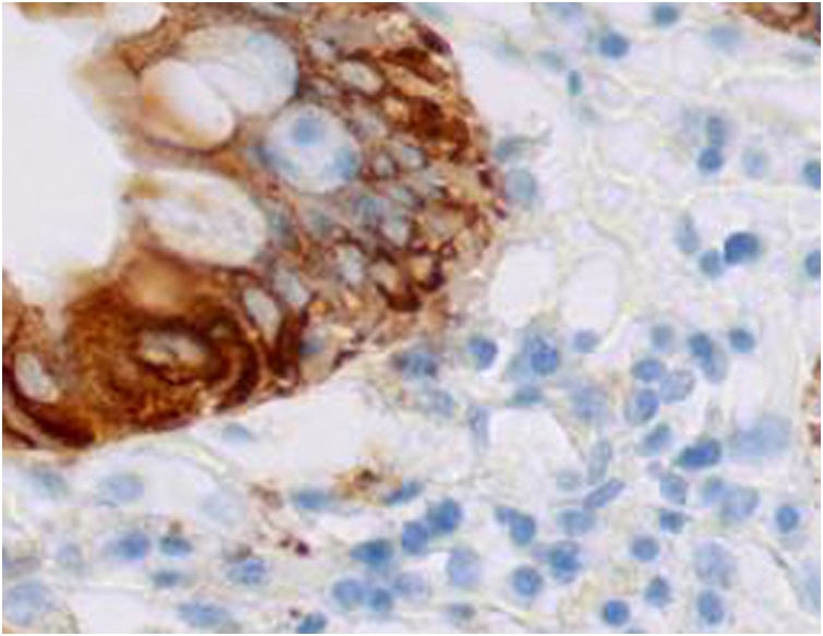
Case presentation: We present the case of a 92-year-old female patient with a massive single nodular skin lesion on her left supraclavicular area, that came back positive for cutaneous metastasis of colon adenocarcinoma.
Discussion: Cutaneous metastasis of colorectal cancer a rare event (2.3%-6%) that usually occur two years after the detection or resection of the primary tumor. It seldom occurs before the identification of the primary tumor and involvement of secondary organs, such as the liver. There are few cases reported with only cutaneous metastases.
Conclusion: In conclusion, dermatological evaluation of patients who are undergoing screening or who have already been diagnosed with cancer is extremely important.
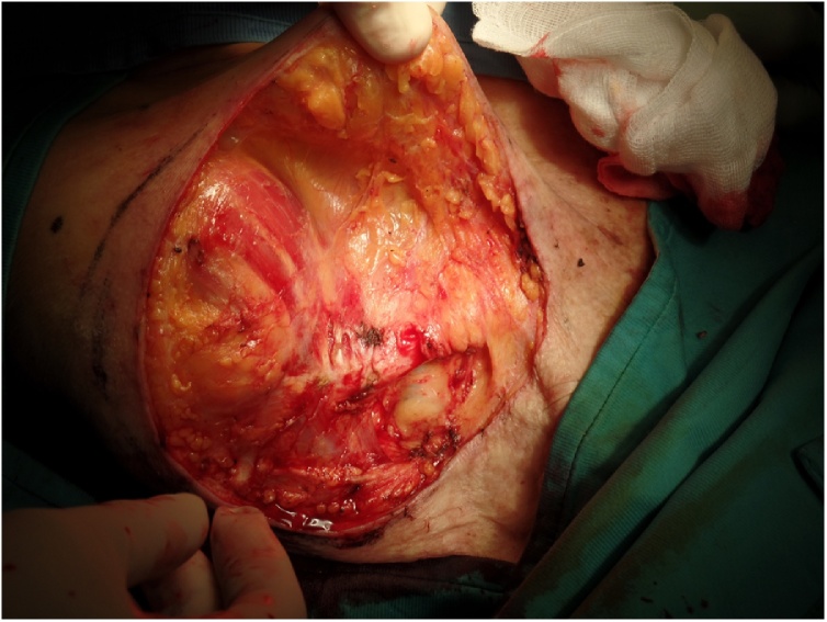
Keywords: Colon Cancer; Cutaneous Metastasis; Fasciocutaneous Flap; Skin cancer.
Copyright © 2019 The Authors. Published by Elsevier Ltd.. All rights reserved.
Figures


References
-
- Rochael M.C., Estrella R.R. Metástases cutâneas. In: Neves R.G., Lupi O., editors. Orgs. Câncer da Pele. MEDSI; Rio de Janeiro: 2001. pp. 393–405.
-
- Krathen R.A., Orengo I.F., Rosen T. Cutaneous metastasis: a meta-analysis of data. South. Med. J. 2003;96:164–167. - PubMed
-
- Hu S.C., Chen G.S., Wu C.S., Chai C.Y., Chen W.T., Lan C.C. Rates of cutaneous metástases from different internal malignancies: experience from a Taiwanese medical center. J. Am. Acad. Dermatol. 2009;60:379–387. - PubMed
-
- Reilly W.T., Nelson H., Schroeder G., Wieand H.S., Bolton J., O’Connell M.J. Wound recurrence following conventional treatment for colorectal cancer: a rare but perhaps underestimated problem. Dis. Colon Rectum. 1996;39:200–207. - PubMed
-
- Saeed S., Keehn C.A., Morgan M.B. Cutaneous metastasis: a clinical, pathological, and immunohistochemical appraisal. J. Cutan. Pathol. 2004;31:419–430. - PubMed
LinkOut - more resources
Full Text Sources
Research Materials
