. 2019 Mar 12;20(5):1235.
doi: 10.3390/ijms20051235.

Detection of Inflammation-Related Melanoma Small Extracellular Vesicle (sEV) mRNA Content Using Primary Melanocyte sEVs as a Reference

Affiliations

Detection of Inflammation-Related Melanoma Small Extracellular Vesicle (sEV) mRNA Content Using Primary Melanocyte sEVs as a Reference

Gina T Bardi et al. Int J Mol Sci. .

Abstract

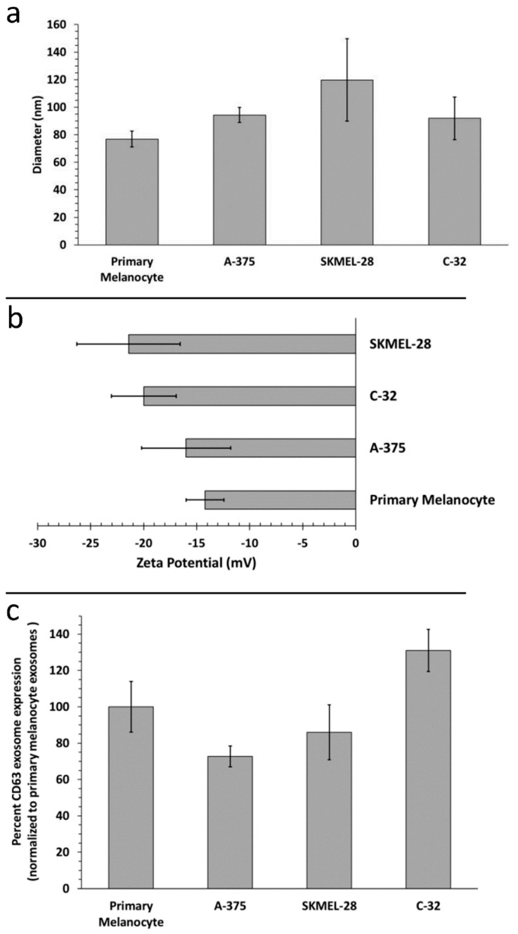
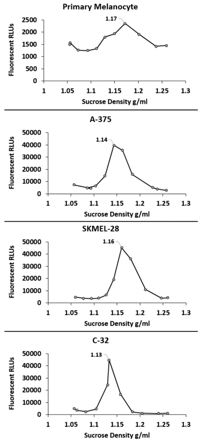
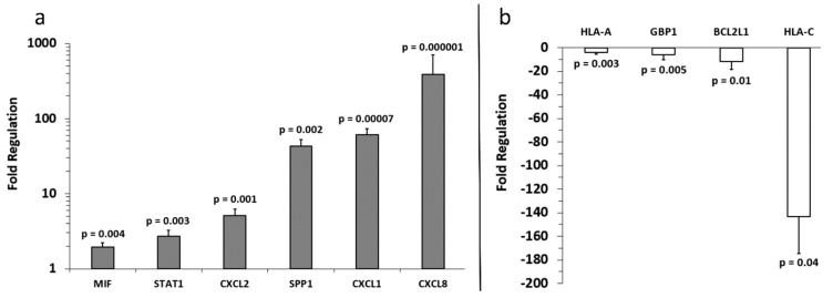
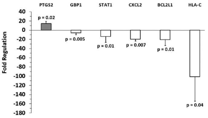
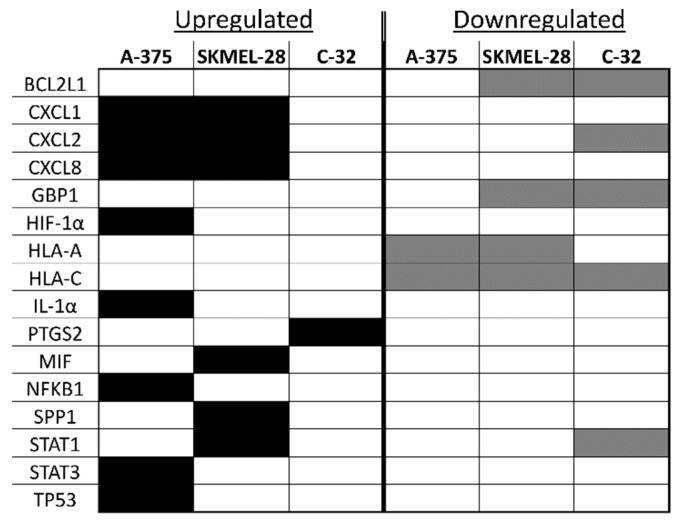
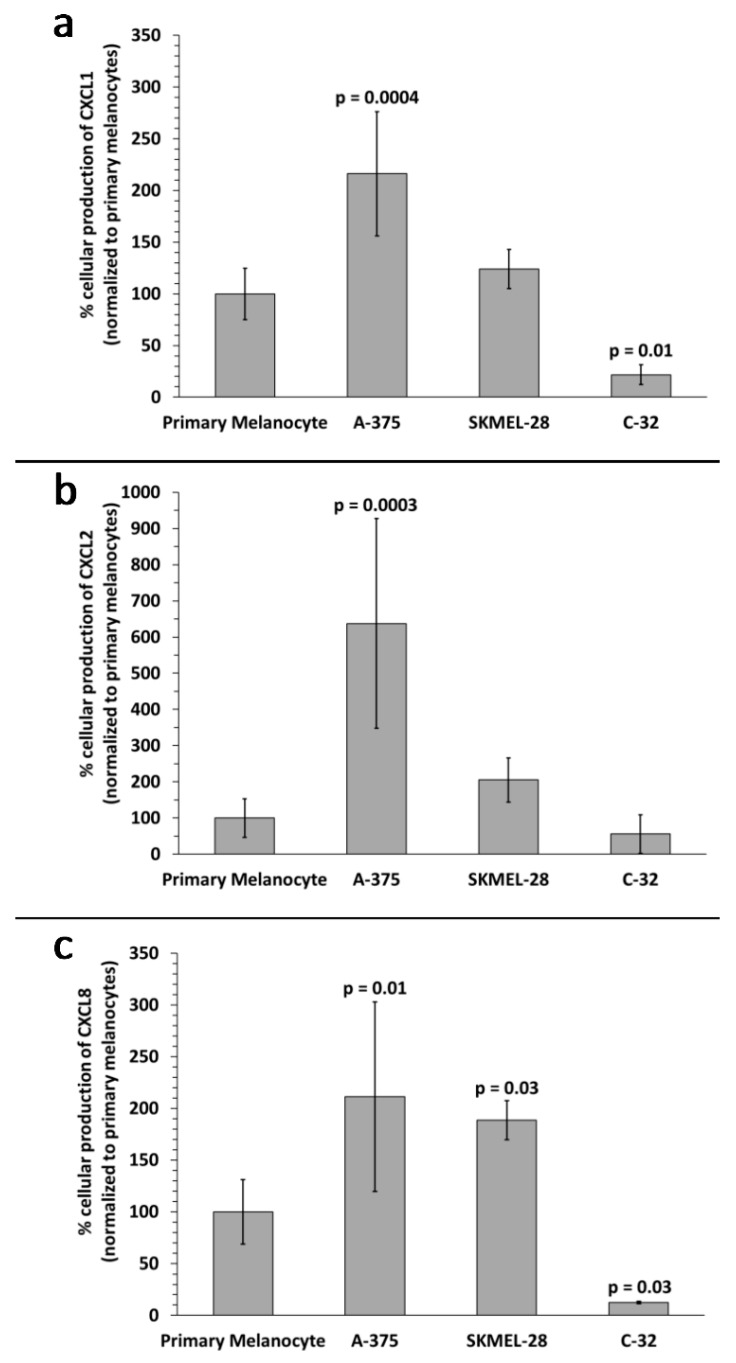
Melanoma-derived small extracellular vesicles (sEVs) participate in tumor pathogenesis. Tumor pathogenesis is highly dependent on inflammatory processes. Given the potential for melanoma sEVs to carry tumor biomarkers, we explored the hypothesis that they may contain inflammation-related mRNA content. Biophysical characterization showed that human primary melanocyte-derived sEVs trended toward being smaller and having less negative (more neutral) zeta potential than human melanoma sEVs (A-375, SKMEL-28, and C-32). Using primary melanocyte sEVs as the control population, RT-qPCR array results demonstrated similarities and differences in gene expression between melanoma sEV types. Upregulation of pro-angiogenic chemokine ligand CXCL1, CXCL2, and CXCL8 mRNAs in A-375 and SKMEL-28 melanoma sEVs was the most consistent finding. This paralleled increased production of CXCL1, CXCL2, and CXCL8 proteins by A-375 and SKMEL-28 sEV source cells. Overall, the use of primary melanocyte sEVs as a control sEV reference population facilitated the detection of inflammation-related melanoma sEV mRNA content.

Keywords: biomarker; exosome; extracellular vesicle; inflammation; mRNA; melanoma.


Conflict of interest statement

The authors declare no conflict of interest.

Figures

Figure 1
Figure 1
Characterization of primary melanocyte small extracellular vesicles (sEVs) and melanoma sEVs. (a) Small extracellular vesicle (EV) sizing by dynamic light scattering (n = 3 independent batch assessments), (b) small EV zeta potential (n = 3 independent batch assessments), (c) primary melanocyte versus melanoma sEV CD63 expression (n = 3 independent batch assessments). Error bars = SD, p values < 0.05 were considered statistically significant and were not detected.
Figure 2
Figure 2
Density characterization of primary melanocyte sEVs and melanoma sEVs. Representative sucrose density gradients are shown. RLU = relative light units corresponding to sEV carbocyanine DiI signal. Peak sEV density is labeled on each gradient.
Figure 3
Figure 3
Fold regulation of A-375 melanoma sEV mRNA versus control primary melanocyte sEV mRNA. (a) Increased and (b) decreased gene expression levels relative to primary melanocytes (normalized to 1) are shown. Three independent sEV batches were pooled and run on (n = 3) replicate arrays, error bars = SD, p values < 0.05 were considered statistically significant.
Figure 4
Figure 4
Fold regulation of SKMEL-28 melanoma sEV mRNA versus control primary melanocyte sEV mRNA. (a) Increased and (b) decreased gene expression levels relative to primary melanocytes (normalized to 1) are shown. Three independent sEV batches were pooled and run on (n = 3) replicate arrays, error bars = SD, p values < 0.05 were considered statistically significant.
Figure 5
Figure 5
Fold regulation of C-32 melanoma sEV mRNA versus control primary melanocyte sEV mRNA. Gene expression levels for primary melanocytes (normalized to 1) are shown. Three independent sEV batches were pooled and run on (n = 3) replicate arrays, error bars = SD, p values < 0.05 were considered statistically significant.
Figure 6
Figure 6
A comparison of inflammation-associated mRNA upregulated or downregulated in melanoma sEVs versus primary melanocyte sEVs. Black shaded boxes indicate upregulated gene expression, and gray shaded boxes indicate downregulated gene expression.
Figure 7
Figure 7
Melanoma cell versus primary melanocyte production of chemokine proteins. Percent protein production levels for (a) CXCL1, (b) CXCL2, and (c) CXCL8 by A-375, SKMEL-28, and C-32 melanoma cells compared to primary melanocytes (control, normalized to 100%) are shown. Data bars represent the average of (n = 4) independent experiments, error bars = SD, p values < 0.05 were considered statistically significant.

References

    1. Rinderknecht M., Detmar M. Tumor lymphangiogenesis and melanoma metastasis. J. Cell. Physiol. 2008;216:347–354. doi: 10.1002/jcp.21494. - DOI - PubMed
    1. Hood J.L. Pre-analytical influences on the population heterogeneity of human extracellular vesicles sourced for nanomedicine uses. Nanomedicine. 2018;13:2669–2674. doi: 10.2217/nnm-2018-0287. - DOI - PubMed
    1. Thery C., Witwer K.W., Aikawa E., Alcaraz M.J., Anderson J.D., Andriantsitohaina R., Antoniou A., Arab T., Archer F., Atkin-Smith G.K., et al. Minimal information for studies of extracellular vesicles 2018 (misev2018): A position statement of the international society for extracellular vesicles and update of the misev2014 guidelines. J. Extracell. Vesicles. 2018;7:1535750. doi: 10.1080/20013078.2018.1535750. - DOI - PMC - PubMed
    1. Kowal J., Arras G., Colombo M., Jouve M., Morath J.P., Primdal-Bengtson B., Dingli F., Loew D., Tkach M., Thery C. Proteomic comparison defines novel markers to characterize heterogeneous populations of extracellular vesicle subtypes. Proc. Natl. Acad. Sci. USA. 2016;113:E968–E977. doi: 10.1073/pnas.1521230113. - DOI - PMC - PubMed
    1. Gould S.J., Raposo G. As we wait: Coping with an imperfect nomenclature for extracellular vesicles. J. Extracell. Vesicles. 2013;2:20389. doi: 10.3402/jev.v2i0.20389. - DOI - PMC - PubMed