. 2019 Mar 12;20(5):1237.
doi: 10.3390/ijms20051237.

CXCR2: A Novel Mediator of Mammary Tumor Bone Metastasis

Affiliations

CXCR2: A Novel Mediator of Mammary Tumor Bone Metastasis

Bhawna Sharma et al. Int J Mol Sci. .

Abstract

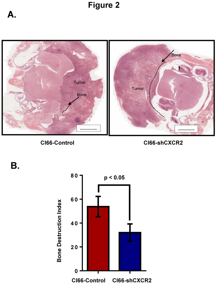
Most breast cancer patients die due to bone metastasis. Although metastasis accounts for 5% of the breast cancer cases, it is responsible for most of the deaths. Sometimes even before the detection of a primary tumor, most of the patients have bone and lymph node metastasis. Moreover, at the time of death, breast cancer patients have the bulk of the tumor burden in their bones. Therapy options are available for the treatment of primary tumors, but there are minimal options for treating breast cancer patients who have bone metastasis. C-X-C motif chemokine receptor type 2 (CXCR2) receptor-mediated signaling has been shown to play a critical role during bone-related inflammations and its ligands C-X-C motif chemokine ligand 6 (CXCL6) and 8 (CXCL8) aid in the resorption of bone during bone metastasis. In this study, we tested the hypothesis that CXCR2 contributes to mammary tumor-induced osteolysis and bone metastasis. In the present study, we examined the role of both tumor cell-derived and host-derived CXCR2 in influencing mammary tumor cell bone metastasis. For understanding the role of tumor cell-derived CXCR2, we utilized Cl66 CXCR2 knockdown (Cl66-shCXCR2) and Cl66-Control cells (Cl66-Control) and observed a significant decrease in tumor growth and tumor-induced osteolysis in Cl66-shCXCR2 cells in comparison with the Cl66-Control cells. Next, for understanding the role of host-derived CXCR2, we utilized mice with genomic knockdown of CXCR2 (Cxcr2-/-) and injected Cl66-Luciferase (Cl66-Luc) or 4T1-Luciferase (4T1-Luc) cells. We observed decreased bone destruction and metastasis in the bone of Cxcr2-/- mice. Our data suggest the importance of both tumor cell- and host-derived CXCR2 signaling in the bone metastasis of breast cancer cells.

Keywords: CXCR2; bone microenvironment; metastasis.


Conflict of interest statement

The authors declare no conflict of interest.

Figures

Figure 1
Figure 1
Downregulation of CXCR2 in tumor cells reduces calvarial tumor growth. (A) Schematic representation of the tumor cell injections in Balb/c mice. Injection of Cl66-Control or Cl66-shCXCR2 cells mixed and suspended in 25 μL of Hank’s Balanced Salt solution and 25 μL of growth factor reduced matrigel on the dorsal side on calvaria of Balb/c mice using a 23 gauge needle is marked as day 1. Mice were monitored for 21 days for tumor growth and sacrificed. (B) The graph shows the growth kinetics of tumor formed by Cl66-Control and Cl66-shCXCR2 cells on the calvaria of Balb/c mice. Statistical analysis was performed using the Mann-Whitney Rank Sum Test with * p = 0.045 and n = 5 per group.
Figure 2
Figure 2
CXCR2 downregulation in Cl66 cells significantly decreased tumor-induced osteolysis. (A) Representative images show H&E staining demonstrating intact cranial bone in Cl66-shCXCR2 group in comparison with severe bone destruction in Cl66-Control group. Scale bar represents 10,000 μm. (B) Bone destruction index was used to measure the severity of the lesions. Bar graph showing significantly lower bone destruction index in Cl66-shCXCR2 group (32 ± 5) in comparison with Cl66-Control group (54 ± 6) (n = 5, p < 0.05).
Figure 3
Figure 3
CXCR2 downregulation in tumor cells lowers activated osteoclast number at the tumor-bone interface. (A) Representative images of osteolytic activity as determined by TRAP staining at the tumor-bone interface from Cl66-Control and Cl66-shCXCR2 tumor-bearing mice. Scale bar represents 10 μm (B) Bar graph showing a significantly lower number of TRAP-positive osteoclasts in Cl66-shCXCR2 (65 ± 9) in comparison with the Cl66-Control tumor group (157 ± 12) (n = 5, p = 0.003) at the tumor-bone interface.
Figure 4
Figure 4
Host CXCR2 promotes tumor cell growth in the bone microenvironment. (A) Schematic diagram of the Cl66-Luc cell injection in calvaria of wild type and Cxcr2−/− mice. (B) The graph shows the kinetics of tumor volume formed by Cl66-Luc cells in the bone microenvironment of the wild type and CXCR2 −/− mice. Statistical analysis was done using the Mann-Whitney Rank Sum Test with * p = 0.036 and n = 5/group.
Figure 5
Figure 5
Host Cxcr2 deficiency reduced tumor-induced osteolysis. (A) Representative images show H&E staining demonstrating the intact cranial bone in Cxcr2−/− mice in comparison with severe bone destruction in wild type mice. Scale bar represents 100 μm (B) Bar graph shows significantly lower bone destruction index in Cxcr2−/− mice in comparison with wild type mice (n = 5, p < 0.05). (C) Representative images show immunohistochemical staining for PCNA cells demonstrating higher cell proliferating cells in the tumor of the wild type mice in comparison with the Cxcr2−/− mice. Scale bar represents 10 μm. (D) Bar graph demonstrates a lower number of proliferating cells in Cxcr2−/− mice in comparison with wild type mice (n = 5, p = 0.0079). (E) Representative images show immunohistochemical staining for isolectin B4 positive cells demonstrating higher microvessel density in the tumor of the wild type mice in comparison with the Cxcr2−/− mice. Scale bar represents 10 μm. (F) Bar graph demonstrates lower microvessel density at the tumor-bone interface in Cxcr2−/− mice in comparison with wild type mice. (n = 5, p = 0.04).
Figure 6
Figure 6
Host Cxcr2 influences mammary tumor bone metastasis. (A) Diagram depicts the schematic representation of intracardiac tumor cell injection in wild type and Cxcr2−/− mice. (B) In vivo luminescent images of the wild type and Cxcr2−/− group showing the higher spread of Cl66-Luc cells in the whole body of the wild type mice in comparison with the Cxcr2−/− mice. (C) Quantification of bone metastasis burden from Cl66-Luc cells injected intracardially in wild type and Cxcr2−/− mice based on whole body BLI imaging. The wild type mice group showed a higher tumor burden than the Cxcr2−/− mice group. The * sign indicates p value < 0.05. (D) Quantification of bone metastasis burden from 4T1-Luc cells injected intracardially in wild type and Cxcr2−/− mice based on whole body BLI imaging in wild type and Cxcr2−/− mice. The wild type mice group showed a higher metastatic tumor burden than Cxcr2−/− mice group. The * sign indicates p value < 0.05.

References

    1. Siegel R.L., Miller K.D., Jemal A. Cancer statistics, 2018. CA Cancer J. Clin. 2018;68:7–30. doi: 10.3322/caac.21442. - DOI - PubMed
    1. Morris P.G., McArthur H.L., Hudis C.A. Therapeutic options for metastatic breast cancer. Expert Opin. Pharmacother. 2009;10:967–981. doi: 10.1517/14656560902834961. - DOI - PubMed
    1. Mundy G.R. Metastasis to bone: Causes, consequences and therapeutic opportunities. Nat. Rev. 2002;2:584–593. doi: 10.1038/nrc867. - DOI - PubMed
    1. Benoy I.H., Salgado R., Van Dam P., Geboers K., Van Marck E., Scharpe S., Vermeulen P.B., Dirix L.Y. Increased Serum Interleukin-8 in Patients with Early and Metastatic Breast Cancer Correlates with Early Dissemination and Survival. Clin. Cancer Res. 2004;10:7157–7162. doi: 10.1158/1078-0432.CCR-04-0812. - DOI - PubMed
    1. Kozlowski L., Zakrzewska I., Tokajuk P., Wojtukiewicz M.Z. Concentration of interleukin-6 (IL-6), interleukin-8 (IL-8) and interleukin-10 (IL-10) in blood serum of breast cancer patients. Rocz. Akad. Med. Bialymst. 2003;48:82–84. - PubMed

MeSH terms

Substances