Review
. 2019 Mar 12;24(5):997.
doi: 10.3390/molecules24050997.

Novel Therapeutics for Epstein⁻Barr Virus

Affiliations
Review

Novel Therapeutics for Epstein⁻Barr Virus

Graciela Andrei et al. Molecules. .

Abstract

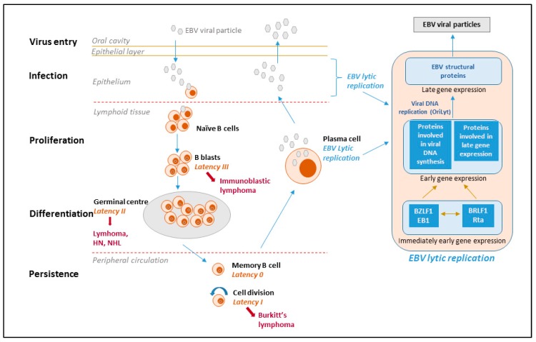
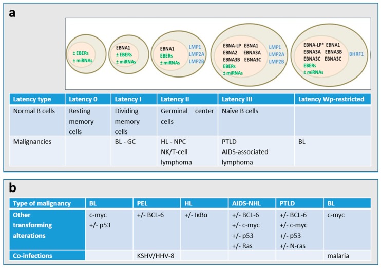
Epstein⁻Barr virus (EBV) is a human γ-herpesvirus that infects up to 95% of the adult population. Primary EBV infection usually occurs during childhood and is generally asymptomatic, though the virus can cause infectious mononucleosis in 35⁻50% of the cases when infection occurs later in life. EBV infects mainly B-cells and epithelial cells, establishing latency in resting memory B-cells and possibly also in epithelial cells. EBV is recognized as an oncogenic virus but in immunocompetent hosts, EBV reactivation is controlled by the immune response preventing transformation in vivo. Under immunosuppression, regardless of the cause, the immune system can lose control of EBV replication, which may result in the appearance of neoplasms. The primary malignancies related to EBV are B-cell lymphomas and nasopharyngeal carcinoma, which reflects the primary cell targets of viral infection in vivo. Although a number of antivirals were proven to inhibit EBV replication in vitro, they had limited success in the clinic and to date no antiviral drug has been approved for the treatment of EBV infections. We review here the antiviral drugs that have been evaluated in the clinic to treat EBV infections and discuss novel molecules with anti-EBV activity under investigation as well as new strategies to treat EBV-related diseases.

Keywords: Epstein–Barr virus; antivirals; cellular targets; nucleoside analogues; nucleotide analogues.


Conflict of interest statement

The authors declare no conflict of interest.

Figures

Figure 1
Figure 1
EBV life cycle, latency stages and derived lymphomas. The viral life cycle includes at least five different stages (virus entry, infection, proliferation, differentiation and persistence), and four of them are associated with EBV diseases. The virus is transmitted through the saliva and infects naïve B-cells in the oropharyngeal mucosa. During primary infection, EBV-infected naïve B-cells express the entire latency gene complex (10 proteins: EBV nuclear antigens (EBNAs), latent membrane protein (LMPs)) as well as EBV-encoded small RNAs (EBERs) and microRNAs. This is called type III latency and this form of latency activates the resting B-cells and drives them to proliferation and transformation. However, these cells are highly immunogenic and are rapidly eliminated by EBV-specific T cells. The virus is able to survive in B-cells because it downregulates its immunogenic proteins. EBV mimics antigen driven B-cell responses and similar to antigen-stimulated blasts, the EBV-infected B-cells enter the follicles, expand, and form germinal centers where they express only three viral proteins (type II latency). Finally, they exit the lymph node expressing only a single viral protein (EBNA1, which ensures that the viral genome divides with the cellular genome) (type I latency). The entry of EBV-infected cells into the peripheral blood results in the shutdown of all viral genes encoding for proteins; this is called latency 0 or latency program where no viral proteins are expressed. Resting memory cells, in which the virus is quiescent, are not attacked by the host immune system and are likely the sites of long-term persistence. Memory B cells occasionally divide to maintain stable number of cells and when a cell that is carrying the virus divides, the viral EBNA1 protein is expressed to allow the viral genome to replicate along with the cell. Memory B-cells may also undergo terminal differentiation into plasma cells and secrete antibodies. If such a cell contains the virus, the EBV lytic program is activated and the infectious virus released from the plasma cells can infect epithelial cells, where the virus can replicate and be shed at high amounts and then be transmitted to other hosts. With the exception of latency type 0, each latency state is found in specific types of EBV-associated malignancies.
Figure 2
Figure 2
(a) Patterns of gene expression during EBV latency. The majority of the endemic BL presents a latency I type and carry a wild-type transformation-competent EBV genome and express only the Epstein–Barr nuclear antigen 1 (EBNA1) from the EBNA1-specific latent promoter Qp, non-coding EBERs (Epstein–Barr virus-encoded small RNAs) and several microRNAs (miRNAs). Around 15% of BL endemic tumors, the so called Wp-restricted BLs, carry an EBNA2 gene-deleted genome and express EBNA1, -3A, -3B, and-3C and the viral Bcl2 homologue BHRF1 from the Wp latent promoter [2,6]. * The EBNA-LP gene is partially deleted in the Wp-restricted latency. A major type of latency in EBV-associated malignancies is latency II, in which the latent membrane proteins LMP1, LMP2A, and LMP2B are expressed in addition to the Latency I genes. The entire EBV latency gene complex, which consists of several EBNA proteins, LMP1, LMP2A, LMP2B, EBERs, and miRNAs are expressed in the type III latency. (b) The cellular genetic alterations and/or co-infections are known to occur in the different types of EBV-associated malignancies. PEL: primary effusion lymphoma; HL: Hodgkin lymphoma; BL: Burkitt lymphoma; NHL: non-Hodgkin lymphoma; PTLD: post-transplant lymphoproliferative disorder; NPC: nasopharyngeal carcinoma; GC: gastric carcinoma.

Similar articles

Cited by

References

    1. Katz B.Z., Shiraishi Y., Mears C.J., Binns H.J., Taylor R. Chronic fatigue syndrome after infectious mononucleosis in adolescents. Pediatrics. 2009;124:189–193. doi: 10.1542/peds.2008-1879. - DOI - PMC - PubMed
    1. Cesarman E. Gammaherpesvirus and lymphoproliferative disorders in immunocompromised patients. Cancer Lett. 2011;305:163–174. doi: 10.1016/j.canlet.2011.03.003. - DOI - PMC - PubMed
    1. Cesarman E. Gammaherpesviruses and lymphoproliferative disorders. Annu. Rev. Pathol. 2014;9:349–372. doi: 10.1146/annurev-pathol-012513-104656. - DOI - PubMed
    1. Murata T., Tsurumi T. Switching of EBV cycles between latent and lytic states. Rev. Med. Virol. 2014;24:142–153. doi: 10.1002/rmv.1780. - DOI - PubMed
    1. Ryan J.L., Morgan D.R., Dominguez R.L., Thorne L.B., Elmore S.H., Mino-Kenudson M., Lauwers G.Y., Booker J.K., Gulley M.L. High levels of Epstein–Barr virus DNA in latently infected gastric adenocarcinoma. Lab. Invest. 2009;89:80–90. doi: 10.1038/labinvest.2008.103. - DOI - PMC - PubMed

MeSH terms

Substances

LinkOut - more resources