. 2019 Mar 13;8(3):62.
doi: 10.3390/antiox8030062.

Hydroxycobalamin Reveals the Involvement of Hydrogen Sulfide in the Hypoxic Responses of Rat Carotid Body Chemoreceptor Cells

Affiliations

Hydroxycobalamin Reveals the Involvement of Hydrogen Sulfide in the Hypoxic Responses of Rat Carotid Body Chemoreceptor Cells

Teresa Gallego-Martin et al. Antioxidants (Basel). .

Abstract

Carotid body (CB) chemoreceptor cells sense arterial blood PO₂, generating a neurosecretory response proportional to the intensity of hypoxia. Hydrogen sulfide (H₂S) is a physiological gaseous messenger that is proposed to act as an oxygen sensor in CBs, although this concept remains controversial. In the present study we have used the H₂S scavenger and vitamin B12 analog hydroxycobalamin (Cbl) as a new tool to investigate the involvement of endogenous H₂S in CB oxygen sensing. We observed that the slow-release sulfide donor GYY4137 elicited catecholamine release from isolated whole carotid bodies, and that Cbl prevented this response. Cbl also abolished the rise in [Ca2+]i evoked by 50 µM NaHS in enzymatically dispersed CB glomus cells. Moreover, Cbl markedly inhibited the catecholamine release and [Ca2+]i rise caused by hypoxia in isolated CBs and dispersed glomus cells, respectively, whereas it did not alter these responses when they were evoked by high [K⁺]e. The L-type Ca2+ channel blocker nifedipine slightly inhibited the rise in CB chemoreceptor cells [Ca2+]i elicited by sulfide, whilst causing a somewhat larger attenuation of the hypoxia-induced Ca2+ signal. We conclude that Cbl is a useful and specific tool for studying the function of H₂S in cells. Based on its effects on the CB chemoreceptor cells we propose that endogenous H₂S is an amplifier of the hypoxic transduction cascade which acts mainly by stimulating non-L-type Ca2+ channels.

Keywords: carotid body; hydrogen sulfide; hydroxycobalamin; hypoxia; oxygen sensing.


Conflict of interest statement

The authors declare no conflict of interest.

Figures

Figure 1
Figure 1
Effect of NaHS and GYY4137 on the release of 3H-CA by CB chemoreceptor cells in the presence and absence of Cbl. (A) Time course of catecholamine release elicited by 200 µM NaHS applied for 10 min. A second application of NaHS was in the presence of 300 µM Cbl (n = 6 and 8, for 0 and 200 µM NaHS, respectively). (B) The black bar shows the magnitude of the release response corresponding to the area under the curve in part A. The empty bar represents the release response obtained for 200 µM NaHS in similar experiments in which the solution rather than the headspace was gassed. The small columns in grey at the right of the figure show that 300 µM Cbl abolished the effect of NaHS, and that when applied alone Cbl caused a modest inhibition of the basal ongoing release of catecholamine (NaHS solution bubbling, n = 2; NaHS gassing surface, n = 8; Cbl, n = 18; Cbl + NaHS, n = 10). (C) Effect of GYY4137 on the release of 3H-CA in the absence and presence of Cbl. 400 µM GYY4137 was applied for 20 min by itself at 30 and 90 min (continuous line); 400 µM GYY4137 was applied for 20 min by itself at 30 min and then again at 90 min, this time in the presence of 300 µM Cbl, which had been applied at 80 min (dashed line). n = 10 in each condition. (D) The bars show the amplitudes of the two successive responses to GYY4137 (area under the curve) when both responses were evoked in the absence of Cbl (empty bars) and when the second response was elicited in the presence of Cbl. Symbols indicate where there was a significant effect.
Figure 2
Figure 2
Effects of Cbl on 3H-CA release response elicited by different levels of hypoxia. The continuous lines in (A), (B), and (C) show the effects on catecholamine release of successive exposures of CBs (at 40–50 min, SI, and 80–90 min, SII) to 2% O2 (A, n = 12 in each group), 5% O2 (B, n = 5 in each group), and 7% O2 (C, n = 5 in each group) under control conditions, whereas the dashed lines illustrate catecholamine release evoked by a similar protocol when 300 µM Cbl was present between 70 and 90 min. (D). Protocol similar to panels A except that Cbl was present both during and after the second hypoxic challenge (from 70 to 110 min), n = 8 and 12, control and experimental group, respectively. Asterisks connote time points where there were significant differences between the amplitudes of 3H-CA release observed in CBs which were exposed or not exposed to Cbl.
Figure 3
Figure 3
Mean effects of 300 µM Cbl on the ratio of the second and first 3H-CA responses to hypoxia shown in Figure 2. Results were calculated separately for 3H-CA release during hypoxia and during the subsequent 20 min of normoxia (Nx) during which release fell back to the baseline. (A)–(C) Mean effects of Cbl on the ratio of the second and first responses to hypoxia for 2, 5, and 7% O2; Cbl was present for 10 min before and during the second hypoxic period. (D) Mean effects of Cbl on the ratio of the second and first responses to hypoxia for 2% O2; Cbl was present for 10 min before and during the second hypoxic period, as well as during the subsequent 20 min of normoxia. Asterisks indicate where there was a significant effect of Cbl. Number of replicates for each condition as is indicated in Figure 2.
Figure 4
Figure 4
Effects of Cbl on the 3H-CA release response elicited by three elevated levels of extracellular K+. (A) The continuous line in shows the effects on catecholamine release of successive exposures of CBs (at 30–40 min, SI, and 80–90 min, SII) to 60 mM K+ (n = 5 in each condition) under control conditions, whereas the dashed lines illustrate catecholamine release evoked by a similar protocol when 300 µM OH- Cbl was present between 70 and 90 min. (B) Bars illustrate the ratio of the second and first responses to high K+ in the presence and absence of Cbl during the 10 min of high K+ and during the subsequent 20 min period in control Tyrode (CT) solution (K+ 5mM) during which catecholamine release remained elevated with 60, 35, and 25 mM (n = 5 in each condition).
Figure 5
Figure 5
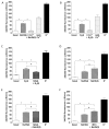
Effects of Cbl on the intracellular Ca2+ responses elicited by 50 µM NaHS, hypoxia, and high external K+. [Ca2+]i was assessed as the F340/F380 fluorescence emission ratio in isolated chemoreceptor cells loaded with fura-2. (A) Mean +/− SEM 340/380 fluorescence ratio (calculated every 8 s) from 74 cells stimulated with NaHS in the absence and presence of 300 µM Cbl. The response to high K+ at the end demonstrates cell viability. The fast Ca2+ transient in response to high K+ at the end of experiments demonstrates that cell was viable. (B) Mean running integrals of the basal and stimulated fluorescence signals (Δ fluorescence/min) obtained from the 74 chemoreceptor cells recorded as in (A). (C) Mean running integrals of the fluorescence signals obtained in 43 cells recorded following the sequence depicted in the figure consisting of the application of three identical hypoxic stimuli (perfusion with 5% CO2/95% N2) except that the middle hypoxic challenge was applied in the presence of 300 µM Cbl. (D) Mean values obtained in 44 cells in a similar ‘sandwich’ style experiment using high (35 mM) external K+ as the stimulus. Symbols connote significant differences between the columns indicated.
Figure 6
Figure 6
Mechanisms of Ca2+ influx induced by hypoxia and 50 µM NaHS. [Ca2+]i was assessed as the F340/F380 fluorescence emission ratio in isolated chemoreceptor cells loaded with fura-2. In all cases the protocols followed the same pattern: a first exposure to hypoxia or NaHS, subsequent application of the experimental condition 0 mM Ca2+ (1 min, A,B), 2 µM nifedipine (3 min, C and E), or 100 nM IBTx (1 min, F) followed by that condition with hypoxia or NaHS and finally high K+ to demonstrate that cells were viable. Bars in all panels indicated the running integrals of the fluorescence signals (Δ fluorescence/min) obtained during the various conditions. (A) Effect of Ca2+ free solution on the Ca2+ signal caused by NaHS (n = 66 cells). (B) Effect of Ca2+ free solution on the Ca2+ signal evoked by hypoxia (n = 44 cells). In both cases, Ca2+ reintroduction caused a rebound increase in the fluorescence emission ratio. (C) Effect of nifedipine on the Ca2+ signal elicited by hypoxia (n = 24 cells). (D) Ca2+ signals evoked by two successive exposures of cells to NaHS under control conditions, data from 51 cells. (E) Ca2+ signals evoked by two successive exposures of cells to NaHS—the first under control conditions and the second in the presence of nifedipine (n = 50 cells). (F) Ca2+ signals evoked by two successive exposures of cells to NaHS; the first under control conditions and the second in the presence of IBTx (n = 47 cells). Symbols connote significant differences between the columns indicated.

Similar articles

Cited by

References

    1. Fidone S., Gonzalez C. Initiation and control of chemoreceptor activity in the carotid body. In: Fishman A.P., Cherniack N.S., Widdicombe J.G., Geiger S.R., editors. Handbook of Physiology, Section 3, The Respiratory System, vol II, Control of Breathing. Volume 2. American Physiological Society; Bethesda, MD, USA: 1986. pp. 247–312.
    1. Gonzalez C., Almaraz L., Obeso A., Rigual R. Carotid body chemoreceptors: From natural stimuli to sensory discharges. Physiol. Rev. 1994;74:829–898. doi: 10.1152/physrev.1994.74.4.829. - DOI - PubMed
    1. Olson K.R., Healy M.J., Qin Z., Skovgaard N., Vulesevic B., Duff D.W., Whitfield N.L., Yang G., Wang R., Perry S.F. Hydrogen sulfide as an oxygen sensor in trout gill chemoreceptors. Am. J. Physiol. Regul. Integr. Comp. Physiol. 2008;295:R669–R680. doi: 10.1152/ajpregu.00807.2007. - DOI - PubMed
    1. Peng Y.J., Nanduri J., Raghuraman G., Souvannakitti D., Gadalla M.M., Kumar G.K., Snyder S.H., Prabhakar N.R. H2S mediates O2 sensing in the carotid body. Proc. Natl. Acad. Sci. USA. 2010;107:10719–10724. doi: 10.1073/pnas.1005866107. - DOI - PMC - PubMed
    1. Li Q., Sun B., Wang X., Jin Z., Zhou Y., Dong L., Jiang L.H., Rong W. A crucial role for hydrogen sulfide in oxygen sensing via modulating large conductance calcium-activated potassium channels. Antioxid. Redox Signal. 2010;12:1179–1189. doi: 10.1089/ars.2009.2926. - DOI - PubMed