Study of the Chronology of Expression of Ten Extracellular Matrix Molecules during the Myogenesis in Cattle to Better Understand Sensory Properties of Meat
- PMID: 30871212
- PMCID: PMC6462999
- DOI: 10.3390/foods8030097
Study of the Chronology of Expression of Ten Extracellular Matrix Molecules during the Myogenesis in Cattle to Better Understand Sensory Properties of Meat
Abstract
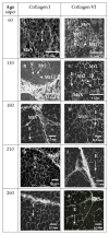
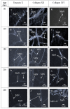
The sensory properties of beef are known to depend on muscle fiber and intramuscular connective tissue composition (IMCT). IMCT is composed of collagens, proteoglycans and glycoproteins. The differentiation of muscle fibers has been extensively studied but there is scarcity in the data concerning IMCT differentiation. In order to be able to control muscle differentiation to improve beef quality, it is essential to understand the ontogenesis of IMCT molecules. Therefore, in this study, we investigated the chronology of appearance of 10 IMCT molecules in bovine Semitendinosus muscle using immunohistology technique at five key stages of myogenesis. Since 60 days post-conception (dpc), the whole molecules were present, but did not have their final location. It seems that they reach it at around 210 dpc. Then, the findings emphasized that since 210 dpc, the stage at which the differentiation of muscle fibers is almost complete, the differentiation of IMCT is almost completed. These data suggested that for the best controlling of the muscular differentiation to improve beef sensory quality, it would be necessary to intervene very early (before the IMCT constituents have acquired their definitive localization and the muscle fibers have finished differentiating), i.e., at the beginning of the first third of gestation.
Keywords: bovine; extracellular matrix; fetus; immunohistology; skeletal muscle.
Conflict of interest statement
The authors declare no conflict of interest.
Figures


Similar articles
-
Role of extracellular matrix in development of skeletal muscle and postmortem aging of meat.Meat Sci. 2015 Nov;109:48-55. doi: 10.1016/j.meatsci.2015.05.015. Epub 2015 May 20. Meat Sci. 2015. PMID: 26141816
-
Are there consistent relationships between major connective tissue components, intramuscular fat content and muscle fibre types in cattle muscle?Animal. 2020 Jun;14(6):1204-1212. doi: 10.1017/S1751731119003422. Epub 2020 Jan 16. Animal. 2020. PMID: 31941561
-
The role of intramuscular connective tissue in meat texture.Anim Sci J. 2010 Feb;81(1):21-7. doi: 10.1111/j.1740-0929.2009.00696.x. Anim Sci J. 2010. PMID: 20163668 Review.
-
The Structure and Role of Intramuscular Connective Tissue in Muscle Function.Front Physiol. 2020 May 19;11:495. doi: 10.3389/fphys.2020.00495. eCollection 2020. Front Physiol. 2020. PMID: 32508678 Free PMC article. Review.
-
Influence of Canadian beef quality grade and method of intramuscular connective tissue isolation on collagen characteristics of the bovine longissimus thoracis.Meat Sci. 2022 Sep;191:108848. doi: 10.1016/j.meatsci.2022.108848. Epub 2022 May 16. Meat Sci. 2022. PMID: 35598426
Cited by
-
Beef Tenderness Prediction by a Combination of Statistical Methods: Chemometrics and Supervised Learning to Manage Integrative Farm-To-Meat Continuum Data.Foods. 2019 Jul 22;8(7):274. doi: 10.3390/foods8070274. Foods. 2019. PMID: 31336646 Free PMC article.
-
Current Advances in Meat Nutritional, Sensory and Physical Quality Improvement.Foods. 2020 Mar 10;9(3):321. doi: 10.3390/foods9030321. Foods. 2020. PMID: 32164289 Free PMC article.
-
Extracellular matrix deposition precedes muscle-tendon integration during murine forelimb morphogenesis.Commun Biol. 2025 Aug 12;8(1):1202. doi: 10.1038/s42003-025-08653-0. Commun Biol. 2025. PMID: 40796656 Free PMC article.
References
-
- Chriki S., Picard B., Jurie C., Reichstadt M., Micol D., Brun J.-P., Journaux L., Hocquette J.-F. Meta-analysis of the comparison of the metabolic and contractile characteristics of two bovine muscles: Longissimus thoracis and Semitendinosus. Meat Sci. 2012;91:423–429. doi: 10.1016/j.meatsci.2012.02.026. - DOI - PubMed
LinkOut - more resources
Full Text Sources

