. 2019 Mar 13;20(6):1271.
doi: 10.3390/ijms20061271.

Hypoxia Induces Astrocyte-Derived Lipocalin-2 in Ischemic Stroke

Affiliations

Hypoxia Induces Astrocyte-Derived Lipocalin-2 in Ischemic Stroke

Fatemeh Ranjbar Taklimie et al. Int J Mol Sci. .

Abstract

Ischemic stroke causes rapid hypoxic damage to the core neural tissue which is followed by graded chronological tissue degeneration in the peri-infarct zone. The latter process is mainly triggered by neuroinflammation, activation of inflammasomes, proinflammatory cytokines, and pyroptosis. Besides microglia, astrocytes play an important role in the fine-tuning of the inflammatory network in the brain. Lipocalin-2 (LCN2) is involved in the control of innate immune responses, regulation of excess iron, and reactive oxygen production. In this study, we analyzed LCN2 expression in hypoxic rat brain tissue after ischemic stroke and in astrocyte cell cultures receiving standardized hypoxic treatment. Whereas no LCN2-positive cells were seen in sham animals, the number of LCN2-positive cells (mainly astrocytes) was significantly increased after stroke. In vitro studies with hypoxic cultured astroglia revealed that LCN2 expression is significantly increased after only 2 h, then further increased, followed by a stepwise decline. The expression pattern of several proinflammatory cytokines mainly followed that profile in wild type (WT) but not in cultured LCN2-deficient astrocytes. Our data revealed that astrocytes are an important source of LCN2 in the peri-infarct region under hypoxic conditions. However, we must also stress that brain-intrinsic LCN2 after the initial hypoxia period might come from other sources such as invaded immune cells and peripheral organs via blood circulation. In any case, secreted LCN2 might have an influence on peripheral organ functions and the innate immune system during brain hypoxia.

Keywords: astrocyte; hypoxia; lipocalin-2; neuroinflammation.


Conflict of interest statement

The authors declare no conflicts of interests.

Figures

Figure 1
Figure 1
Regulation of LCN2 at (A) mRNA and (B) protein levels at various time points after tMCAO in the peri-infarct zone of the rat brain. (A) mRNA levels increased stepwise and changes became first significant after 24 h. (B) LCN2 protein amounts increased in a temporally delayed way as shown in representative WB samples and by densitometric evaluation of immune-labeled bands. (C) LCN2 ELISA determinations in the blood serum of rats show a significant increase of LCN2 coinciding well with the above described expression levels in the damaged cortical brain area. * p ≤ 0.05, ** p ≤ 0.01, and *** p ≤ 0.001.
Figure 2
Figure 2
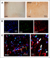
Immunohistochemical (A) and double-immunofluorescence (B) staining of LCN2 after stroke in the cerebral cortex of tMCAO rats. (A) Numbers of LCN2-positive cells massively increased after tMCAO. In sham-operated animals, almost no LCN2-positive cells were visible. (C) Double-labeling revealed that many GFAP-positive (red) astrocytes are also positive for LCN2 (turquoise-white). In (C), a blood vessel (stars) crosses the section, and thin elongated endothelial cells (yellow arrows) can be identified. At higher magnification, two LCN2-positive (white arrows) and a LCN2-negative (white arrowhead) can be seen.
Figure 3
Figure 3
Effect of hypoxia on (A) LCN2 gene expression and (B) protein levels of LCN2 in cultured primary astrocytes isolated from the cerebral cortex. (A) RT-qPCR analysis reveals a significant increase in LCN2 mRNA levels at 2 h post hypoxia. (B) LCN2 protein concentrations measured in supernatant of cultured hypoxic astrocytes at 2 h post hypoxia also shows a significant upregulation of LCN2. * p ≤ 0.05, ** p ≤ 0.01, and *** p ≤ 0.001. Norm, normoxia; Hypo, Hypoxia.
Figure 4
Figure 4
Effect of reoxygenation after an initial 2 h hypoxia on the mRNA expression profiles of LCN2 and various pro- and anti-inflammatory cytokines in mouse astrocyte cultures from the cerebral cortex of WT and LCN2-deficient animals. (A) LCN2 expression is significantly but temporarily induced in response in WT astrocytes 3 and 6 h after reoxygenation and returned to control level at 12 h reoxygenation. (BD) The profiles of HIF-1α, TNF-α, and IL-6 mRNA expression mainly follow the LCN2 pattern in WT astrocytes. Such an induction of HIF-1α, TNF-α and IL-6 expression was not observed in LCN2-deficient astrocytes. (E) In contrast, the anti-inflammatory cytokine IL-10 was significantly elevated in LCN2-deficient astroglia but only moderately elevated in WT glia. Rep, reoxygenation (reperfusion). * p ≤ 0.05, ** p ≤ 0.01, and *** p ≤ 0.001. The number of animals included for reoxygenation was n = 5 per time point. Norm, normoxia; Hypo, Hypoxia.
Figure 5
Figure 5
Schematic illustration of the in vitro hypoxia model used in this study.

Similar articles

Cited by

References

    1. Sarkar S., Chakraborty D., Bhowmik A., Ghosh M.K. Cerebral ischemic stroke: Cellular fate and therapeutic opportunities. Front. Biosci. (Landmark Ed.) 2019;24:435–450. - PubMed
    1. Zhang Q.Y., Wang Z.J., Sun D.M., Wang Y. Novel therapeutic effects of leonurine on ischemic stroke: New mechanisms of bbb integrity. Oxidative Med. Cell. Longev. 2017;2017:7150376. doi: 10.1155/2017/7150376. - DOI - PMC - PubMed
    1. Liddelow S.A., Guttenplan K.A., Clarke L.E., Bennett F.C., Bohlen C.J., Schirmer L., Bennett M.L., Munch A.E., Chung W.S., Peterson T.C., et al. Neurotoxic reactive astrocytes are induced by activated microglia. Nature. 2017;541:481–487. doi: 10.1038/nature21029. - DOI - PMC - PubMed
    1. Sofroniew M.V. Astrocyte barriers to neurotoxic inflammation. Nat. Rev. Neurosci. 2015;16:249–263. doi: 10.1038/nrn3898. - DOI - PMC - PubMed
    1. Gabryel B., Kasprowska D., Kost A., Labuzek K., Urbanek T. Astrocytes in ischemic stroke—A potential target for neuroprotective strategies. Postepy Hig. Med. Dosw. (Online) 2015;69:384–397. doi: 10.5604/17322693.1147866. - DOI - PubMed