

Doxorubicin Exposure Causes Subacute Cardiac Atrophy Dependent on the Striated Muscle-Specific Ubiquitin Ligase MuRF1
- PMID: 30871347
- PMCID: PMC6422170
- DOI: 10.1161/CIRCHEARTFAILURE.118.005234
Doxorubicin Exposure Causes Subacute Cardiac Atrophy Dependent on the Striated Muscle-Specific Ubiquitin Ligase MuRF1
Abstract
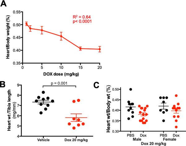
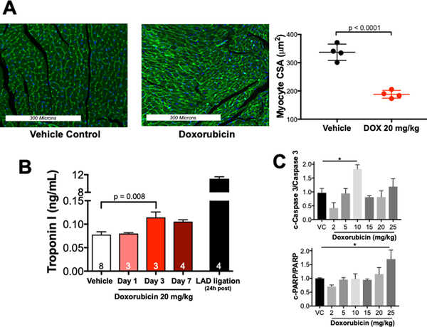
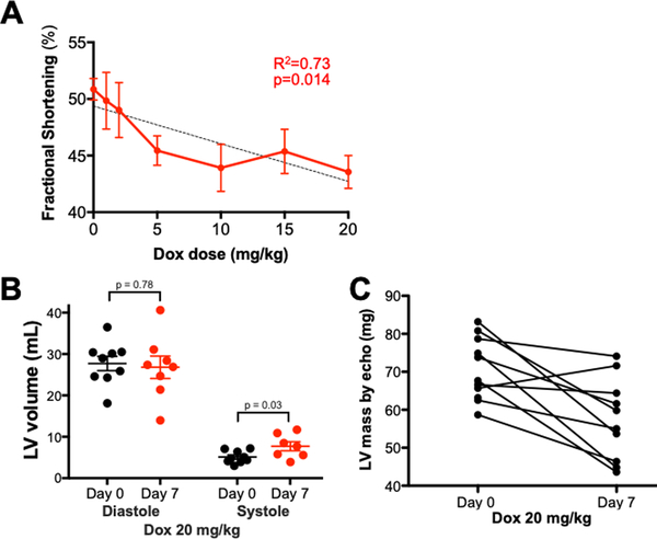
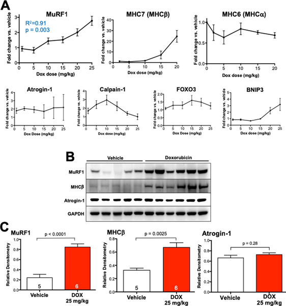
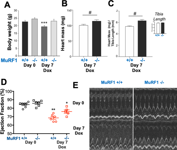
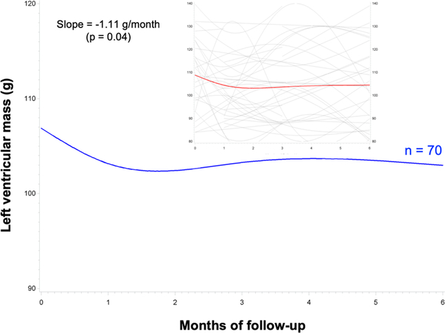
Background Anthracycline chemotherapeutics, such as doxorubicin, are used widely in the treatment of numerous malignancies. The primary dose-limiting adverse effect of anthracyclines is cardiotoxicity that often presents as heart failure due to dilated cardiomyopathy years after anthracycline exposure. Recent data from animal studies indicate that anthracyclines cause cardiac atrophy. The timing of onset and underlying mechanisms are not well defined, and the relevance of these findings to human disease is unclear. Methods and Results Wild-type mice were sacrificed 1 week after intraperitoneal administration of doxorubicin (1-25 mg/kg), revealing a dose-dependent decrease in cardiac mass ( R2=0.64; P<0.0001) and a significant decrease in cardiomyocyte cross-sectional area (336±29 versus 188±14 µm2; P<0.0001). Myocardial tissue analysis identified a dose-dependent upregulation of the ubiquitin ligase, MuRF1 (muscle ring finger-1; R2=0.91; P=0.003) and a molecular profile of muscle atrophy. To investigate the determinants of doxorubicin-induced cardiac atrophy, we administered doxorubicin 20 mg/kg to mice lacking MuRF1 (MuRF1-/-) and wild-type littermates. MuRF1-/- mice were protected from cardiac atrophy and exhibited no reduction in contractile function. To explore the clinical relevance of these findings, we analyzed cardiac magnetic resonance imaging data from 70 patients in the DETECT-1 cohort and found that anthracycline exposure was associated with decreased cardiac mass evident within 1 month and persisting to 6 months after initiation. Conclusions Doxorubicin causes a subacute decrease in cardiac mass in both mice and humans. In mice, doxorubicin-induced cardiac atrophy is dependent on MuRF1. These findings suggest that therapies directed at preventing or reversing cardiac atrophy might preserve the cardiac function of cancer patients receiving anthracyclines.
Keywords: anthracycline; atrophy; cardiotoxicity; doxorubicin; heart failure; mice; ubiquitin.
Figures
Comment in
-
Anthracycline Cardiotoxicity.Circ Heart Fail. 2019 Mar;12(3):e005910. doi: 10.1161/CIRCHEARTFAILURE.119.005910. Circ Heart Fail. 2019. PMID: 30871350 No abstract available.
References
-
- Bloom MW, Hamo CE, Cardinale D, Ky B, Nohria A, Baer L, Skopicki H, Lenihan DJ, Gheorghiade M, Lyon AR and Butler J. Cancer Therapy-Related Cardiac Dysfunction and Heart Failure: Part 1: Definitions, Pathophysiology, Risk Factors, and Imaging. Circulation Heart failure. 2016;9:e002661. - PMC - PubMed
-
- Mukku RB, Fonarow GC, Watson KE, Ajijola OA, Depasquale EC, Nsair A, Baas AS, Deng MC and Yang EH. Heart Failure Therapies for End-Stage Chemotherapy-Induced Cardiomyopathy. Journal of cardiac failure. 2016;22:439–48. - PubMed
-
- Luminari S, Montanini A, Caballero D, Bologna S, Notter M, Dyer MJ, Chiappella A, Briones J, Petrini M, Barbato A, Kayitalire L and Federico M. Nonpegylated liposomal doxorubicin (MyocetTM) combination (R-COMP) chemotherapy in elderly patients with diffuse large B-cell lymphoma (DLBCL): results from the phase II EUR018 trial. Annals of oncology : official journal of the European Society for Medical Oncology / ESMO. 2010;21:1492–9. - PubMed
-
- Tirelli U, Errante D, Van Glabbeke M, Teodorovic I, Kluin-Nelemans JC, Thomas J, Bron D, Rosti G, Somers R, Zagonel V and Noordijk EM. CHOP is the standard regimen in patients > or = 70 years of age with intermediate-grade and high-grade non-Hodgkin’s lymphoma: results of a randomized study of the European Organization for Research and Treatment of Cancer Lymphoma Cooperative Study Group. Journal of clinical oncology : official journal of the American Society of Clinical Oncology. 1998;16:27–34. - PubMed
-
- Cardinale D, Sandri MT, Martinoni A, Tricca A, Civelli M, Lamantia G, Cinieri S, Martinelli G, Cipolla CM and Fiorentini C. Left ventricular dysfunction predicted by early troponin I release after high-dose chemotherapy. Journal of the American College of Cardiology. 2000;36:517–22. - PubMed
Publication types
MeSH terms
Substances
Grants and funding
LinkOut - more resources
Full Text Sources
Medical
