

Immunohistochemical localization of NGF, BDNF, and their receptors in a normal and AMD-like rat retina
- PMID: 30871541
- PMCID: PMC6417162
- DOI: 10.1186/s12920-019-0493-8
Immunohistochemical localization of NGF, BDNF, and their receptors in a normal and AMD-like rat retina
Abstract
Background: Age-related macular degeneration (AMD) is a major cause of blindness in developed countries, and the molecular pathogenesis of AMD is poorly understood. A large body of evidence has corroborated the key role of neurotrophins in development, proliferation, differentiation, and survival of retinal cells. Neurotrophin deprivation has been proposed to contribute to retinal-cell death associated with neurodegenerative diseases. Little is known about the expression of the immature form of neurotrophins (proneurotrophins) and their mature form [e.g., nerve growth factor (proNGF and mNGF) and brain-derived neurotrophic factor (proBDNF and mBDNF)] in the retina during physiological aging and against the background of AMD. In addition, cell-specific localization of proteins NGF and BDNF in the retina during AMD development is not clear. Here, we evaluated contributions of the age-related alterations in the neurotrophin system to the development of AMD-like retinopathy in OXYS rats.
Methods: Male OXYS rats at preclinical (20 days), early (3 months), and late (18 months) stages of the disease and age-matched male Wistar rats (as controls) were used. We performed immunohistochemical localization of NGF, BDNF, and their receptors TrkA, TrkB, and p75NTR by fluorescence microscopy in retinal sections from OXYS and Wistar rats.
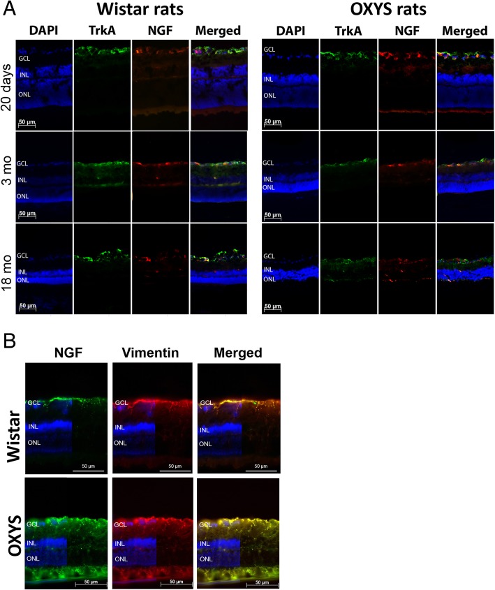
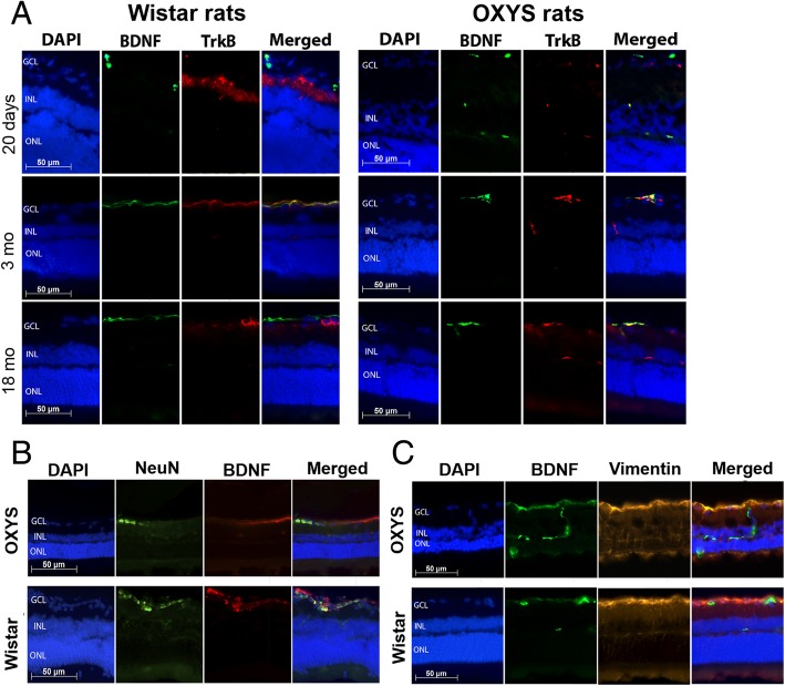
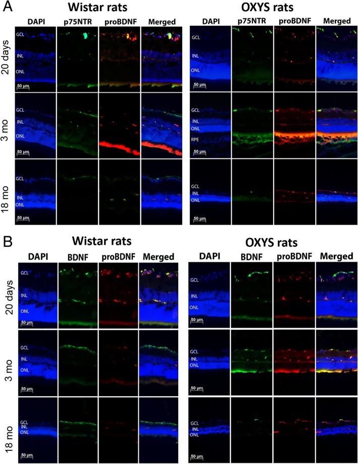
Results: We found increased NGF staining in Muller cells in 18-month-old OXYS rats (progressive stage of retinopathy). In contrast, we observed only subtle changes in the labeling of mature BDNF (mBDNF) and TrkB during the development of AMD-like retinopathy in OXYS rats. Using colocalization with vimentin and NeuN, we detected a difference in the cell type-specific localization of mBDNF between OXYS and Wistar rats. We showed that the mBDNF protein was located in Muller cells in OXYS rats, whereas in the Wistar retina, mBDNF immunoreactivity was detected in Muller cells and ganglion cells. During the development of AMD-like retinopathy, proBDNF dominated over mBDNF during increasing cell loss in the OXYS retina.
Conclusions: These data indicate that alterations in the balance of neurotrophic factors in the retina are involved in the development of AMD-like retinopathy in OXYS rats and confirm their participation in the pathogenesis of AMD in humans.
Keywords: Age-related macular degeneration; BDNF; NGF; OXYS rats; Retina; Retinopathy.
Conflict of interest statement
Ethics approval and consent to participate
All the animal procedures and experimental protocols were approved by the Institutional Review Board of the Institute of Cytology and Genetics, according to The Guidelines for Manipulations of Experimental Animals.
Consent for publication
Not applicable.
Competing interests
The authors declare that they have no competing interests.
Publisher’s Note
Springer Nature remains neutral with regard to jurisdictional claims in published maps and institutional affiliations.
Figures
References
Publication types
MeSH terms
Substances
LinkOut - more resources
Full Text Sources
Medical
