. 2019 Mar 12;16(1):60.
doi: 10.1186/s12974-019-1446-z.

Loss of NF-κB p50 function synergistically augments microglial priming in the middle-aged brain

Affiliations

Loss of NF-κB p50 function synergistically augments microglial priming in the middle-aged brain

Thomas Taetzsch et al. J Neuroinflammation. .

Abstract

Background: While NF-κB p50 function is impaired in central nervous system disease, aging in non-CNS tissues, and response to reactive oxygen species, the role of NF-κB p50 in aging-associated microglial pro-inflammatory priming is poorly understood.

Methods: Male NF-κB p50+/+ and NF-κB p50-/- mice at three different ages (1.5-3.0 month old, 8.0-11.0 month old, and 16.0-18.0 month old) were treated with LPS (5 mg/kg, IP) to trigger peripheral inflammation, where circulating cytokines, neuroinflammation, microglia morphology, and NF-κB p50/p65 function in brain tissue were determined 3 h later.

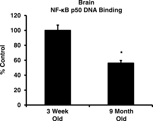
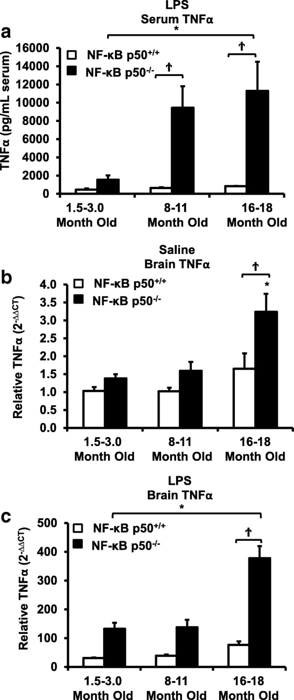
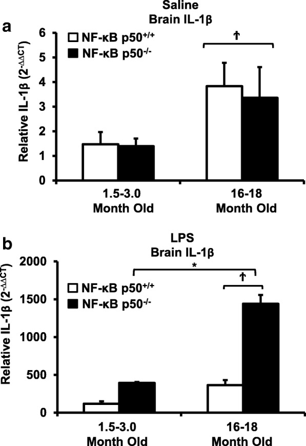
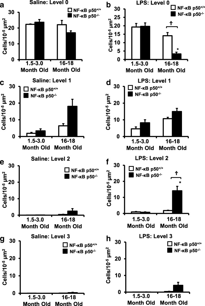
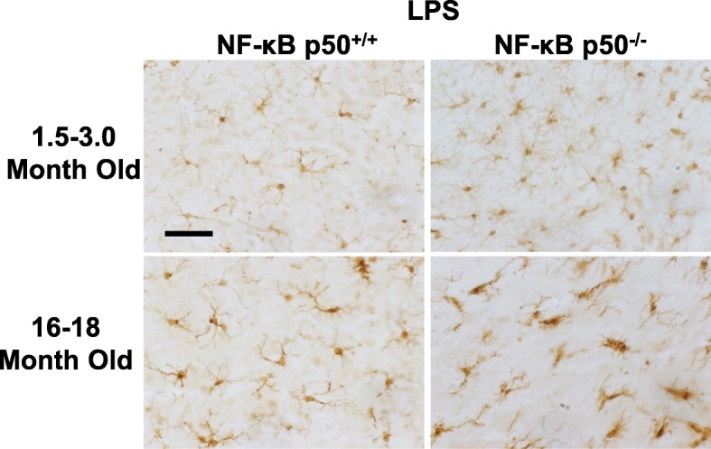
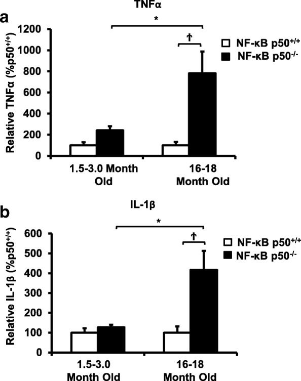
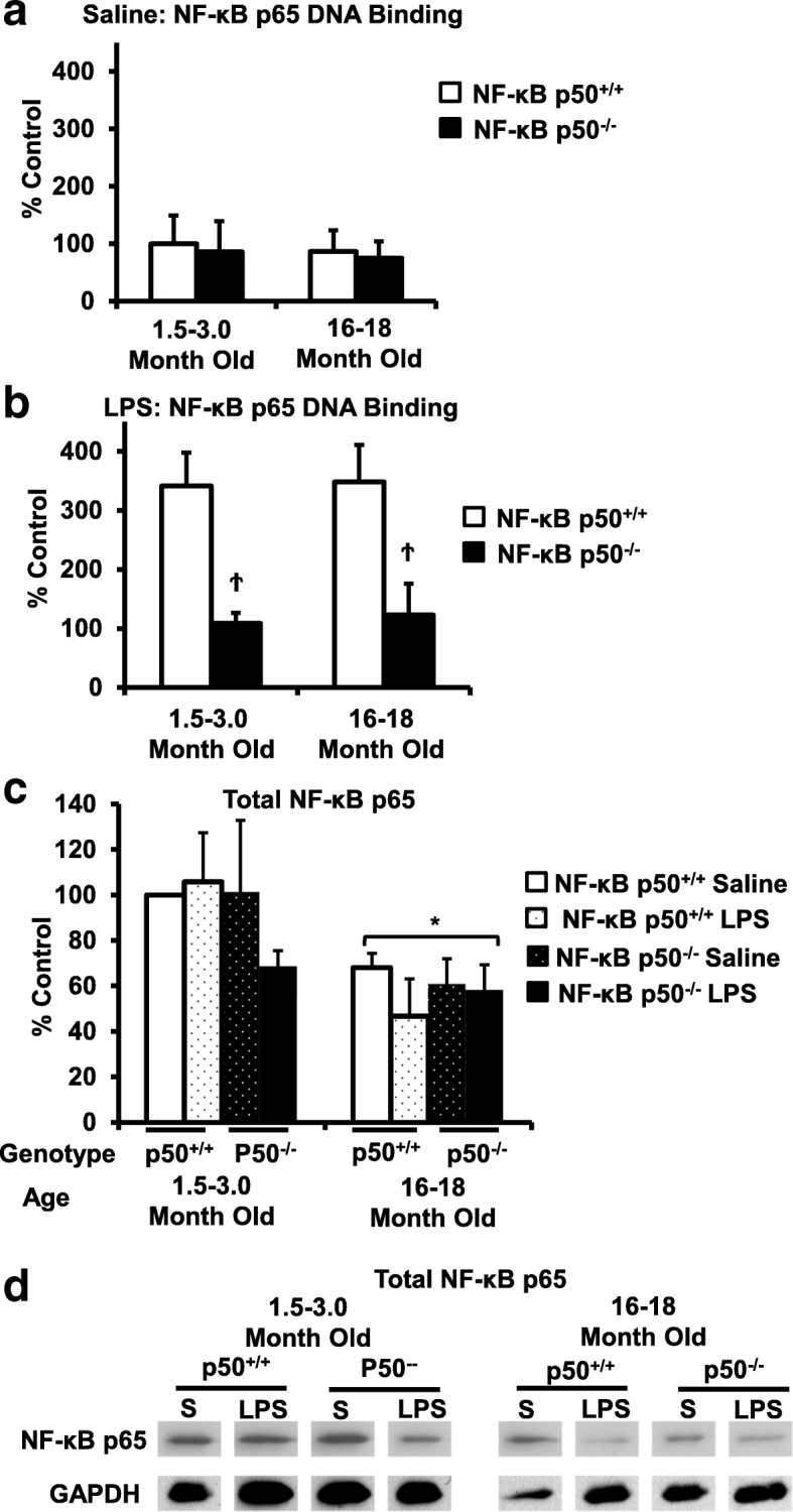
Results: Peripheral LPS injection in 9-month-old C57BL/6 mice resulted in lower NF-κB p50 DNA binding of nuclear extracts from the whole brain, when compared to 3-week-old C57BL/6 mice, revealing differences in LPS-induced NF-κB p50 activity in the brain across the mouse lifespan. To examine the consequences of loss NF-κB p50 function with aging, NF-κB p50+/+ and NF-κB p50-/- mice of three different age groups (1.5-3.0 month old, 8.0-11.0 month old, and 16.0-18.0 month old) were injected with LPS (5 mg/kg, IP). NF-κB p50-/- mice showed markedly elevated circulating, midbrain, and microglial TNFα when compared to NF-κB p50+/+ mice at all ages. Notably, the 16.0-18.0-month-old (middle aged) NF-κB p50-/- mice exhibited synergistically augmented LPS-induced serum and midbrain TNFα when compared to the younger (1.5-3.0 month old, young adult) NF-κB p50-/- mice. The 16.0-18.0-month-old LPS-treated NF-κB p50-/- mice also had the highest midbrain IL-1β expression, largest number of microglia with changes in morphology, and greatest elevation of pro-inflammatory factors in isolated adult microglia. Interestingly, aging NF-κB p50-/- mice exhibited decreased brain NF-κB p65 expression and activity.

Conclusions: These findings support that loss of NF-κB p50 function and aging in middle-aged mice may interact to excessively augment peripheral/microglial pro-inflammatory responses and point to a novel neuroinflammation signaling mechanism independent the NF-κB p50/p65 transcription factor in this process.

Keywords: Aging; Microglia; NF-κB; Priming.


Conflict of interest statement

Ethics approval and consent to participate

This research was performed within the strict NIH ethics and guidelines with the approval of the IUSM IACUC committee.

Consent for publication

Not applicable.

Competing interests

The authors declare that they have no competing interests.

Publisher’s Note

Springer Nature remains neutral with regard to jurisdictional claims in published maps and institutional affiliations.

Figures

Fig. 1
Fig. 1
LPS-induced NF-κB p50 DNA binding changes across the mouse lifespan. NF-κB p50 activity was assessed by determining NF-κB p50 DNA binding in whole brain nuclear extract from 9-month-old and 3-week-old C57BL/6 mice injected with LPS (5 mg/kg, IP). Whole brain nuclear extract from the older mice injected with LPS showed reduced DNA binding when compared to the younger mice. Values are reported as mean percent of 3 week saline control ± SEM. An asterisk indicates significant difference (P < 0.05) from control. n = 4
Fig. 2
Fig. 2
Loss of NF-κB p50 function and aging synergistically interact to augment serum and brain TNFα in middle-aged mice. NF-κB p50+/+ and NF-κB p50−/− mice from three different age groups (1.5–3.0 month old, 8.0–11.0 month old, and 16.0–18.0 month old) were injected with saline or LPS (5 mg/kg, IP) to determine age-related effects of loss of NF-κB p50 function on peripheral and neuro-inflammation at 3 h post-injection. a Circulating TNFα protein was measured by ELISA. Values are reported as mean expression ± SEM. Midbrain TNFα mRNA levels were assessed following b saline or c LPS treatment by quantitative RT-PCR. Values were normalized to GAPDH using the 2−ΔΔCT method and are reported as mean percent of saline control ± SEM. An asterisk indicates significant difference (P < 0.05) from the 1.5–3.0-month-old group and a dagger indicates a significant difference between mouse strains. n = 3–7
Fig. 3
Fig. 3
Loss of NF-κB p50 function and aging synergistically interact to augment midbrain IL-1β in middle-aged mice. Young adult (1.5–3.0 month old) and middle-aged (16.0–18.0 month old) NF-κB p50+/+ and NF-κB p50−/− mice were treated with a saline or b LPS (5 mg/kg, IP), and brain tissue was collected at 3 h post-injection. IL-1β expression in the brain was evaluated by quantitative RT-PCR. Values are normalized to GAPDH using the 2−ΔΔCT method and are reported as mean percent of NF-κB p50+/+ young adult saline control ± SEM. An asterisk indicates significant difference (P < 0.05) from the 1.5–3.0-month old group and a dagger indicates a significant difference between mouse strains. n = 3
Fig. 4
Fig. 4
Aging NF-κB p50−/− mice have enhanced activated microglia morphology in response to peripheral LPS injection. Young adult (1.5–3.0 month old) and middle-aged (16.0–18.0 month old) NF-κB p50+/+ and NF-κB p50−/− mice were injected with saline or LPS (5 mg/kg, IP) to evaluate loss of NF-κB p50 function on microglia morphology in vivo. IBA1 stained microglia within the substantia nigra pars compacta (in the midbrain) were categorized into stages of activation ranging from resting (stage 0) to highly activated (stage 3). The relative number of microglia at 3 h post-injection within stage 0 (a, b), stage 1(c, d), stage 2(e, f), and g & h stage 3(g, h) was quantified by the fractionator method. Values are reported as mean cells/μm2 ± SEM of 3 coronal sections (40 μm) per animal. An asterisk indicates significant difference (P < 0.05) from control and a dagger indicates a difference between mouse strains. n = 3
Fig. 5
Fig. 5
Representative images demonstrating augmented changes in microglia morphology in middle-aged NF-κB p50−/− mice. Young adult (1.5–3.0 month old) and middle-aged (16.0–18.0 month old) NF-κB p50+/+ and NF-κB p50−/− mice were injected with saline or LPS (5 mg/kg, IP) to evaluate loss of NF-κB p50 function on microglia morphology in vivo. Three coronal sections (40 μm) per animal containing the substantia nigra pars compacta were stained with IBA1. Representative images are shown from the substantia nigra pars compacta at × 40 and the scale bar depicts 50 μM. n = 3
Fig. 6
Fig. 6
Loss of NF-κB p50 function and aging synergistically interact to amplify TNFα and IL-1β in isolated microglia. Young adult (1.5–3.0 month old) and middle-aged (16.0–18.0 month old) NF-κB p50+/+ and NF-κB p50−/− mice were injected with LPS (5 mg/kg, IP), and isolation of microglia from the whole brain with CD11b microbeads was performed at 3 h post-injection. Differences in a TNFα and b IL-1β were evaluated by quantitative RT-PCR. Values are normalized to GAPDH using the 2−ΔΔCT method and are reported as mean percent of NF-κB p50+/+ young adult saline control ± SEM. An asterisk indicates significant difference (P < 0.05) from the 1.5–3-month group and a dagger indicates a difference between mouse strains. n = 3
Fig. 7
Fig. 7
NF-κB p65 DNA expression and activity in aging and NF-κBp50−/− mice. Young adult (1.5–3.0 month old) and middle-aged (16.0–18.0 month old) NF-κB p50+/+ and NF-κB p50−/− mice were injected with saline or LPS (5 mg/kg, IP). Nuclear extract collected from whole brain tissue at 3 h following a saline or b LPS injection was evaluated for NF-κB p65 DNA binding with ELISA. c Total NF-κB p65 protein in brain tissue at 3 h post-injection was evaluated by western blot and normalized to GAPDH. d Representative image of NF-κB p65 and GAPDH western blot bands used to calculate the total NF-κB p65 brain protein. Values are reported as mean percent of saline control ± SEM. An asterisk indicates a significant age effect (P < 0.05) and a dagger indicates a significant difference between mouse strains (P < 0.05). n = 3

References

    1. Hickman SE, Kingery ND, Ohsumi TK, Borowsky ML, Wang LC, Means TK, El Khoury J. The microglial sensome revealed by direct RNA sequencing. Nat Neurosci. 2013;16:1896–1905. doi: 10.1038/nn.3554. - DOI - PMC - PubMed
    1. Sun Q, Wu W, Hu YC, Li H, Zhang D, Li S, Li W, Li WD, Ma B, Zhu JH, et al. Early release of high-mobility group box 1 (HMGB1) from neurons in experimental subarachnoid hemorrhage in vivo and in vitro. J Neuroinflammation. 2014;11:106. doi: 10.1186/1742-2094-11-106. - DOI - PMC - PubMed
    1. Rosenberger K, Derkow K, Dembny P, Kruger C, Schott E, Lehnardt S. The impact of single and pairwise toll-like receptor activation on neuroinflammation and neurodegeneration. J Neuroinflammation. 2014;11:166. doi: 10.1186/s12974-014-0166-7. - DOI - PMC - PubMed
    1. Aguzzi A, Barres BA, Bennett ML. Microglia: scapegoat, saboteur, or something else? Science. 2013;339:156–161. doi: 10.1126/science.1227901. - DOI - PMC - PubMed
    1. Block ML, Zecca L, Hong JS. Microglia-mediated neurotoxicity: uncovering the molecular mechanisms. Nat Rev Neurosci. 2007;8:57–69. doi: 10.1038/nrn2038. - DOI - PubMed

MeSH terms