. 2019 Mar 14;7(1):74.
doi: 10.1186/s40425-019-0553-9.

Cocultures of human colorectal tumor spheroids with immune cells reveal the therapeutic potential of MICA/B and NKG2A targeting for cancer treatment

Affiliations

Cocultures of human colorectal tumor spheroids with immune cells reveal the therapeutic potential of MICA/B and NKG2A targeting for cancer treatment

Tristan Courau et al. J Immunother Cancer. .

Abstract

Background: Immunotherapies still fail to benefit colorectal cancer (CRC) patients. Relevant functional assays aimed at studying these failures and the efficacy of cancer immunotherapy in human are scarce. 3D tumor cultures, called tumor organoids or spheroids, represent interesting models to study cancer treatments and could help to challenge these issues.

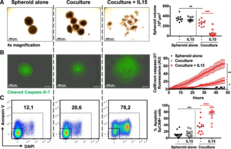
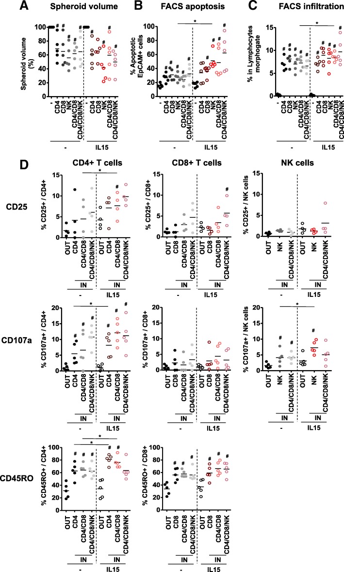
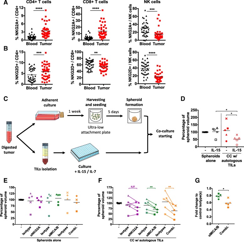
Methods: We analyzed heterotypic cocultures of human colon tumor-derived spheroids with immune cells to assess the infiltration, activation and function of T and NK cells toward human colorectal tumors in vitro.

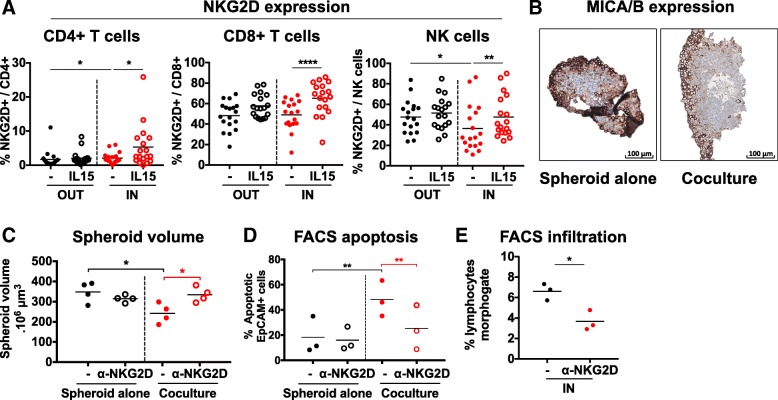
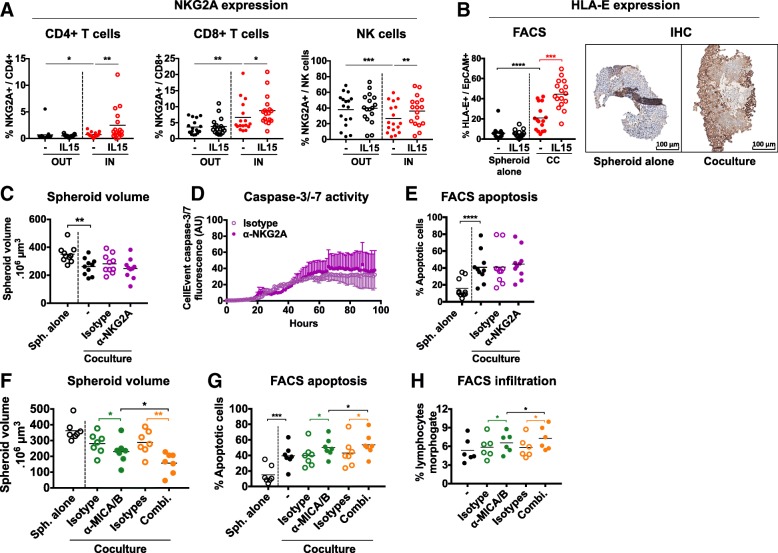
Results: We showed that allogeneic T and NK cells rapidly infiltrated cell line-derived spheroids, inducing immune-mediated tumor cell apoptosis and spheroid destruction. NKG2D, a key activator of cytotoxic responses, was engaged on infiltrating cells. We thus assessed the therapeutic potential of an antibody targeting the specific ligands of NKG2D, MICA and MICB, in this system. Anti-MICA/B enhanced immune-dependent destruction of tumor spheroid by driving an increased NK cells infiltration and activation. Interestingly, tumor cells reacted to immune infiltration by upregulating HLA-E, ligand of the inhibitory receptor NKG2A expressed by CD8 and NK cells. NKG2A was increased after anti-MICA/B treatment and, accordingly, combination of anti-MICA/B and anti-NKG2A was synergistic. These observations were ultimately confirmed in a clinical relevant model of coculture between CRC patients-derived spheroids and autologous tumor-infiltrating lymphocytes.

Conclusions: Altogether, we show that tumor spheroids represent a relevant tool to study tumor-lymphocyte interactions on human tissues and revealed the antitumor potential of immunomodulatory antibodies targeting MICA/B and NKG2A.

Keywords: Colorectal cancer; Immunotherapy; MICA/B; NKG2A; Spheroids.


Conflict of interest statement

Ethics approval and consent to participate

This study was approved by the French ethical committee (approval n°2016/45), and all subjects gave written informed consent.

Consent for publication

Not applicable

Competing interests

RR, LAM and MB are employees of Innate Pharma. This work was partly supported by a collaborative grant obtained from Innate Pharma by TA, MA and LLB.

Publisher’s Note

Springer Nature remains neutral with regard to jurisdictional claims in published maps and institutional affiliations.

Figures

Fig. 1
Fig. 1
Allogeneic activated/memory T and NK cells are able to infiltrate HT29 tumor spheroids. a Scheme of the coculture (CC) protocol between HT29 spheroids and CD19-CD14- sorted PBMCs from healthy donors. b Immunofluorescence (n = 2 independent experiments) and flow cytometry (n = 19 independent experiments) analyses of spheroid immune infiltration in the presence or not of IL-15 at 24 h. c Flow cytometry analyses of T and NK cells (respectively gated CD3+ and CD3-CD56+ among live single cells lymphocytes) as well as CD4+ and CD8+ T cells subsets (respectively gated CD4 + CD8- and CD4-CD8+ among CD3+) percentages in the IN and OUT compartments, in the presence or not of IL-15 at 24 h. n = 19 independent experiments. d Flow cytometry analyses of CD25, CD107a and CD45RO expression by CD4+ T cells, CD8+ T cells and NK cells in the IN and OUT compartments in the presence or not of IL-15 at 24 h. n = 8 to 18 independent experiments. Statistical significance of immunofluorescence experiments was analyzed using the Mann-Whitney test, the others using Wilcoxon matched-pairs signed rank test (* p < 0.05; ** p < 0.005, *** p < 0.001, **** p < 0.0001)
Fig. 2
Fig. 2
Immune infiltration and activation in HT29 spheroids lead to tumor cell apoptosis and spheroid destruction. HT29 spheroids were cocultured or not with CD19-CD14- PBMCs and IL-15 and we monitored a spheroid volume using microscopic pictures, b the dynamic cleavage of caspase-3/− 7 in the spheroids using Incucyte live imaging system, and c the staining by Annexin V and DAPI dyes of EpCAM+ cells by flow cytometry at 48 h. n = 4 to 12 independent experiments. Statistical significance of A panel was analyzed using 2-way ANOVA, the other panels using the Wilcoxon matched-pairs signed rank test (* p < 0.05; ** p < 0.005, *** p < 0.001, **** p < 0.0001)
Fig. 3
Fig. 3
Specific effects of CD4, CD8 and NK cells cocultured with HT29 spheroids alone or combined. HT29 spheroids were cocultured with sorted CD4 T cells, CD8 T cells and NK cells at a 1:1 ratio, alone or in combination and with or without IL-15. We analyzed a spheroid destruction through analysis of macroscopic pictures and b tumor cell apoptosis using flow cytometry after 48 h as well as c immune infiltration and d expression of CD25, CD107a and CD45RO by CD4 T cells, CD8 T cells and NK cells in IN and OUT compartments after 24 h. n = 4 to 5 independent experiments. Statistical significance was analyzed using the Wilcoxon matched-pairs signed rank test (# p = 0.06, as compared to spheroid alone in A to C panels and to OUT cells in D panels)
Fig. 4
Fig. 4
NKG2D-MICA/B pathway is engaged during the cocultures. a NKG2D expression by CD4 T cells, CD8 T cells and NK cells in the IN and OUT compartments, in the presence or not of IL-15, as measured by flow cytometry at 24 h. n = 18 independent experiments b MICA/B expression by tumor cells in the spheroids cocultured or not with CD19-CD14- PBMCs, as measured by immunohistochemistry at 24 h. Representative pictures of 1 experiment. c to e Analyses of c spheroid volume, d tumor cell apoptosis and e spheroid infiltration 48 h after coculturing HT29 spheroids with CD19-CD14- PBMCs in the presence or not of anti-NKG2D blocking antibodies. n = 3 to 4 independent experiments. Statistical significance of A panel was analyzed using the Wilcoxon matched-pairs signed rank test, C to E panels using paired t test (* p < 0.05; ** p < 0.005, *** p < 0.001, **** p < 0.0001)
Fig. 5
Fig. 5
Anti-MICA/B induce immune-mediated anti-tumor effects through increased NK cell infiltration and activation in HT29 spheroids. HT29 spheroids were cocultured or not with CD19-CD14- PBMCs in the presence or not of anti-MICA/B antibodies or corresponding control isotype. We analyzed tumor cell death and spheroid destruction using (a, n = 10) microscopic pictures at 48 h, (b, n = 4) Incucyte live imaging or (c, n = 10) flow cytometry at 48 h, as well as (d and e) overall or (f and g) particular T and NK cells infiltration using flow cytometry (d, f and g, n = 10) and immunofluorescence (e, n = 1) at 24 h. We also analyzed by flow cytometry the expression of (h, n = 8) NKG2D, (i, n = 6) CD137, (j, n = 7) CD16 and (k, n = 8) NKG2A by NK cells in the IN and OUT compartments at 24 h. Statistical significance of immunofluorescence experiment was analyzed using the Mann-Whitney test, A panel using 2-way ANOVA and the other panels using the Wilcoxon matched-pairs signed rank test (* p < 0.05; ** p < 0.005, *** p < 0.001, **** p < 0.0001)
Fig. 6
Fig. 6
NKG2A-HLA-E pathway is engaged during the cocultures and NKG2A blockade synergizes with anti-MICA/B antibodies. a NKG2A expression by CD4 T cells, CD8 T cells and NK cells in the IN and OUT compartments and in the presence or not of IL-15, analyzed by flow cytometry at 24 h. n = 17 independent experiments. b HLA-E expression by tumor cells in the spheroids cocultured or not with CD19-CD14- PBMCs, analyzed by flow cytometry (n = 16 independent experiments) and immunohistochemistry (representative pictures of 1 experiment) at 24 h. c to e Analyses of (c, n = 4) caspase-3/− 7 activity, (d, n = 10) spheroid volume, and (e, n = 10) spheroid infiltration 48 h after coculturing HT29 spheroids with CD19-CD14- PBMCs in the presence or not of anti-NKG2A blocking antibodies or corresponding control isotype. f to h Analyses of (f, n = 7) spheroid volume, (g, n = 7) tumor cell apoptosis, and (h, n = 7) spheroid infiltration 48 h after coculturing HT29 spheroids with CD19-CD14- PBMCs in the presence or not of anti-MICA/B antibodies alone or combined with anti-NKG2A blocking antibodies, or with corresponding control isotype antibodies alone or combined. Statistical significance of c panel was analyzed using 2-way ANOVA, the other panels using the Wilcoxon matched-pairs signed rank test (* p < 0.05; ** p < 0.005, *** p < 0.001, **** p < 0.0001)
Fig. 7
Fig. 7
Cocultures between patients-derived spheroids and autologous TILs confirm therapeutic potential of MICA/B and NKG2A targeting. a and b Flow cytometry analyses of a NKG2A and b NKG2D expression by CD4 T cells, CD8 T cells and NK cells in the blood and tumor of colorectal cancer patients. n = 41 independent experiments. c Scheme of the protocol used to create primary colon tumor-derived spheroids and maintain autologous TILs in culture before cocultures. d Representation of the change in spheroid volume 48 h after coculturing or not primary CRC-derived spheroids and autologous TILs in the presence or not of IL-15, measured using microscopic pictures. e to g Change in spheroid volume 48 h after culturing primary CRC-derived spheroids with (f and g) or without (e) autologous TILs, in the presence or not of anti-MICA/B, anti-NKG2A or corresponding control isotypes. In d to f panels, spheroid volumes are normalized to the culture condition without stimulation. g panel represents fold changes between anti-MICA/B and its isotype, and between combination and combination of isotypes. In d to g panels, each patient is represented by a specific symbol. n = 5 independent experiments. Statistical significance of a and b panels was analyzed Wilcoxon matched-pairs signed rank test, the other panels using paired t test (* p < 0.05; ** p < 0.005, *** p < 0.001, **** p < 0.0001)

References

    1. Malmberg K-J, Carlsten M, Björklund A, Sohlberg E, Bryceson YT, Ljunggren H-G. Natural killer cell-mediated immunosurveillance of human cancer. Semin Immunol. 2017;31:20–29. doi: 10.1016/j.smim.2017.08.002. - DOI - PubMed
    1. Golstein P, Griffiths GM. An early history of T cell-mediated cytotoxicity. Nat Rev Immunol. 2018;18(8):527–35. - PubMed
    1. Melero I, Rouzaut A, Motz GT, Coukos G. T-cell and NK-cell infiltration into solid tumors: a key limiting factor for efficacious Cancer immunotherapy. Cancer Discov. 2014;4:522–526. doi: 10.1158/2159-8290.CD-13-0985. - DOI - PMC - PubMed
    1. Sharma P, Allison JP. The future of immune checkpoint therapy. Science. 2015;348:56–61. doi: 10.1126/science.aaa8172. - DOI - PubMed
    1. Pitt JM, Vétizou M, Daillère R, Roberti MP, Yamazaki T, Routy B, et al. Resistance mechanisms to immune-checkpoint blockade in cancer: tumor-intrinsic and -extrinsic factors. Immunity. 2016;44:1255–1269. doi: 10.1016/j.immuni.2016.06.001. - DOI - PubMed

Publication types

MeSH terms

Substances