. 2019 Mar 14;17(1):82.
doi: 10.1186/s12967-019-1822-6.

Production of a cellular product consisting of monocytes stimulated with Sylatron® (Peginterferon alfa-2b) and Actimmune® (Interferon gamma-1b) for human use

Affiliations

Production of a cellular product consisting of monocytes stimulated with Sylatron® (Peginterferon alfa-2b) and Actimmune® (Interferon gamma-1b) for human use

Daniel S Green et al. J Transl Med. .

Abstract

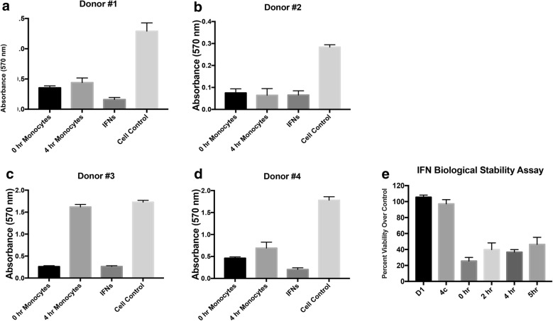
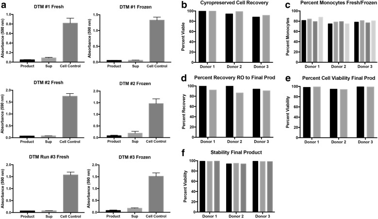
Background: Monocytes are myeloid cells that reside in the blood and bone marrow and respond to inflammation. At the site of inflammation, monocytes express cytokines and chemokines. Monocytes have been shown to be cytotoxic to tumor cells in the presence of pro-inflammatory cytokines such as Interferon Alpha, Interferon Gamma, and IL-6. We have previously shown that monocytes stimulated with both interferons (IFNs) results in synergistic killing of ovarian cancer cells. We translated these observations to an ongoing clinical trial using adoptive cell transfer of autologous monocytes stimulated ex vivo with IFNs and infused into the peritoneal cavity of patients with advanced, chemotherapy resistant, ovarian cancer. Here we describe the optimization of the monocyte elutriation protocol and a cryopreservation protocol of the monocytes isolated from peripheral blood.

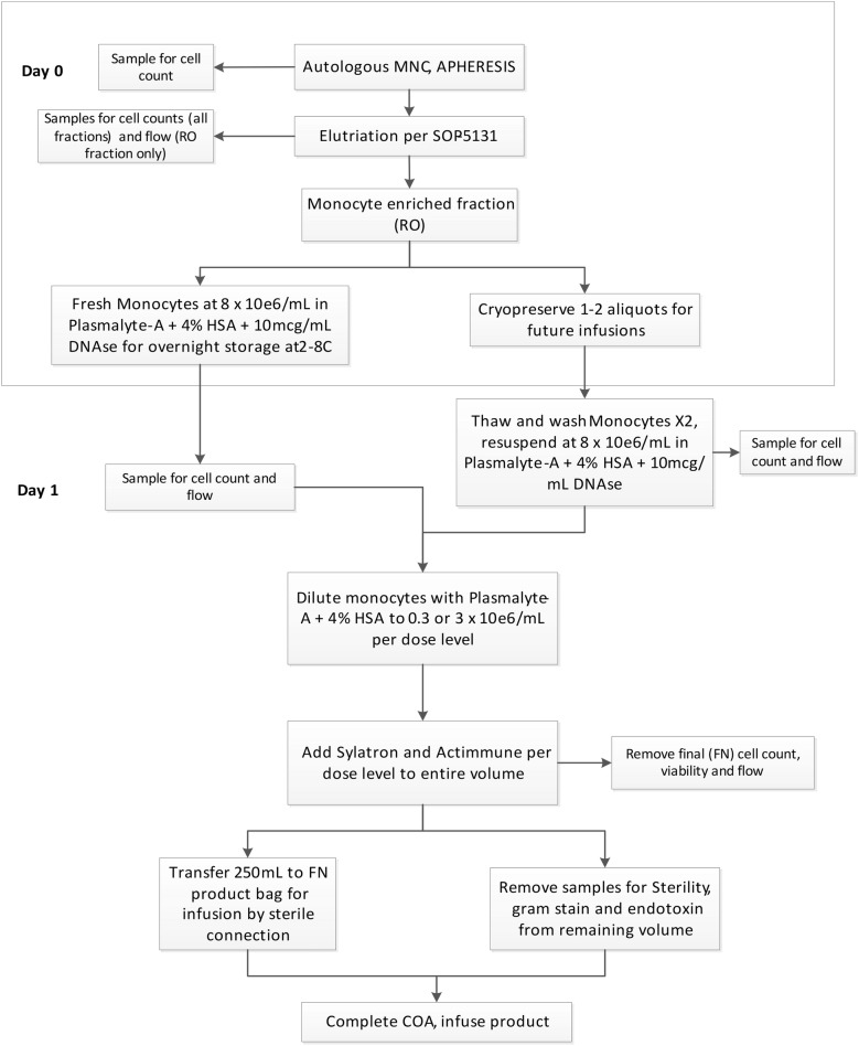
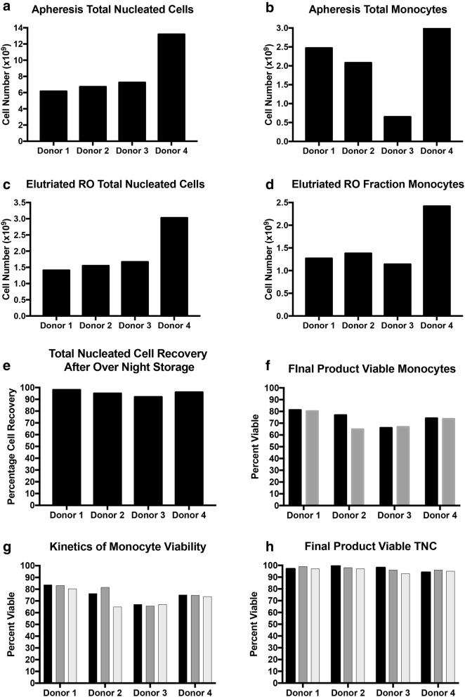
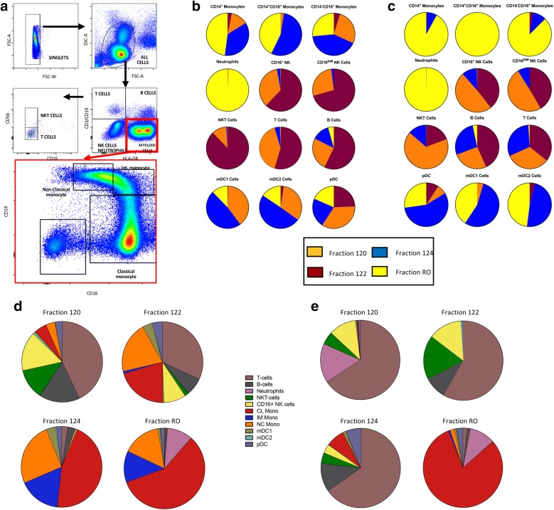
Methods: Counter flow elutriation was performed on healthy donors or women with ovarian cancer. The monocyte-containing, RO-fraction was assessed for total monocyte number, purity, viability, and cytotoxicity with and without a cryopreservation step. All five fractions obtained from the elutriation procedure were also assessed by flow cytometry to measure the percent of immune cell subsets in each fraction.

Results: Both iterative monocyte isolation using counter flow elutriation or cryopreservation following counter flow elutriation can yield over 2 billion monocytes for each donor with high purity. We also show that the monocytes are stable, viable, and retain cytotoxic functions when cultured with IFNs.

Conclusion: Large scale isolation of monocytes from both healthy donors and patients with advanced, chemotherapy resistant ovarian cancer, can be achieved with high total number of monocytes. These monocytes can be cryopreserved and maintain viability and cytotoxic function. All of the elutriated cell fractions contain ample immune cells which could be used for other cell therapy-based applications.

Keywords: Cell therapy; Cellular immunotherapy; Innate immunity; Interferons; Monocytes.


Conflict of interest statement

The authors declare that they have no competing interests.

Figures

Fig. 1
Fig. 1
Work flow of product creation from for fresh and frozen monocytes. This schematic shows the work flow from apheresis to either creation of the final product using fresh or cryopreserved monocytes
Fig. 2
Fig. 2
Quantification of cell numbers and viability. Apheresis followed by counter-flow elutriation was performed on 4 healthy donors. a Total nucleated cells and total monocytes from the bullk apheresis shown for all donors as total cell number. b Total monocytes from the bulk apheresis. c Total Nucleated Cells in the RO fraction. d Total monocytes in the RO fraction. e Recovery of total nucleated cells from overnight storage was measured. f Viability of the final product monocytes at 0 h (black bars) and 4 h (grey bars) was measured. g Kinetics monocyte viability the on day 1 (black bars), day 2 (grey bars) and the final product were measured. h Total nucleated cell count was performed on the final product at 0 h (black bars), 2 h (grey bars), and 4 h (white bars)
Fig. 3
Fig. 3
Flow cytometric analysis and quantification of immune subtypes in the four fractions of counter-flow elutriation. a A condensed work flow of flow cytometry of of cells from counter flow elutriation, with a focus on the monocyte population (red box). b Percentages of immune subtypes in all fractions from healthy donor 1. c Percentages of immune subtypes in all fractions from healthy donor 2. d Proportion of each immune subtype by fraction of elutriation for healthy donor 1. e Proportion of immune subtypes by fraction of elutriation for healthy donor 2 (complete work-flow analysis in Additional file 1: Table S3, Additional file 2: Figure S1, Additional file 3: Figure S2, Additional file 4: Figure S3 and Additional file 5: Figure S4)
Fig. 4
Fig. 4
Workflow for elutriated fraction RO for the quantification of percent monocytes. a Cells from the final product from a healthy donor after the addition of interferons were gated on singlets, followed by CD45+ cells, live cells, and then the CD15Lin population. Total monocytes were counted as the combination of Classical (CD14+), intermediate (CD14+CD16+), and non-classical CD14/dimCD16+. b Shows the backgating of the monocyte population. c Analysis of the same product from a after 4 h incubation at room temperature. d Shows the backgating strategy as in b
Fig. 5
Fig. 5
Cytotoxicity and IFN stability. Apheresis and counter-flow elutriation was performed on four donors. A final product was generated combining the monocytes with IFNs at the second dose level (a, b) and the fourth dose level (c, d). The products were assayed for cytotoxicity to OVCAR3 ovarian cancer cells. Time 0 h product (black bars), product stored 4 h at room temperature (light grey), IFNs alone (dark grey), and cell medium supernatant without IFNs (white bar) were measured. e IFN stability was measured after incubation at room temperature for 0, 2, 4, and 5 h; initial (D1) and overnight (4c) monocyte supernatants are used as controls, for cytotoxicity with OVCAR3 cells
Fig. 6
Fig. 6
Cytotoxicity of the final product before and after cryopreservation. Three separate apheresis and elutriation runs were performed. a At the time of product release both final product (IFNs and monocytes) and the IFNs alone were assayed for toxicity using OVCAR3 cells as the target. b Percent viable total nucleated cells (black bars) and the final product (grey bars). c Percent fresh monocytes in the RO fraction (black bars), the RO cryopreserved (light grey bars), final product fresh (dark grey bars), and final product cryopreserved (white bars). d Percent recovery from the fresh RO fraction (black bars) and cryopreserved RO fraction (grey bars). e Percent viability of the fresh final product (black bars) and cryopreserved product (grey bars). f Stability of the product measured by monocyte viability at 0 h (black bars), 2 h (light grey bars), and 4 h (dark grey bars)
Fig. 7
Fig. 7
Toxicity screen of Sylatron and Actimmune on primary human cells. a Myofibroblasts, b renal epithelial cells, c human vascular endothelial cells (HUVEC), d primary hepatocytes were assayed with Sylatron (light green bars), Actimmune (light blue bars), Sylatron and Actimmune (light red bars) or Sylatron and monocytes (dark green bars), Actimmune and monocytes (dark blue bars), Sylatron, Actimmune, and monocytes (dark red bars) for 3 days and cell viability was measured using trypan blue. Staurosporine (black bars) was used as a positive control for cell toxicity. Experiments were performed three times with three healthy donor monocytes, except for the hepatocytes which were repeated three times with three separate primary hepatocyte donors. Yellow asterisks indicate comparison to cell control; purple indicate comparison to monocyte-only control. Numbers of asterisks indicate p-value as follows: * < 0.05; ** < 0.01; *** < 0.001; **** < 0.0001

References

    1. Yang JC, Rosenberg SA. Current approaches to the adoptive immunotherapy of cancer. Adv Exp Med Biol. 1988;233:459–467. doi: 10.1007/978-1-4899-5037-6_50. - DOI - PubMed
    1. Draper LM, Kwong ML, Gros A, Stevanovic S, Tran E, Kerkar S, et al. Targeting of HPV-16+ epithelial cancer cells by TCR gene engineered T cells directed against E6. Clin Cancer Res. 2015;21(19):4431–4439. doi: 10.1158/1078-0432.CCR-14-3341. - DOI - PMC - PubMed
    1. June CH, Sadelain M. Chimeric antigen receptor therapy. N Engl J Med. 2018;379(1):64–73. doi: 10.1056/NEJMra1706169. - DOI - PMC - PubMed
    1. Atilla E, Kilic P, Gurman G. Cellular therapies: day by day, all the way. Transfus Apher Sci. 2018;57(2):187–196. doi: 10.1016/j.transci.2018.04.019. - DOI - PubMed
    1. Popovic A, Jaffee EM, Zaidi N. Emerging strategies for combination checkpoint modulators in cancer immunotherapy. J Clin Invest. 2018;128(8):3209–3218. doi: 10.1172/JCI120775. - DOI - PMC - PubMed

Publication types

MeSH terms